
1 July 2010 - Case #182
All cases are archived on our website. To view them sorted by case number, diagnosis or category, visit our main Case of the Month page. To subscribe or unsubscribe to Case of the Month or our other email lists, click here.
Thanks to Dr. Ankur Sangoi, El Camino Hospital, Mountain View, California for contributing this case.
Case #182
Clinical history:
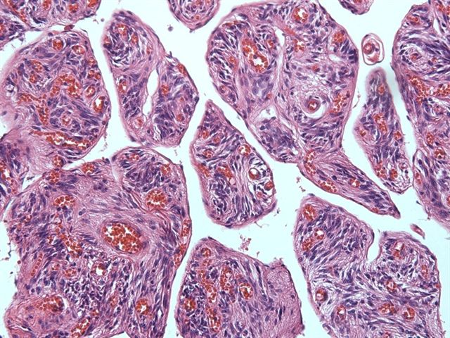
A 55 year old man presented with a 3.5 cm mass in his neck, which was excised.
Microscopic images:


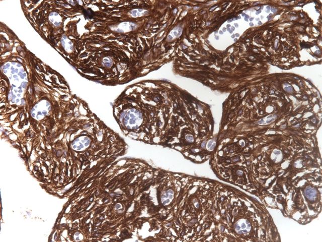
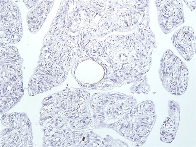
Immunohistochemistry images:
What is your diagnosis?
Diagnosis: Spindle cell lipoma, pseudoangiomatous variant
Discussion:
Spindle cell lipomas are benign tumors usually found in the neck, shoulders or back of middle aged men. They are composed of parallel arrays of CD34+ bland spindle cells with eosinophilic cytoplasm, mixed with myxoid matrix and ropey collagen. The background has adipose tissue and blood vessels. There are no mitotic figures.
The pseudoangiomatous variant is rare and has branching, irregular spaces with villiform connective tissue projections, giving a striking angiomatoid appearance (Histopathology 1994;24:565). The authors in a recent study demonstrated that the lining cells are positive for D2-40, a lymphatic marker, and recommend that this variant be called angiomatous (Pathol Int 2007;57:26).
Wide local excision of spindle cell lipoma is usually curative, with very rare recurrences (Cancer 1975;36:1852).
The differential diagnosis includes:
All cases are archived on our website. To view them sorted by case number, diagnosis or category, visit our main Case of the Month page. To subscribe or unsubscribe to Case of the Month or our other email lists, click here.
Thanks to Dr. Ankur Sangoi, El Camino Hospital, Mountain View, California for contributing this case.
Website news:
(1) We have updated these chapters / topics:
(2) Thanks to the following contributors of images: Dr. Semir Vranic, University of Sarajevo, for contributing images of Paget's disease and Basal-like Breast Cancer for the Breast-malignant chapter; Dr. Asmaa Gaber, Egypt, for contributing images of Spongiotic Dermatitis for the Skin-nontumor chapter.
(3) We had record traffic again in May 2010 of 274,366 total visits or 8,850 visits per day. Thanks for your support and assistance in making our website more useful to pathologists and laboratory personnel.
Visit and follow our Blog to see recent updates to the website.
(1) We have updated these chapters / topics:
- Penis / scrotum by Drs. Alcides Chaux and Antonio L. Cubilla; it now has 90 topics, 311 pages, 684 images and 448 references
- Stains topic - Topoisomerase II alpha (TOP2A), reviewed by Dr. Semir Vranic, University of Sarajevo
(2) Thanks to the following contributors of images: Dr. Semir Vranic, University of Sarajevo, for contributing images of Paget's disease and Basal-like Breast Cancer for the Breast-malignant chapter; Dr. Asmaa Gaber, Egypt, for contributing images of Spongiotic Dermatitis for the Skin-nontumor chapter.
(3) We had record traffic again in May 2010 of 274,366 total visits or 8,850 visits per day. Thanks for your support and assistance in making our website more useful to pathologists and laboratory personnel.
Visit and follow our Blog to see recent updates to the website.
Case #182
Clinical history:
A 55 year old man presented with a 3.5 cm mass in his neck, which was excised.
Microscopic images:
Immunohistochemistry images:
CD34
S100
What is your diagnosis?
Click here for diagnosis and discussion:
Diagnosis: Spindle cell lipoma, pseudoangiomatous variant
Discussion:
Spindle cell lipomas are benign tumors usually found in the neck, shoulders or back of middle aged men. They are composed of parallel arrays of CD34+ bland spindle cells with eosinophilic cytoplasm, mixed with myxoid matrix and ropey collagen. The background has adipose tissue and blood vessels. There are no mitotic figures.
The pseudoangiomatous variant is rare and has branching, irregular spaces with villiform connective tissue projections, giving a striking angiomatoid appearance (Histopathology 1994;24:565). The authors in a recent study demonstrated that the lining cells are positive for D2-40, a lymphatic marker, and recommend that this variant be called angiomatous (Pathol Int 2007;57:26).
Wide local excision of spindle cell lipoma is usually curative, with very rare recurrences (Cancer 1975;36:1852).
The differential diagnosis includes:
- Lipomatous hemangiopericytoma: adipose tissue, CD34+ spindle cells, variable myxoid stroma; vasculature is prominent but has large gaping sinusoidal spaces (staghorn)
- Myxoid lipoma: no prominent spindle cells, no vascular appearance, CD34 negative
