
17 May 2006 - Case #46
All cases are archived on our website. To view them sorted by case number, diagnosis or category, visit our main Case of the Month page. To subscribe or unsubscribe to Case of the Month or our other email lists, click here.
This case was contributed by Dr. David Cohen, Herzliyah Medical Center, Israel and Dr. Victor Lee, KK Women's and Children's Hospital, Singapore for contributing these cases (each independently submitted a similar case 3 weeks apart).
Case #46
Case #1:
Clinical history:
A 55 year old woman had a subscapular soft tissue mass. Grossly, it was 7 x 6 x 3 cm with areas of fat, gray-white fibrous tissue and peripheral skeletal muscle. No gritty, mucoid or hemorrhagic areas were identified.
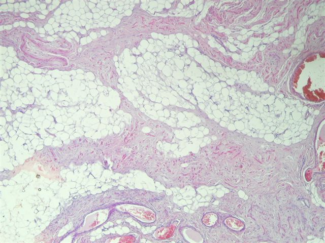
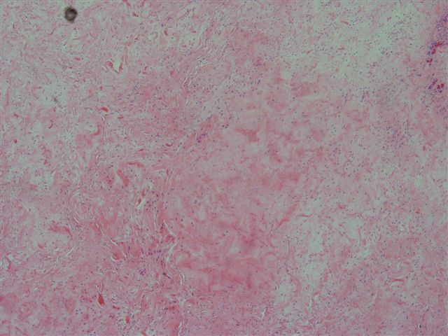
Microscopic images:
Case #2:
Clinical history:
An 85 year old man had a scapular mass, which was excised.
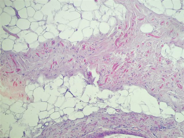
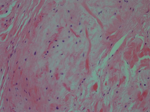
Microscopic images:
What is your diagnosis?
Diagnosis: Elastofibroma
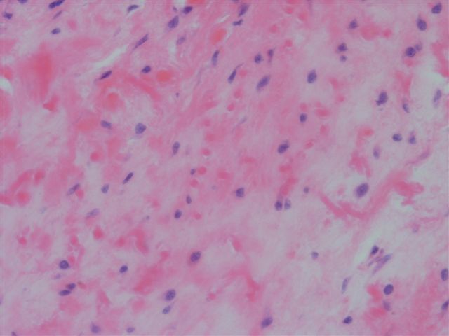
Stain image (case #2):

Discussion:
Elastofibroma typically presents as a poorly circumscribed tumor of the subscapular region in the elderly. At this site, it is also called elastofibroma dorsi. It represents a reactive hyperplasia due to abnormal elastogenesis. It occasionally occurs in other areas, such as the deltoid muscle, hip, thigh and stomach and rarely occurs as multiple subcutaneous nodules (J Am Acad Dermatol 2004;50:126). Imaging and autopsy studies suggest that it may be more common than previously thought and that bilateral or multiple involvement is often subclinical.
Elastofibroma is associated with a history of intense manual labor and may be due to mechanical friction between the scapula and chest wall that causes degeneration of elastic fibers. The elastic fibers also contain type II collagen, normally only found in articular cartilage and ocular structures.
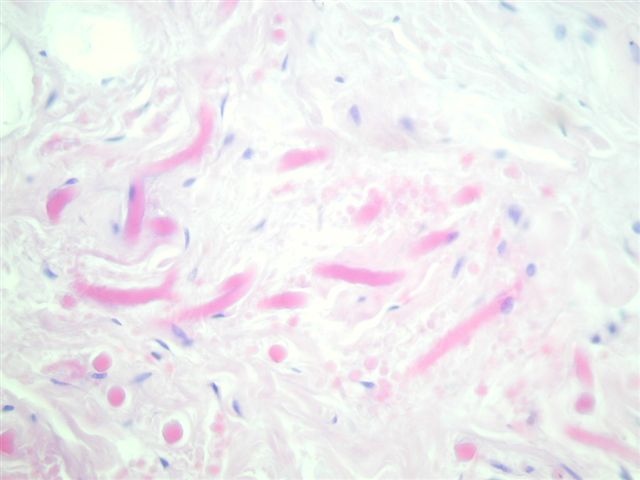
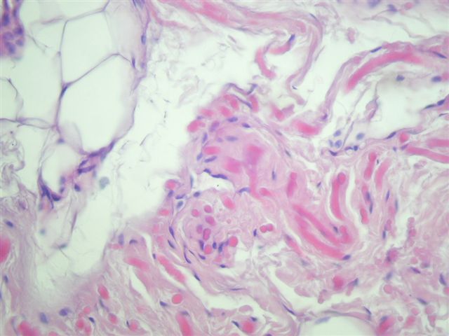
Microscopically, there are bland spindle cells, collagen bundles and acidophilic, refractile cylinders with a dense central core. The cylinders are strongly positive with elastic stains. FNA shows a hypocellular smear with diagnostic aggregates of globules within a collagenous matrix. The altered elastic fibers have a green-yellow autofluorescence with ultraviolet light (Diagn Cytopathol 2002;26:310).
The spindled cells are immunoreactive for vimentin and are frequently immunoreactive for CD34 (Virchows Arch 2006;448:195). They are negative for alpha smooth muscle actin, desmin and S100. Electron microscopy shows cylinders composed of immature amorphous elastic tissue, with a central core of mature elastic fibers.
Excision is curative but only necessary for symptomatic patients. The differential diagnosis is limited. An elastofibrolipoma is similar but well circumscribed with a fibrous capsule and has more conspicuous adipose tissue. Focal areas of an elastofibroma may resemble amyloid.
References: J Electron Microsc (Tokyo) 2006;55:89
All cases are archived on our website. To view them sorted by case number, diagnosis or category, visit our main Case of the Month page. To subscribe or unsubscribe to Case of the Month or our other email lists, click here.
This case was contributed by Dr. David Cohen, Herzliyah Medical Center, Israel and Dr. Victor Lee, KK Women's and Children's Hospital, Singapore for contributing these cases (each independently submitted a similar case 3 weeks apart).
Case #46
Case #1:
Clinical history:
A 55 year old woman had a subscapular soft tissue mass. Grossly, it was 7 x 6 x 3 cm with areas of fat, gray-white fibrous tissue and peripheral skeletal muscle. No gritty, mucoid or hemorrhagic areas were identified.
Microscopic images:
Case #2:
Clinical history:
An 85 year old man had a scapular mass, which was excised.
Microscopic images:
What is your diagnosis?
Click here for diagnosis and discussion:
Diagnosis: Elastofibroma
Stain image (case #2):
Orcein (elastic) stain
Discussion:
Elastofibroma typically presents as a poorly circumscribed tumor of the subscapular region in the elderly. At this site, it is also called elastofibroma dorsi. It represents a reactive hyperplasia due to abnormal elastogenesis. It occasionally occurs in other areas, such as the deltoid muscle, hip, thigh and stomach and rarely occurs as multiple subcutaneous nodules (J Am Acad Dermatol 2004;50:126). Imaging and autopsy studies suggest that it may be more common than previously thought and that bilateral or multiple involvement is often subclinical.
Elastofibroma is associated with a history of intense manual labor and may be due to mechanical friction between the scapula and chest wall that causes degeneration of elastic fibers. The elastic fibers also contain type II collagen, normally only found in articular cartilage and ocular structures.
Microscopically, there are bland spindle cells, collagen bundles and acidophilic, refractile cylinders with a dense central core. The cylinders are strongly positive with elastic stains. FNA shows a hypocellular smear with diagnostic aggregates of globules within a collagenous matrix. The altered elastic fibers have a green-yellow autofluorescence with ultraviolet light (Diagn Cytopathol 2002;26:310).
The spindled cells are immunoreactive for vimentin and are frequently immunoreactive for CD34 (Virchows Arch 2006;448:195). They are negative for alpha smooth muscle actin, desmin and S100. Electron microscopy shows cylinders composed of immature amorphous elastic tissue, with a central core of mature elastic fibers.
Excision is curative but only necessary for symptomatic patients. The differential diagnosis is limited. An elastofibrolipoma is similar but well circumscribed with a fibrous capsule and has more conspicuous adipose tissue. Focal areas of an elastofibroma may resemble amyloid.
References: J Electron Microsc (Tokyo) 2006;55:89
