
16 August 2006 - Case #56
All cases are archived on our website. To view them sorted by case number, diagnosis or category, visit our main Case of the Month page. To subscribe or unsubscribe to Case of the Month or our other email lists, click here.
This case was contributed by Dr. Ankur Sangoi, Stanford University, Stanford, California, USA.
Case #56
Clinical history:
The patient is an 85 year old Mexican woman with a 1 month history of very pruritic and scaly eroded plaques on her trunk with a few on her face. There were no mucosal lesions or intact vesicles.
Microscopic images:

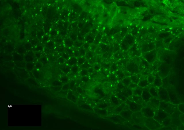
Immunofluorescence images:
What is your diagnosis?
Diagnosis: Pemphigus foliaceus
Discussion:
Pemphigus is a collection of rare, nonhereditary, chronic, autoimmune diseases with flaccid blisters and denuded skin. They typically affect men and women equally, usually ages 30 to 59 years.
Pemphigus foliaceus is a chronic blistering disease that typically spares the mucous membranes but involves healthy appearing skin with blisters that form after rubbing (Nikolsky sign). It typically has a mild course because the blisters are superficial. Often the lesions do not appear bullous because crusts and erosions replace the bullae. Histologically, there is acantholysis of the granular cell layer of the epidermis, with rounded keratinocytes and a few inflammatory cells. Eosinophils may be present.
The blisters in pemphigus foliaceus are due to IgG antibodies against desmoglein 1, a desmosomal glycoprotein expressed within the granular cell layer. IgG immunostains for this antibody show a net-like pattern. C3 deposition has also been described (Br J Dermatol 1997;136:249).
An endemic form, called fogo selvagem (wild fire in Portugese) occurs in Brazil (Br J Dermatol 2006;155:446, Int J Dermatol 2005;44:293).
Treatment is typically corticosteroids or immunosuppressive agents.
The differential diagnosis includes other forms of pemphigus (erythematosus: also superficial cleavage but typically involves the face; vulgaris and vegetans: deep cleavage planes) and other acantholytic diseases, such as Darier disease, Hailey-Hailey disease, viral vesicles, drug related pemphigus-like lesions and transient acantholytic dermatosis. Patients with pemphigus foliaceus may also have other autoimmune disorders or thymoma.
References: Medscape: Pemphigus Foliaceus [Accessed 29 March 2024]
All cases are archived on our website. To view them sorted by case number, diagnosis or category, visit our main Case of the Month page. To subscribe or unsubscribe to Case of the Month or our other email lists, click here.
This case was contributed by Dr. Ankur Sangoi, Stanford University, Stanford, California, USA.
Case #56
Clinical history:
The patient is an 85 year old Mexican woman with a 1 month history of very pruritic and scaly eroded plaques on her trunk with a few on her face. There were no mucosal lesions or intact vesicles.
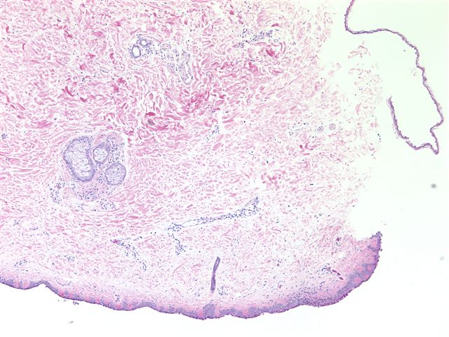
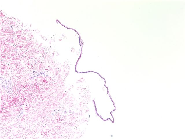
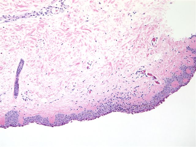
Microscopic images:
Low power
Low power
High power
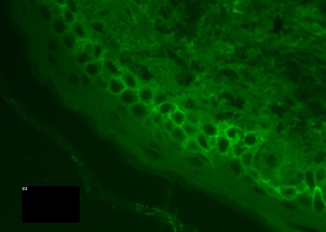
Immunofluorescence images:
C3
IgG
What is your diagnosis?
Click here for diagnosis and discussion:
Diagnosis: Pemphigus foliaceus
Discussion:
Pemphigus is a collection of rare, nonhereditary, chronic, autoimmune diseases with flaccid blisters and denuded skin. They typically affect men and women equally, usually ages 30 to 59 years.
Pemphigus foliaceus is a chronic blistering disease that typically spares the mucous membranes but involves healthy appearing skin with blisters that form after rubbing (Nikolsky sign). It typically has a mild course because the blisters are superficial. Often the lesions do not appear bullous because crusts and erosions replace the bullae. Histologically, there is acantholysis of the granular cell layer of the epidermis, with rounded keratinocytes and a few inflammatory cells. Eosinophils may be present.
The blisters in pemphigus foliaceus are due to IgG antibodies against desmoglein 1, a desmosomal glycoprotein expressed within the granular cell layer. IgG immunostains for this antibody show a net-like pattern. C3 deposition has also been described (Br J Dermatol 1997;136:249).
An endemic form, called fogo selvagem (wild fire in Portugese) occurs in Brazil (Br J Dermatol 2006;155:446, Int J Dermatol 2005;44:293).
Treatment is typically corticosteroids or immunosuppressive agents.
The differential diagnosis includes other forms of pemphigus (erythematosus: also superficial cleavage but typically involves the face; vulgaris and vegetans: deep cleavage planes) and other acantholytic diseases, such as Darier disease, Hailey-Hailey disease, viral vesicles, drug related pemphigus-like lesions and transient acantholytic dermatosis. Patients with pemphigus foliaceus may also have other autoimmune disorders or thymoma.
References: Medscape: Pemphigus Foliaceus [Accessed 29 March 2024]
