
23 July 2014 - Case #320
All cases are archived on our website. To view them sorted by case number, diagnosis or category, visit our main Case of the Month page. To subscribe or unsubscribe to Case of the Month or our other email lists, click here.
Thanks to Dr. Syeddah Mujtaba, Dallah Hospital (Saudi Arabia), for contributing this case.


New books for July 2014
Advertisement
Case #320
Clinical history:
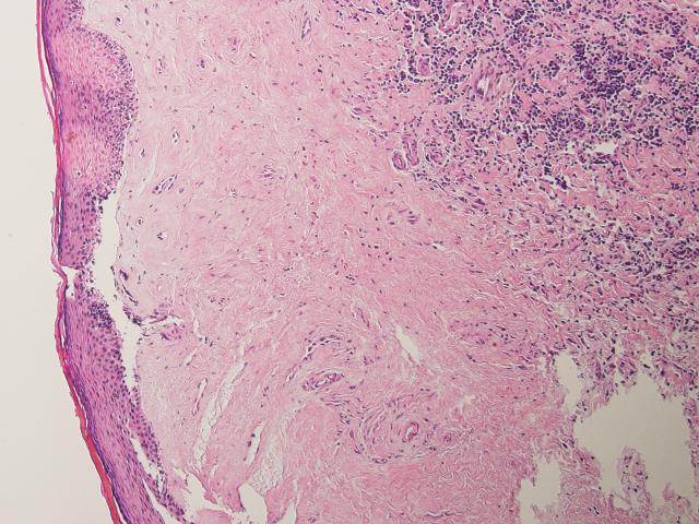
A 30 year old man presented with phimosis. A white plaque lesion was noted on his glans penis, measuring 0.7 x 0.6 cm in size, which was biopsied.
Microscopic images:

What is your diagnosis?
Diagnosis: Balanitis xerotica obliterans
Discussion:
Balanitis xerotica obliterans, also known as lichen sclerosus of genitalia, is the male equivalent of lichen sclerosus et atrophicus of the vulva, a chronic and atrophic mucocutaneous condition. It is the most common cause of pathological phimosis in boys and may also cause narrowing of the urethral meatus and paraphimosis. It also affects middle aged men.
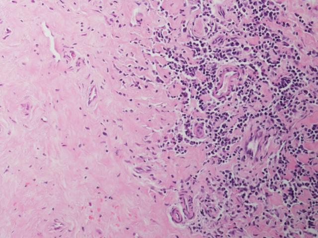
Clinically, it presents as a gray-white, irregular geographic foci of atrophy in the inner foreskin, glans or perimeatal area, with variable erosion, ulceration and raised pearly white areas. Microscopy shows a thinning or thickening of epidermis with orthokeratotic hyperkeratosis. There is also lamina propria thickening and loss of structures due to edema, sclerosis or hyalinization. The basal layer shows vacuolar degeneration, accompanied by diffuse fibrosis and a deep lymphocytic infiltrate. Atypical cases may have epithelial changes of penile intraepithelial neoplasia (PeIN), usually differentiated.
Due to the association with low grade keratinizing squamous cell carcinoma, biopsy is recommended if not resolved after circumcision and by other authors in all cases (Am J Surg Pathol 2003;27:1448, Am J Surg Pathol 2004;28:895, BJU Int 2013;111:970, BJU Int 2006;98:74, J Urol 2007;178:2268).
All cases are archived on our website. To view them sorted by case number, diagnosis or category, visit our main Case of the Month page. To subscribe or unsubscribe to Case of the Month or our other email lists, click here.
Thanks to Dr. Syeddah Mujtaba, Dallah Hospital (Saudi Arabia), for contributing this case.
New books for July 2014
- Bardales: The Invasive Cytopathologist: Ultrasound Guided Fine-Needle Aspiration of Superficial Masses by Ricardo Bardales, provides a comprehensive review of the cytology of superficial neoplastic and non-neoplastic disease processes obtained by percutaneous ultrasound guidance, particularly of thyroid, parathyroid, lymph nodes, salivary glands, breast, and soft tissue with ultrasound image and histopathology correlation.
- Bibbo: Comprehensive Cytopathology, 4th ed. by Can Baykal and Kurtulus Didem Yazganoglu, is a superb atlas that presents an unrivalled wealth of original high-quality clinical photographs of almost all benign and malignant skin tumors.
For more information visit our New Books page!
Website news:
(1) We are currently working on a new website infrastructure. The improvements will include a new topic search feature, more consistent templates for each chapter and topic, a cleaner look to the Home Page and topics and an easier process to update topics with an anticipated faster turn-around-time. We will keep you advised.
(2) Our Feature page for July is Digital / Imaging / Photography, and highlights Leica Biosystems, Milestone Medical, OPTRONICS and Photodyne Technologies. It also contains an original short article, "Digital Pathology in Microscopy: Looking Beyond the Slide", by Jaleh Mansouri, M.D.
(3) Don't forget to check out the Books of the Month on the left side of the Home Page, and the Mystery Case on the right side of the Home Page.
Visit and follow our Blog to see recent updates to the website.
Case #320
Clinical history:
A 30 year old man presented with phimosis. A white plaque lesion was noted on his glans penis, measuring 0.7 x 0.6 cm in size, which was biopsied.
Microscopic images:
What is your diagnosis?
Click here for diagnosis and discussion:
Diagnosis: Balanitis xerotica obliterans
Discussion:
Balanitis xerotica obliterans, also known as lichen sclerosus of genitalia, is the male equivalent of lichen sclerosus et atrophicus of the vulva, a chronic and atrophic mucocutaneous condition. It is the most common cause of pathological phimosis in boys and may also cause narrowing of the urethral meatus and paraphimosis. It also affects middle aged men.
Clinically, it presents as a gray-white, irregular geographic foci of atrophy in the inner foreskin, glans or perimeatal area, with variable erosion, ulceration and raised pearly white areas. Microscopy shows a thinning or thickening of epidermis with orthokeratotic hyperkeratosis. There is also lamina propria thickening and loss of structures due to edema, sclerosis or hyalinization. The basal layer shows vacuolar degeneration, accompanied by diffuse fibrosis and a deep lymphocytic infiltrate. Atypical cases may have epithelial changes of penile intraepithelial neoplasia (PeIN), usually differentiated.
Due to the association with low grade keratinizing squamous cell carcinoma, biopsy is recommended if not resolved after circumcision and by other authors in all cases (Am J Surg Pathol 2003;27:1448, Am J Surg Pathol 2004;28:895, BJU Int 2013;111:970, BJU Int 2006;98:74, J Urol 2007;178:2268).
