
17 September 2014 - Case #326
All cases are archived on our website. To view them sorted by case number, diagnosis or category, visit our main Case of the Month page. To subscribe or unsubscribe to Case of the Month or our other email lists, click here.
Thanks to Dr. Ankur Sangoi, El Camino Hospital, California (USA), for contributing this case. This case was reviewed in May 2020 by Dr. Jennifer Bennett, University of Chicago and Dr. Carlos Parra-Herran, University of Toronto.
Case #326
Clinical history:
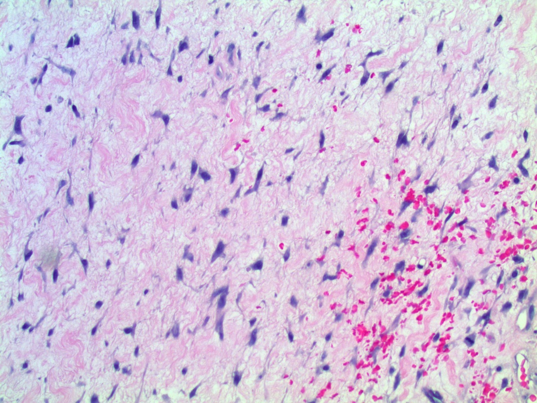
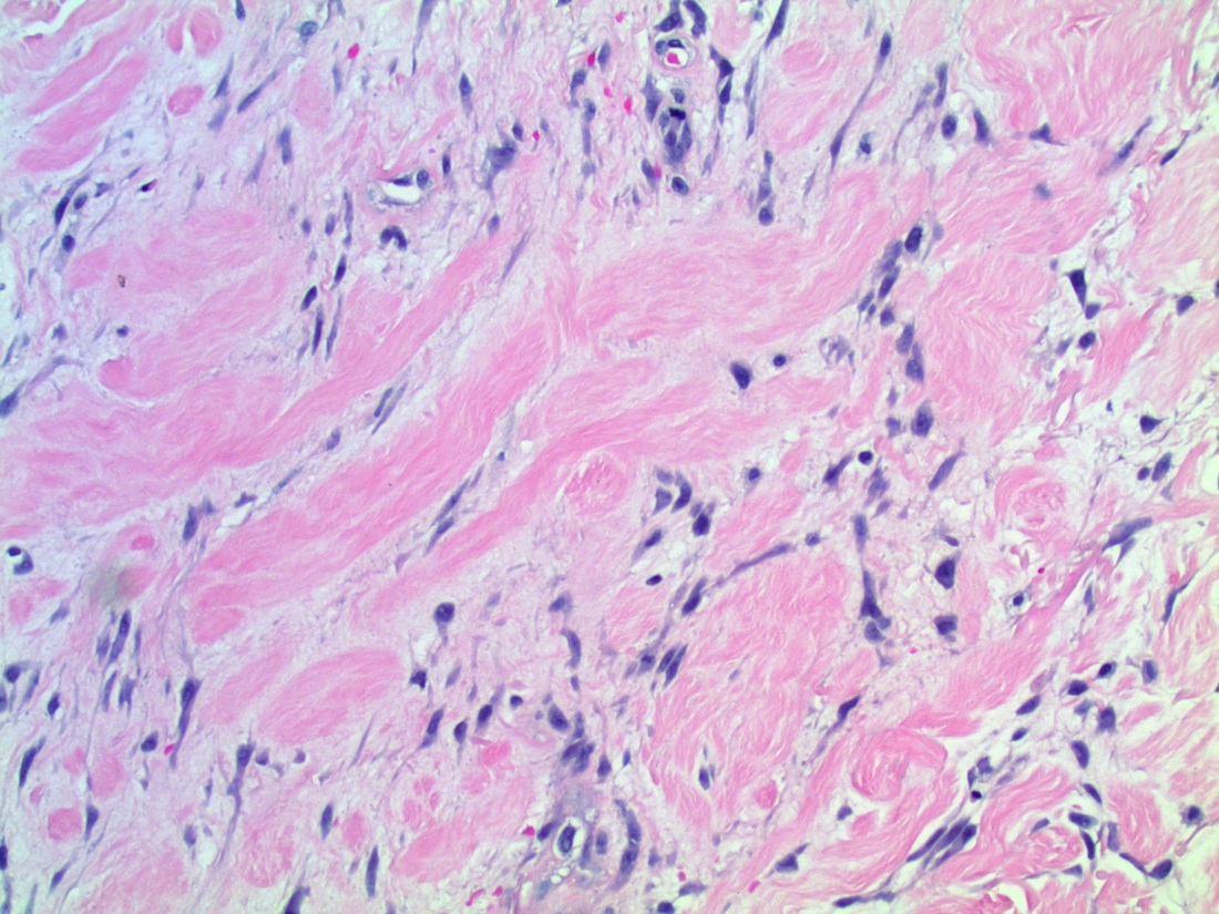
A 34 year old woman had perineal cysts that were biopsied. She was an avid cyclist.
Microscopic images:
What is your diagnosis?
Diagnosis: Perineal nodular induration
Discussion:
Perineal nodules are rare reactive lesions reported in male and female cyclists and equestrians, due to repetitive perineal trauma (Hautarzt 2000;51:763, Int J Gynecol Pathol 2010;29:398). They have also been termed cyclist's nodule, reactive fibroblastic and myofibroblastic proliferation of the vulva and third testicle of the cyclist (Histopathology 2003;42:615). In women, a recent report describes 4 cases of unilateral nodules or swellings of the labium major in competitive cyclists ages 15 - 45 years (Am J Surg Pathol 2011;35:110). Histologically, there was a mixture of adipose tissue, hyalinized tissue with bland spindled fibroblasts, blood vessels and nerve fibers, with variable perivascular lymphocytic infiltrate, thick cords of fibrous tissue, elastic fibers and epithelioid, plasmacytoid or ganglion-like cells.
The spindle cells cells were immunoreactive for estrogen receptor and plump mesenchymal cells were positive for smooth muscle actin, consistent with myofibroblasts. These cells were negative for desmin, S100, CD34 and HMGA2 (Int J Surg Pathol 2014;22:71).
The differential diagnosis includes various other mesenchymal lesions, which have a different clinical setting.
These lesions are benign but may recur. In this case, after cessation of cycling and rest, the nodule went away.
All cases are archived on our website. To view them sorted by case number, diagnosis or category, visit our main Case of the Month page. To subscribe or unsubscribe to Case of the Month or our other email lists, click here.
Thanks to Dr. Ankur Sangoi, El Camino Hospital, California (USA), for contributing this case. This case was reviewed in May 2020 by Dr. Jennifer Bennett, University of Chicago and Dr. Carlos Parra-Herran, University of Toronto.
Website news:
(1) Do you use Amazon.com? Support PathologyOutlines! If you buy a book, or anything else, from Amazon.com by clicking on the banners / links located on our website, Amazon pays us a small percentage from their profits - without raising consumer prices. Or click here. Thanks!
(2) We enjoy seeing and talking with the pathology community at the conferences we attend and CAP '14 is no different. Thanks again to everyone who stopped by. Pictures are available on our Blog and Facebook page.
(3) Our Feature page for September is Antibodies / Biomarkers and highlights our advertisers Advanced Cell Diagnostics, Inc. (ACD) and Leica Biosystems. It also contains an original short article, Antibodies in Pathology: The Role of Multiplex Immunohistochemistry, by Jaleh Mansouri, M.D. This past August was the best August we've ever had in terms of traffic - up 32% from last year!
(4) Did you know that PathologyOutlines.com offers you an opportunity to quickly and easily compare products and services offered by various companies? Check out all of the Surveys we have posted for Computer Systems-AP/LIS, Imaging / Digital / Photography and Reference Laboratories to help find what you're looking for. We plan on adding additional surveys in the next few months.
Visit and follow our Blog to see recent updates to the website.
Case #326
Clinical history:
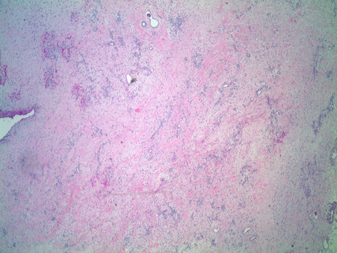
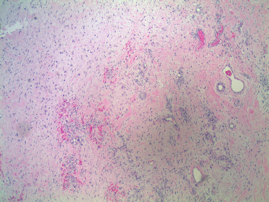
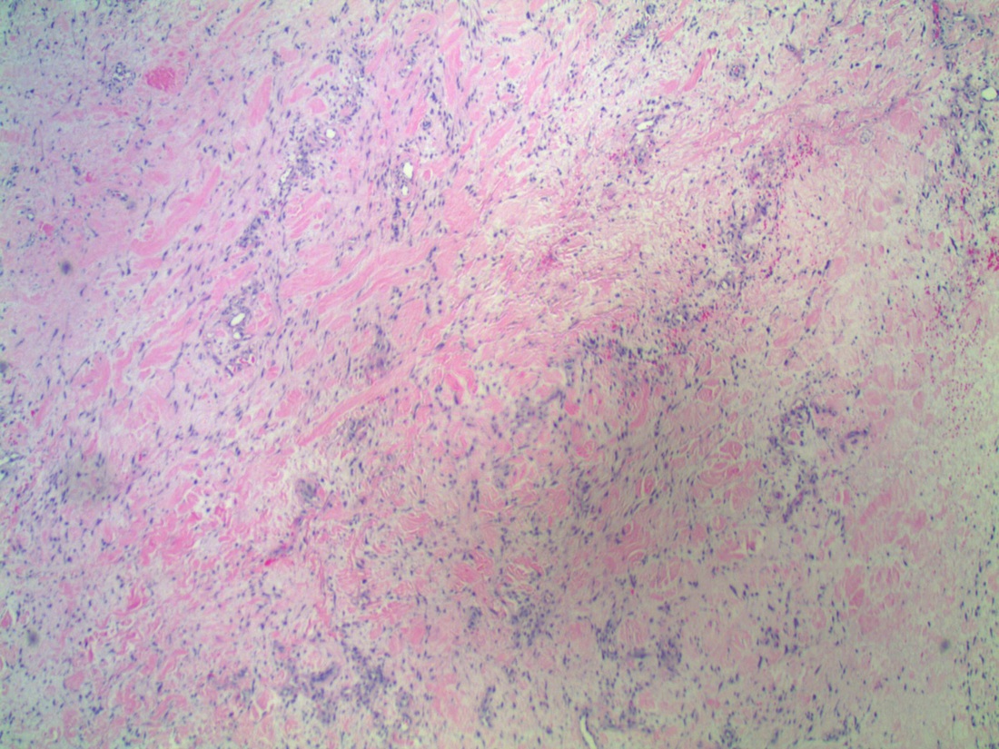
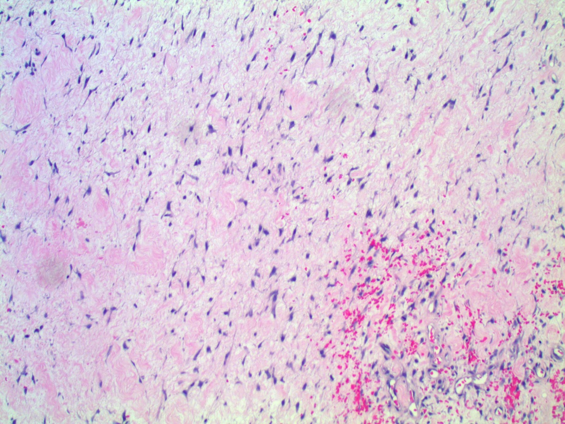
A 34 year old woman had perineal cysts that were biopsied. She was an avid cyclist.
Microscopic images:
H&E images
What is your diagnosis?
Click here for diagnosis and discussion:
Diagnosis: Perineal nodular induration
Discussion:
Perineal nodules are rare reactive lesions reported in male and female cyclists and equestrians, due to repetitive perineal trauma (Hautarzt 2000;51:763, Int J Gynecol Pathol 2010;29:398). They have also been termed cyclist's nodule, reactive fibroblastic and myofibroblastic proliferation of the vulva and third testicle of the cyclist (Histopathology 2003;42:615). In women, a recent report describes 4 cases of unilateral nodules or swellings of the labium major in competitive cyclists ages 15 - 45 years (Am J Surg Pathol 2011;35:110). Histologically, there was a mixture of adipose tissue, hyalinized tissue with bland spindled fibroblasts, blood vessels and nerve fibers, with variable perivascular lymphocytic infiltrate, thick cords of fibrous tissue, elastic fibers and epithelioid, plasmacytoid or ganglion-like cells.
The spindle cells cells were immunoreactive for estrogen receptor and plump mesenchymal cells were positive for smooth muscle actin, consistent with myofibroblasts. These cells were negative for desmin, S100, CD34 and HMGA2 (Int J Surg Pathol 2014;22:71).
The differential diagnosis includes various other mesenchymal lesions, which have a different clinical setting.
These lesions are benign but may recur. In this case, after cessation of cycling and rest, the nodule went away.
