
5 February 2015 - Case #341
All cases are archived on our website. To view them sorted by case number, diagnosis or category, visit our main Case of the Month page. To subscribe or unsubscribe to Case of the Month or our other email lists, click here.
Thanks to Dr. Reema Jaffar, AmeriPath, Indiana (USA), for contributing this case and the discussion.

Advertisement
Case #341
Clinical history:
A 60 year old woman was found to have multiple, slow growing liver lesions and underwent a liver biopsy.
Microscopic images:

What is your diagnosis?
Diagnosis: Epithelioid hemangioendothelioma
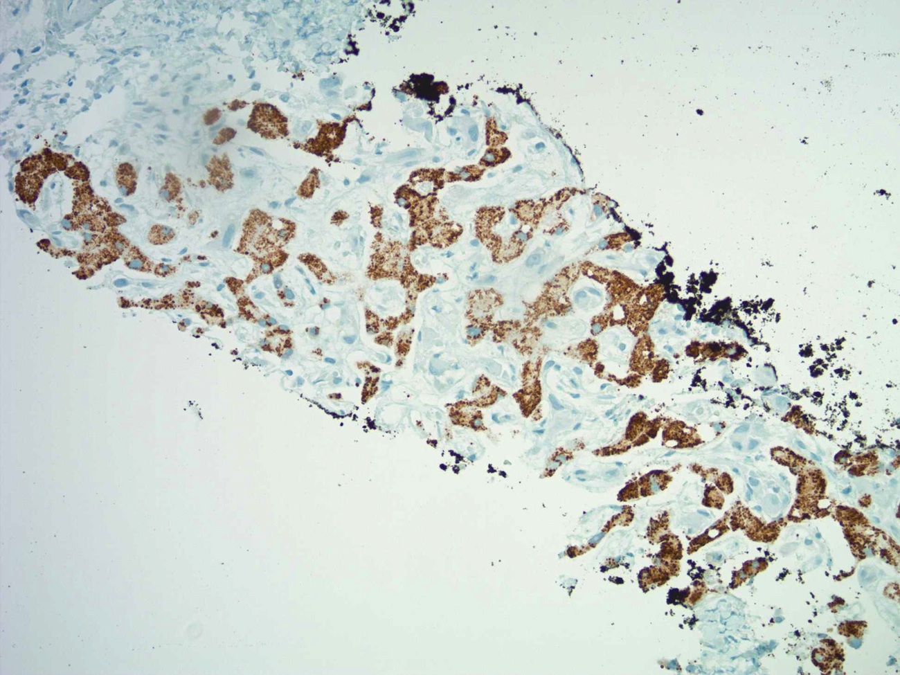
Immunostain images:

Discussion:
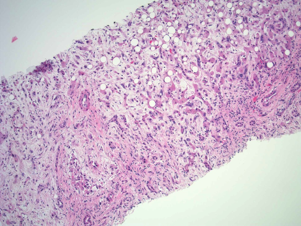
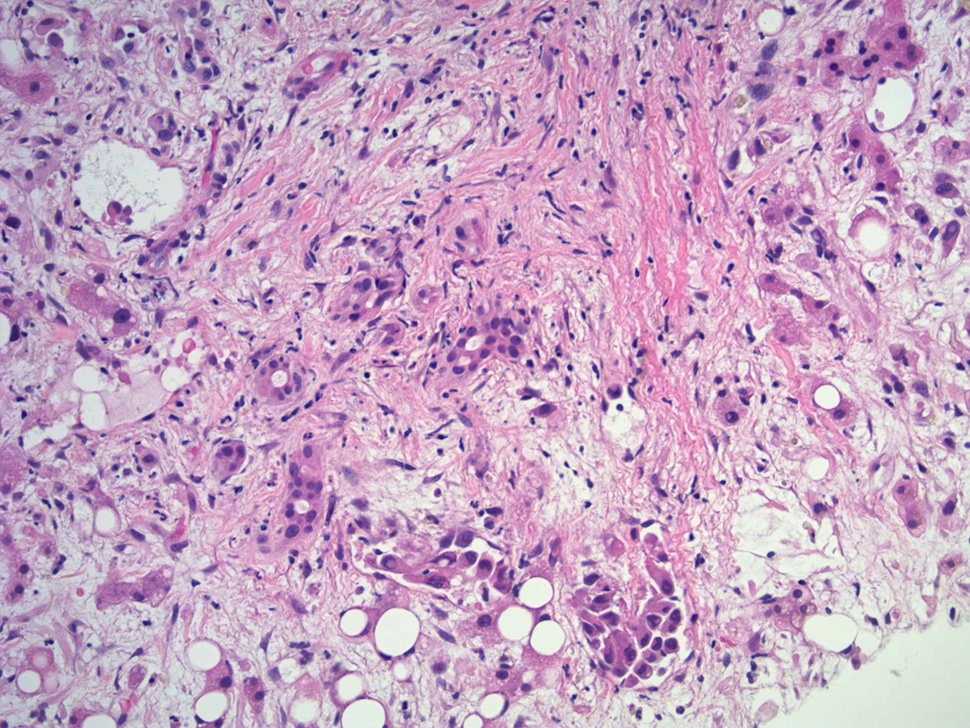
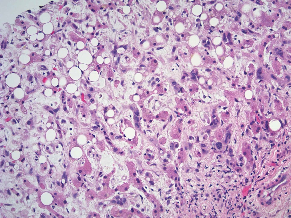
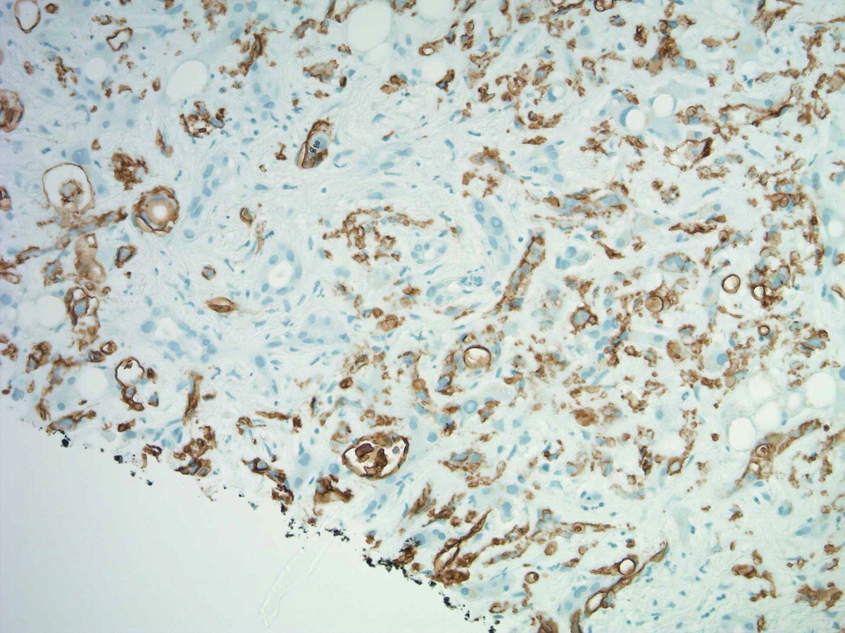
The H&E sections show an intrasinusoidal and intravascular infiltrative growth pattern (figure 1) with intraluminal tufting (figure 2) of epithelioid and spindle cells with surrounding compressed hepatocytes with steatosis (figure 3). Figure 4 shows epithelioid cells with intracytoplasmic vacuoles ("signet ring cells"). The immunostains show that the epithelioid cells are strongly positive for CD34 and CD31, and although the hepatocytes are immunoreactive for HepPar1, the intrasinusoidal proliferation is HepPar1 negative.
Epithelioid hemangioendothelioma is a low grade, slow growing, malignant endothelial neoplasm which typically occurs in middle aged patients and is more common in women (60%). Most cases are due to t(1;3)(p36.3;q25), which produces the fusion gene WWTR1::CAMTA1 (Am J Surg Pathol 2001;25:684, Genes Chromosomes Cancer 2011;50:644). These tumors rarely produce the YAP1::TFE3 fusion gene (Genes Chromosomes Cancer 2013;52:775).
Grossly, epithelioid hemangioendothelioma is often multifocal, grey-white and firm, with variable calcification. Histologically, it has a characteristic zonal pattern with peripheral tumor cells growing along preexisting sinusoids and terminal hepatic venules, with sinusoidal proliferation and intraluminal polypoid or tuft-like projections. Some epithelioid cells show rudimentary intracytoplasmic lumina. Cytologically, the atypical cells have moderate eosinophilic cytoplasm, relatively large hyperchromatic and irregular nuclei and small inconspicuous nucleoli. The surrounding hepatocytes show compression and focal steatosis. The midzone shows sinusoidal obliteration with marked atrophic hepatocyte plates in a fibrous and myxochondroid stroma, with variable calcification (Clin Mol Hepatol 2013;19:315). Tumor cells are immunoreactive for vascular markers CD31, CD34 and D2-40. They are also positive for CD10.
The differential diagnosis includes intrahepatic cholangiocarcinoma, angiosarcoma (rapid growth, highly cellular with ectatic neoplastic vessels exhibiting congestion and hemorrhage, usually marked atypia, extramedullary hematopoiesis, usually no sclerosis), sclerotic hepatocellular carcinoma and sclerosed hemangioma (well circumscribed, no venous invasion, no atypia).
Epithelioid hemangioendothelioma typically has indolent behavior. Treatment options are surgical resection with curative intent or liver transplantation if multiple tumors are present (World J Gastroenterol 2014;20:7049, HPB (Oxford) 2010;12:546).
All cases are archived on our website. To view them sorted by case number, diagnosis or category, visit our main Case of the Month page. To subscribe or unsubscribe to Case of the Month or our other email lists, click here.
Thanks to Dr. Reema Jaffar, AmeriPath, Indiana (USA), for contributing this case and the discussion.
Advertisement
Website news:
(1) We have added information about Copyright and PathologyOutlines.com to the bottom of the Home Page with a link, or you can click here.
(2) We have now posted the Jobs report for the 4th quarter of 2014. There were 121 job postings for full time or part time pathologists at PathologyOutlines.com, which is a 68.1% increase from the 72 postings in the fourth quarter of 2013. For all of 2014, there were 351 postings, a 31.5% increase from the 267 postings for 2013 (a full report for 2014 will be posted separately). Click here for the full report.
(3) Are you looking for information on CME, Apps or Board Review Questions? Click here for this page.
(4) On 22 January 2015, we had record traffic of 37,254 visits and 135,670 page views. We will have to see if this is an "outlier", but in any event, thanks for your support.
Visit and follow our Blog to see recent updates to the website.
Case #341
Clinical history:
A 60 year old woman was found to have multiple, slow growing liver lesions and underwent a liver biopsy.
Microscopic images:
H&E
What is your diagnosis?
Click here for diagnosis and discussion:
Diagnosis: Epithelioid hemangioendothelioma
Immunostain images:
Left to right: CD34, CD31, HepPar1
Discussion:
The H&E sections show an intrasinusoidal and intravascular infiltrative growth pattern (figure 1) with intraluminal tufting (figure 2) of epithelioid and spindle cells with surrounding compressed hepatocytes with steatosis (figure 3). Figure 4 shows epithelioid cells with intracytoplasmic vacuoles ("signet ring cells"). The immunostains show that the epithelioid cells are strongly positive for CD34 and CD31, and although the hepatocytes are immunoreactive for HepPar1, the intrasinusoidal proliferation is HepPar1 negative.
Epithelioid hemangioendothelioma is a low grade, slow growing, malignant endothelial neoplasm which typically occurs in middle aged patients and is more common in women (60%). Most cases are due to t(1;3)(p36.3;q25), which produces the fusion gene WWTR1::CAMTA1 (Am J Surg Pathol 2001;25:684, Genes Chromosomes Cancer 2011;50:644). These tumors rarely produce the YAP1::TFE3 fusion gene (Genes Chromosomes Cancer 2013;52:775).
Grossly, epithelioid hemangioendothelioma is often multifocal, grey-white and firm, with variable calcification. Histologically, it has a characteristic zonal pattern with peripheral tumor cells growing along preexisting sinusoids and terminal hepatic venules, with sinusoidal proliferation and intraluminal polypoid or tuft-like projections. Some epithelioid cells show rudimentary intracytoplasmic lumina. Cytologically, the atypical cells have moderate eosinophilic cytoplasm, relatively large hyperchromatic and irregular nuclei and small inconspicuous nucleoli. The surrounding hepatocytes show compression and focal steatosis. The midzone shows sinusoidal obliteration with marked atrophic hepatocyte plates in a fibrous and myxochondroid stroma, with variable calcification (Clin Mol Hepatol 2013;19:315). Tumor cells are immunoreactive for vascular markers CD31, CD34 and D2-40. They are also positive for CD10.
The differential diagnosis includes intrahepatic cholangiocarcinoma, angiosarcoma (rapid growth, highly cellular with ectatic neoplastic vessels exhibiting congestion and hemorrhage, usually marked atypia, extramedullary hematopoiesis, usually no sclerosis), sclerotic hepatocellular carcinoma and sclerosed hemangioma (well circumscribed, no venous invasion, no atypia).
Epithelioid hemangioendothelioma typically has indolent behavior. Treatment options are surgical resection with curative intent or liver transplantation if multiple tumors are present (World J Gastroenterol 2014;20:7049, HPB (Oxford) 2010;12:546).
