
16 January 2018 - Case #473 (revised 15 June 2020)
All cases are archived on our website. To view them sorted by case number, diagnosis or category, visit our main Case of the Week page. To subscribe or unsubscribe to Case of the Week or our other email lists, click here.
Thanks to Dr. Cheo Fan Foon, KK Women & Children Hospital (Singapore) for contributing this case and Dr. Nat Pernick, PathologyOutlines.com, Inc. for writing the discussion. This case was reviewed in May 2020 by Dr. Jennifer Bennett, University of Chicago and Dr. Carlos Parra-Herran, University of Toronto.

Advertisement
Case of the Week #473
Clinical history:
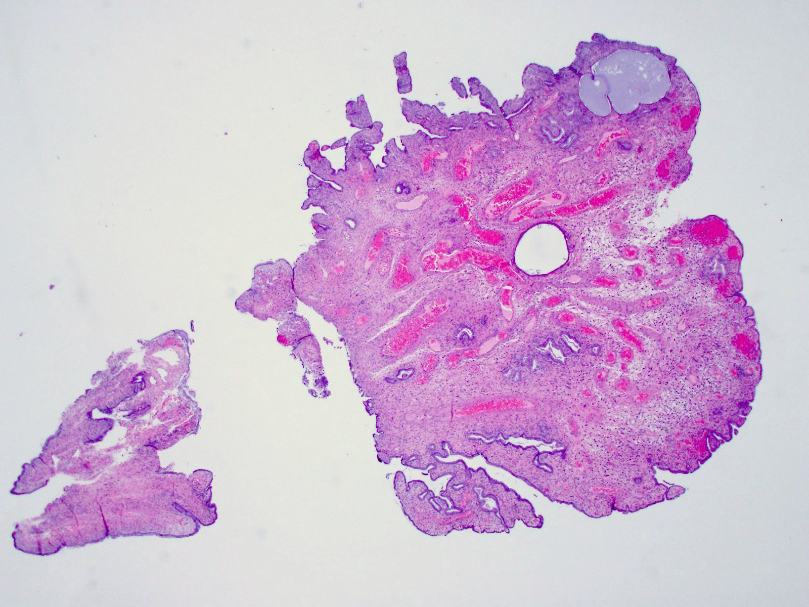
A 58 year old woman presented with postmenopausal bleeding. On examination an endocervical polyp was noted and excised.
Histopathology images:
What is your diagnosis?
Diagnosis:
Endocervical polyp with bizarre stromal cells
Test question (answer at the end): Which statement is true about cervical polyps with bizarre stromal cells?
A. The stromal cells are cytokeratin positive
B. The stromal cells are mitotically active
C. The stromal cells are a negative prognostic indicator
D. They are indolent, unless separate foci or dysplasia or carcinoma are found
Special stains:
Discussion:
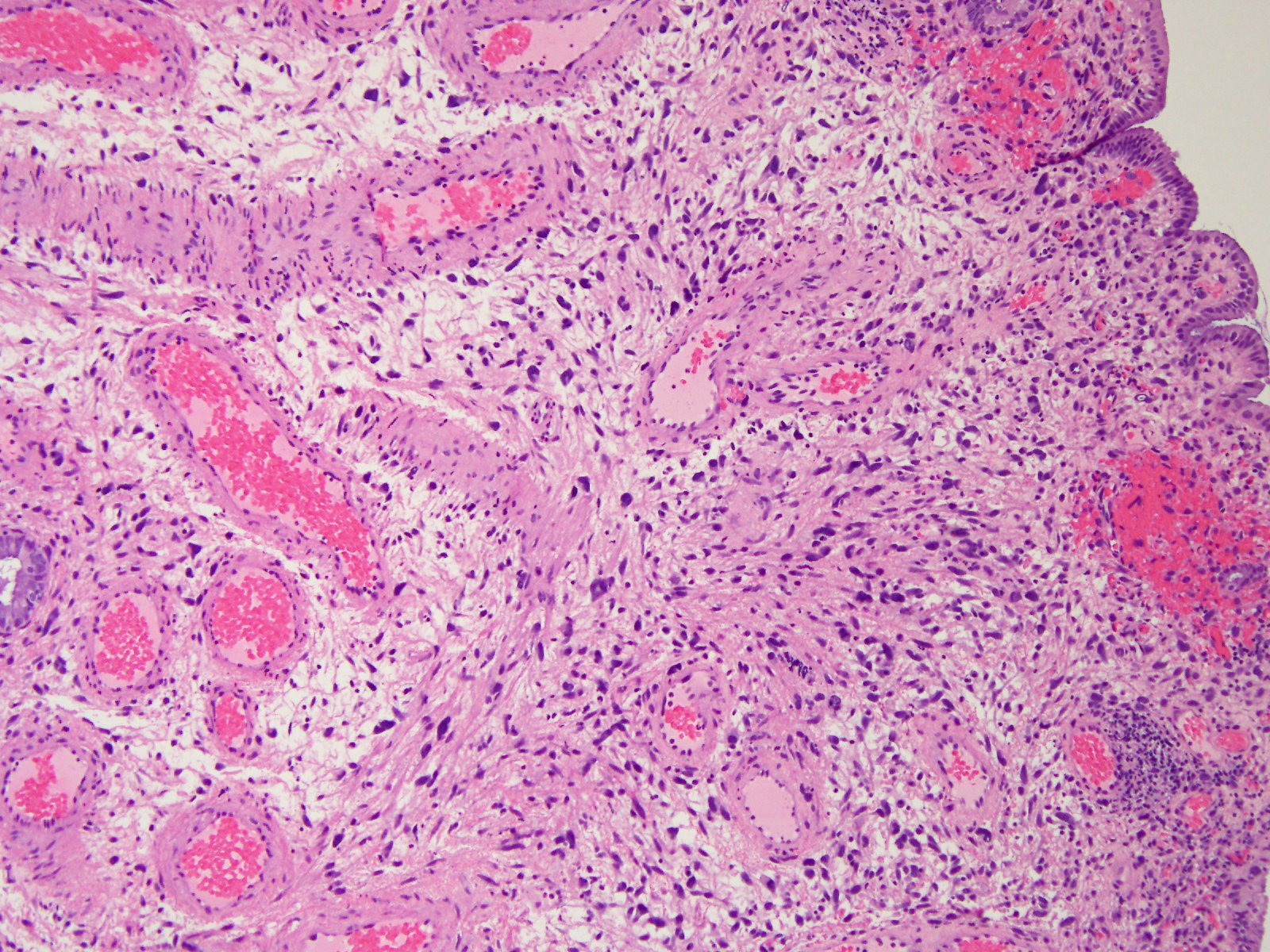
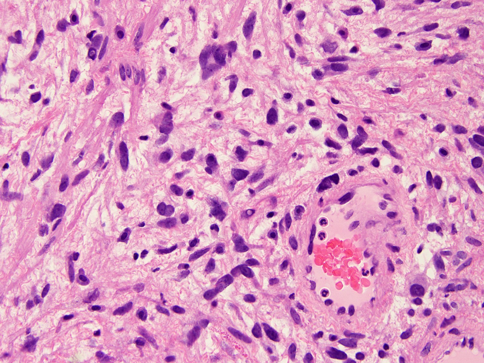
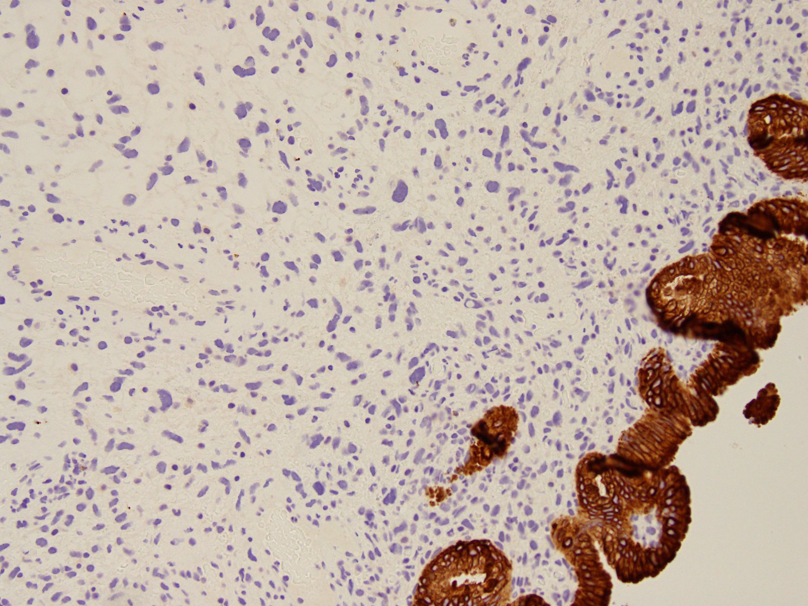
Histologic examination shows polypoid cervical tissue lined by benign endocervical glands. One fragment shows enlarged, pleomorphic stellate and spindle cells in the stroma with smudgy hyperchromatic nuclei. There is no necrosis or mitotic activity. The enlarged stromal cells are negative for AE1 / AE3 and Ki67. Although not shown above, they were also negative for S100 and myogenin.
Endocervical polyps are seen in 2-5% of adult women, usually ages 30-59 years, after examination for bleeding or a mucoid discharge. They are typically indolent but rarely contain foci of dysplasia or carcinoma (J Womens Health (Larchmt) 2007;16:1317). Although the presence of bizarre or atypical stromal cells in endometrial polyps and non-polypoid endometrium in the uterus has been extensively documented, there are few cases reports in the cervix (Am J Clin Pathol 1986;85:633).
The differential diagnosis includes endocervical atypical polypoid adenoma and adenosarcoma. Both are biphasic tumors. Atypical polypoid adenoma has hyperplastic and atypical endometrial glands separated by fascicles of bland smooth muscle and fibrous stroma, with squamous metaplasia almost always present; it is often extensive or has central necrosis (J Minim Invasive Gynecol 2016;23:130). Adenosarcoma typically has phyllodes-like architecture, periglandular stromal condensation and rigid cystic dilation, and an obvious malignant stromal component with increased mitotic activity (Case Rep Obstet Gynecol 2012;2012:358302).
Test question answer:
D. These polyps are indolent, unless separate foci or dysplasia or carcinoma are found. The stromal cells are cytokeratin negative, not mitotically active and have no prognostic significance.
All cases are archived on our website. To view them sorted by case number, diagnosis or category, visit our main Case of the Week page. To subscribe or unsubscribe to Case of the Week or our other email lists, click here.
Thanks to Dr. Cheo Fan Foon, KK Women & Children Hospital (Singapore) for contributing this case and Dr. Nat Pernick, PathologyOutlines.com, Inc. for writing the discussion. This case was reviewed in May 2020 by Dr. Jennifer Bennett, University of Chicago and Dr. Carlos Parra-Herran, University of Toronto.
Advertisement
Case of the Week #473
Clinical history:
A 58 year old woman presented with postmenopausal bleeding. On examination an endocervical polyp was noted and excised.
Histopathology images:
What is your diagnosis?
Diagnosis:
Endocervical polyp with bizarre stromal cells
Test question (answer at the end): Which statement is true about cervical polyps with bizarre stromal cells?
A. The stromal cells are cytokeratin positive
B. The stromal cells are mitotically active
C. The stromal cells are a negative prognostic indicator
D. They are indolent, unless separate foci or dysplasia or carcinoma are found
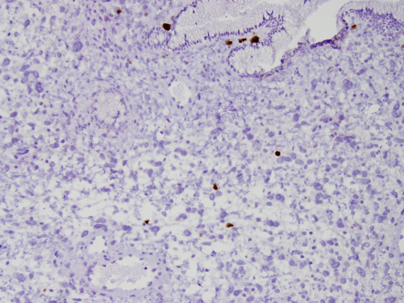
Special stains:
AE1 / AE3
Ki67
Discussion:
Histologic examination shows polypoid cervical tissue lined by benign endocervical glands. One fragment shows enlarged, pleomorphic stellate and spindle cells in the stroma with smudgy hyperchromatic nuclei. There is no necrosis or mitotic activity. The enlarged stromal cells are negative for AE1 / AE3 and Ki67. Although not shown above, they were also negative for S100 and myogenin.
Endocervical polyps are seen in 2-5% of adult women, usually ages 30-59 years, after examination for bleeding or a mucoid discharge. They are typically indolent but rarely contain foci of dysplasia or carcinoma (J Womens Health (Larchmt) 2007;16:1317). Although the presence of bizarre or atypical stromal cells in endometrial polyps and non-polypoid endometrium in the uterus has been extensively documented, there are few cases reports in the cervix (Am J Clin Pathol 1986;85:633).
The differential diagnosis includes endocervical atypical polypoid adenoma and adenosarcoma. Both are biphasic tumors. Atypical polypoid adenoma has hyperplastic and atypical endometrial glands separated by fascicles of bland smooth muscle and fibrous stroma, with squamous metaplasia almost always present; it is often extensive or has central necrosis (J Minim Invasive Gynecol 2016;23:130). Adenosarcoma typically has phyllodes-like architecture, periglandular stromal condensation and rigid cystic dilation, and an obvious malignant stromal component with increased mitotic activity (Case Rep Obstet Gynecol 2012;2012:358302).
Test question answer:
D. These polyps are indolent, unless separate foci or dysplasia or carcinoma are found. The stromal cells are cytokeratin negative, not mitotically active and have no prognostic significance.
