
22 June 2022 - Case of the Month #516
All cases are archived on our website. To view them sorted by case number, diagnosis or category, visit our main Case of the Month page. To subscribe or unsubscribe to Case of the Month or our other email lists, click here.
Thanks to Drs. Hyunkyu Shin and Christian M. Schürch, University Hospital and Comprehensive Cancer Center, Tübingen, Germany, for contributing this case and discussion and to Dr. Borislav Alexiev, Northwestern University Feinberg School of Medicine, Chicago, Illinois, USA for reviewing the discussion.

Advertisement
Case of the Month #516
Clinical history:
A 27 year old man presented with multiple peritoneal masses, up to 3.5 cm in size, with extension into the retroperitoneum and abdominal lymphadenopathy with necrosis.
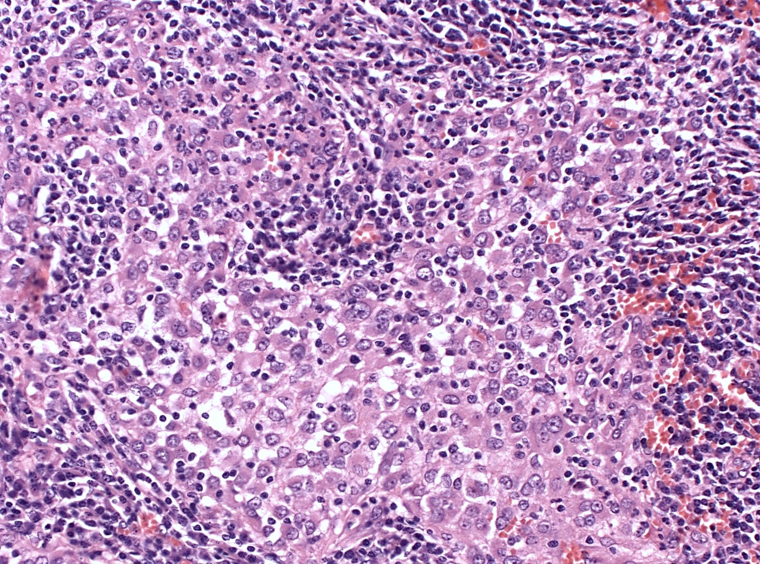
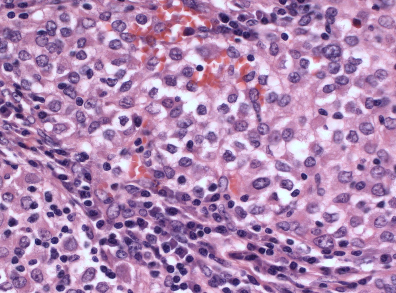
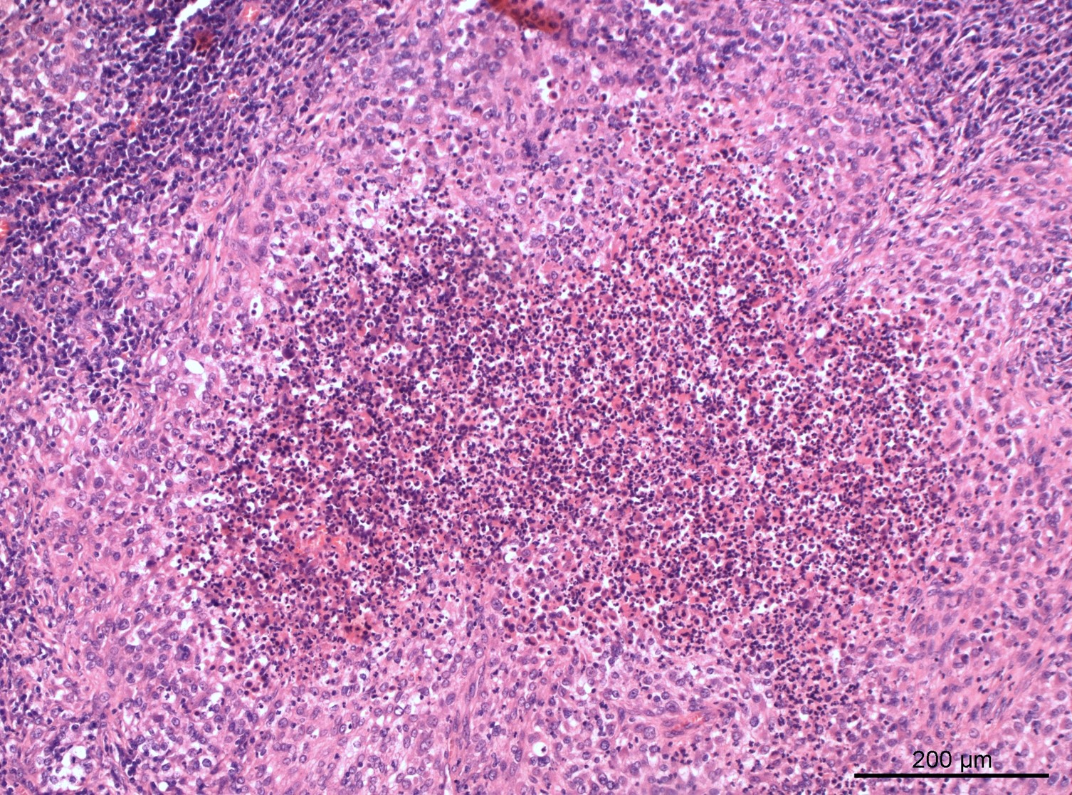
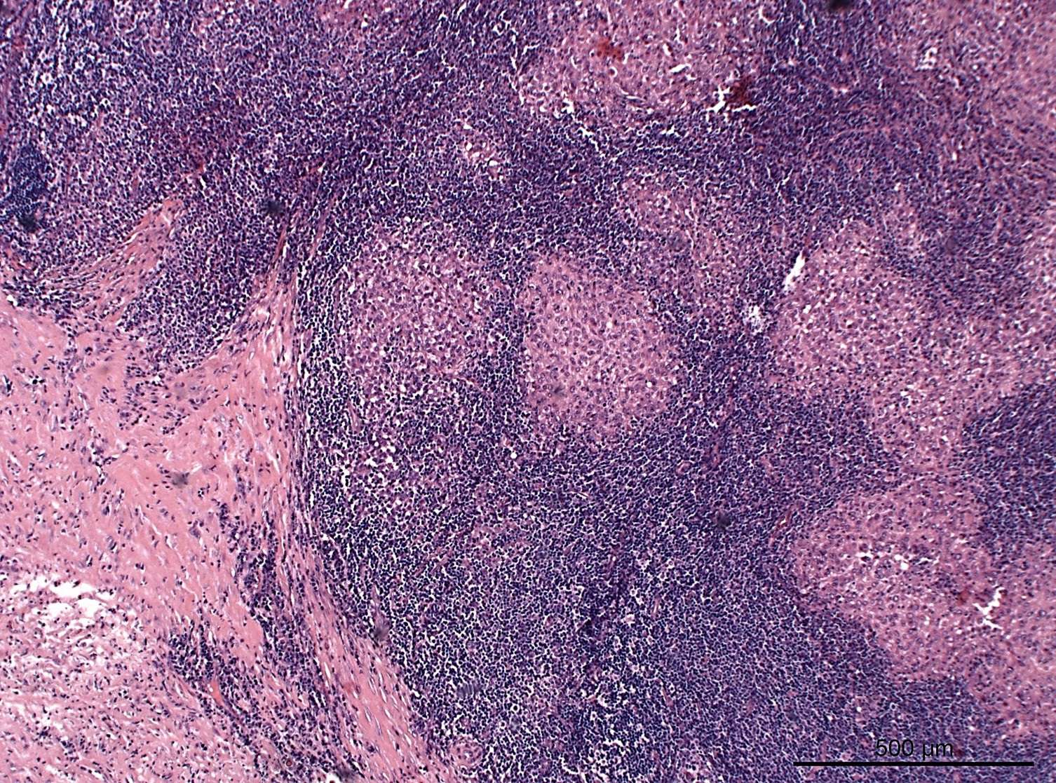
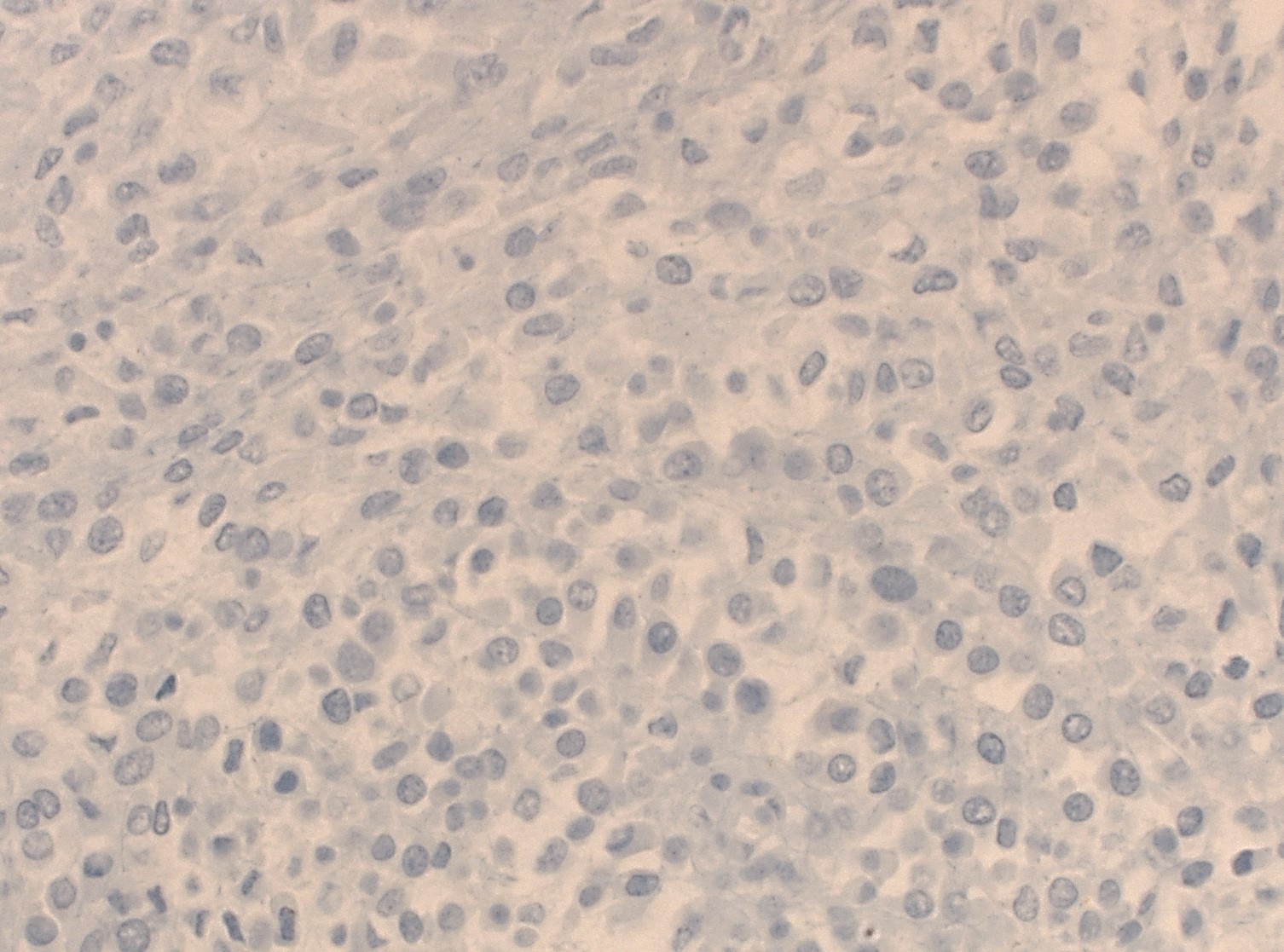
Histopathology images:


What is your diagnosis?
Diagnosis: Malignant epithelioid neoplasm with EWSR1/FUS::CREB fusion
Test question (answer at the end):
Which is a true feature of malignant epithelioid neoplasm with EWSR1/FUS::CREB fusion?
A. A confirmed gene fusion EWSR1/FUS::CREB is considered pathognomonic for this entity
B. Predilection sites are visceral organs
C. This tumor generally shows excellent prognosis without metastasis
D. This tumor is positive for at least one of the epithelial markers, such as AE1/3
E. This tumor should not exhibit morphologic features of a mesenchymal neoplasm
Stains
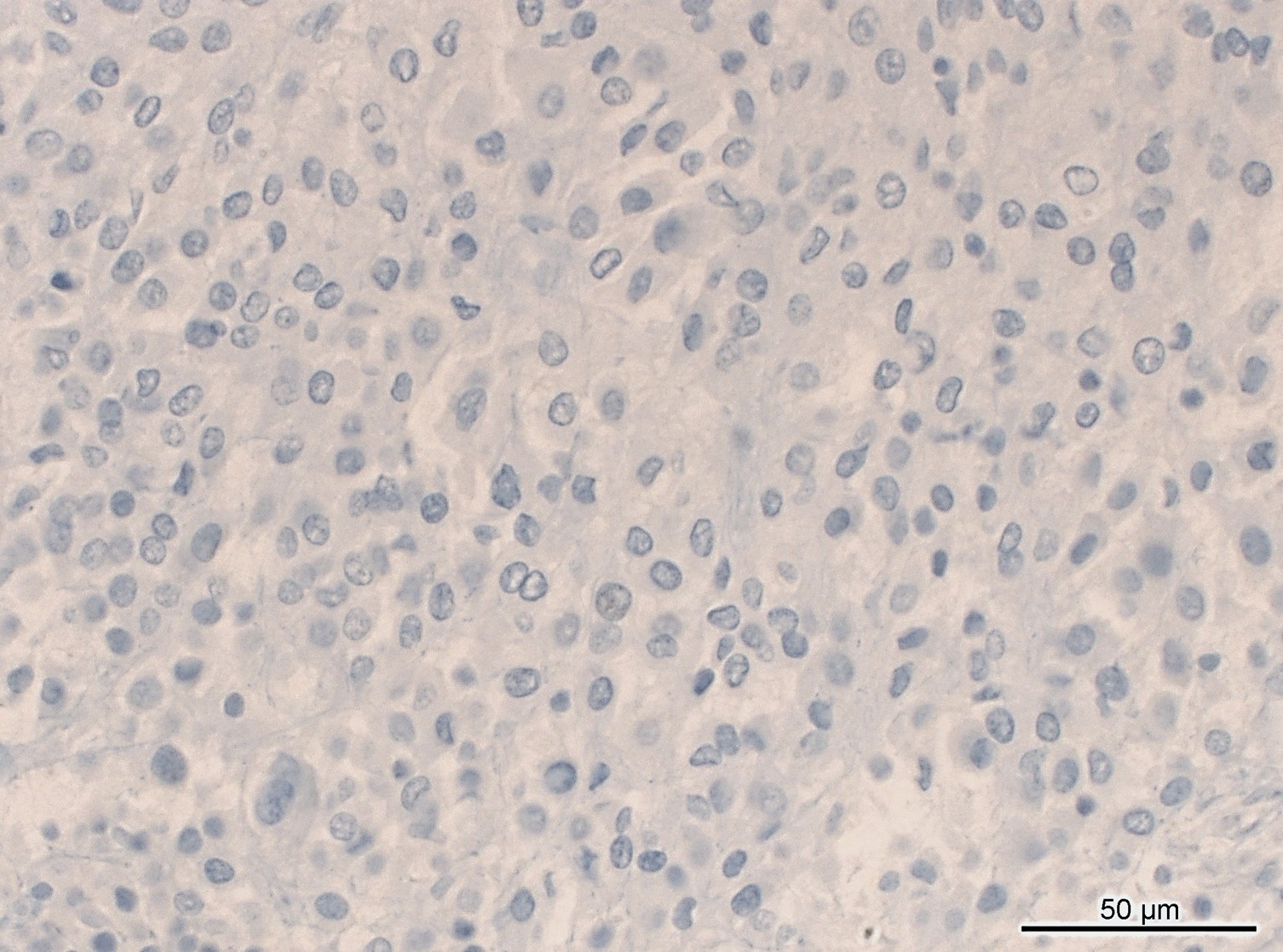
Discussion:
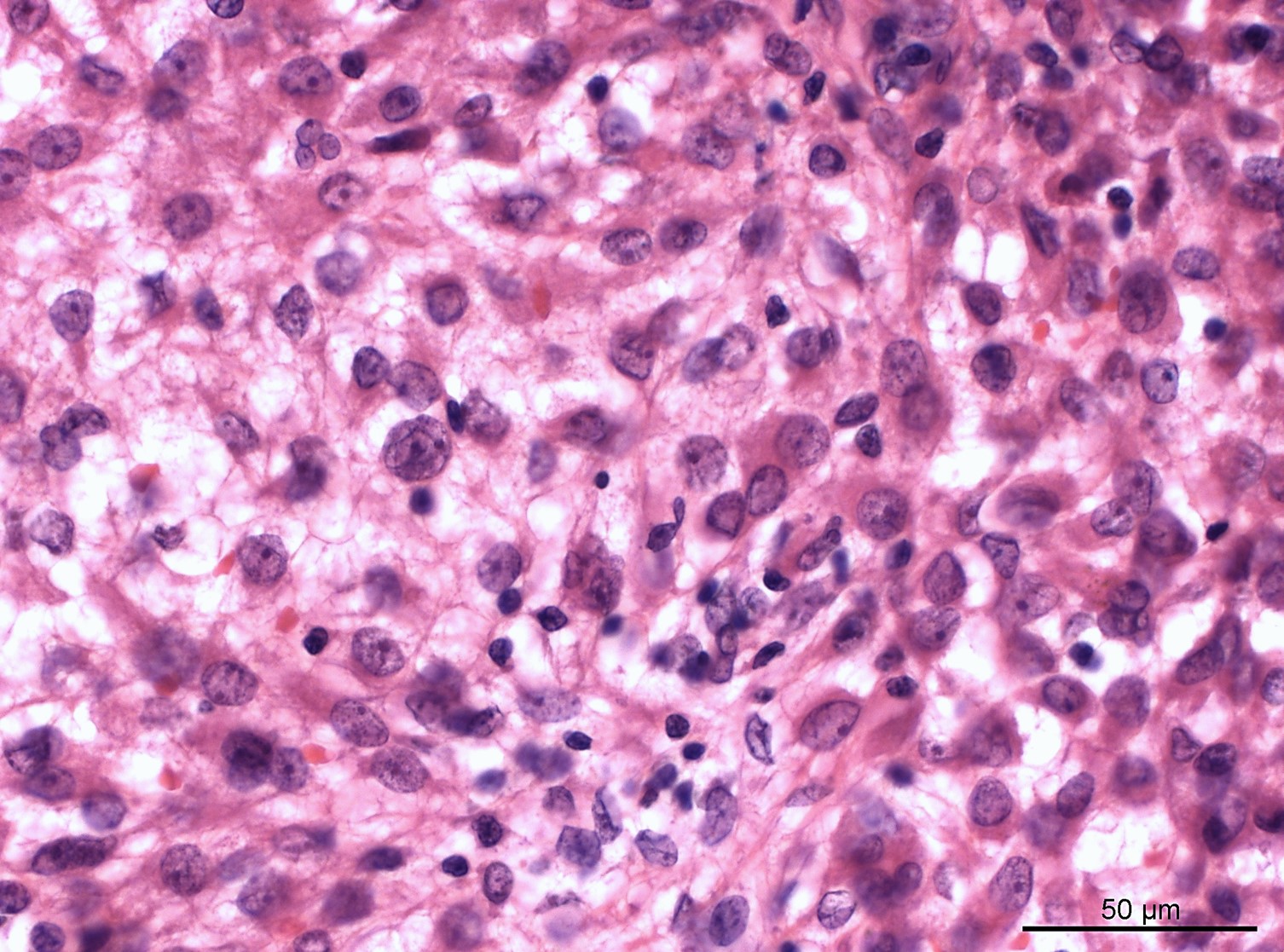
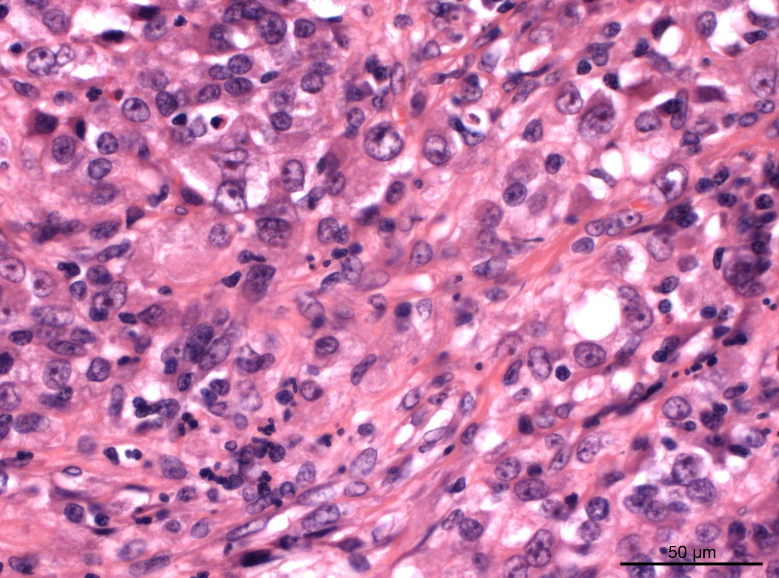
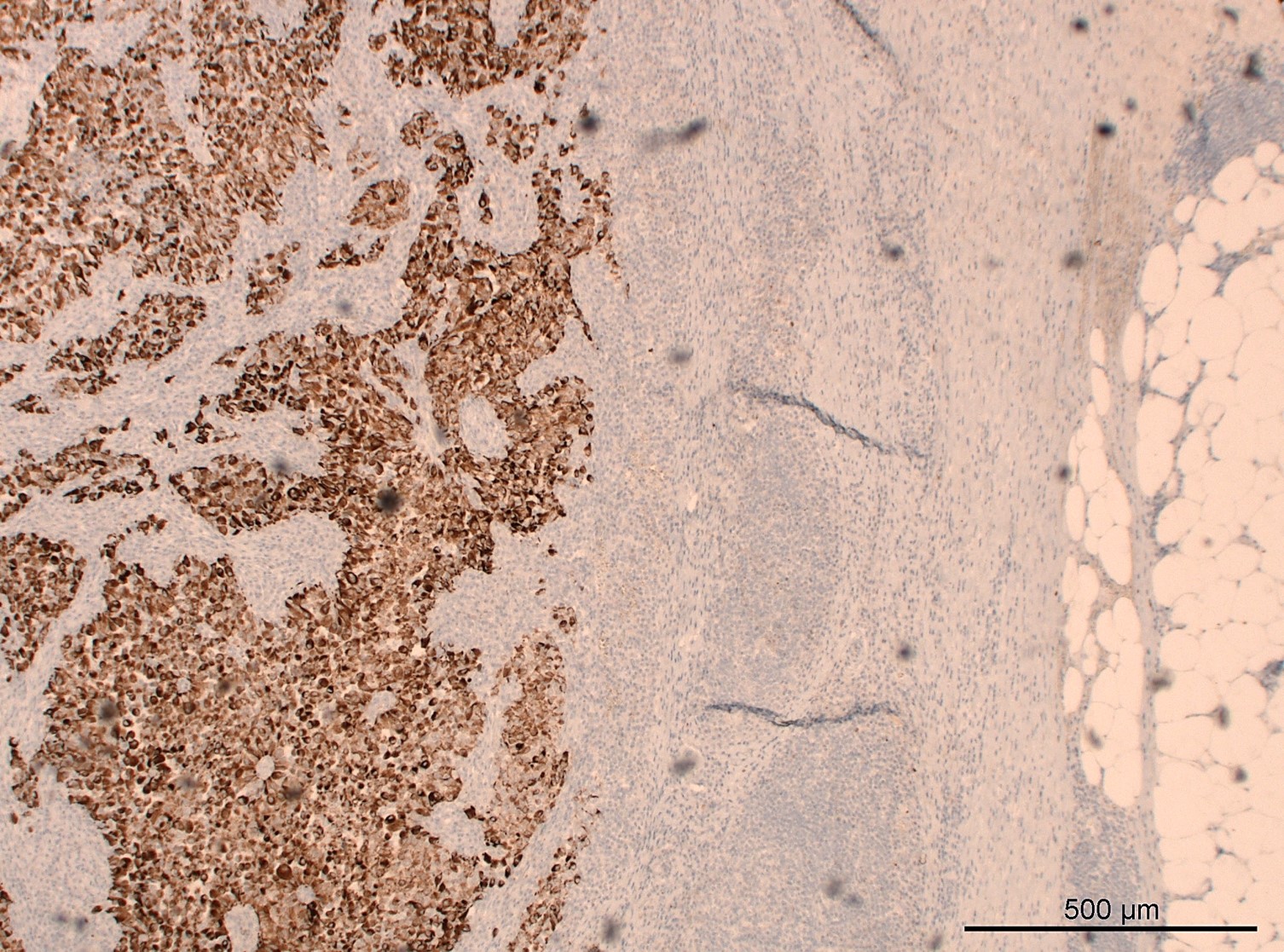
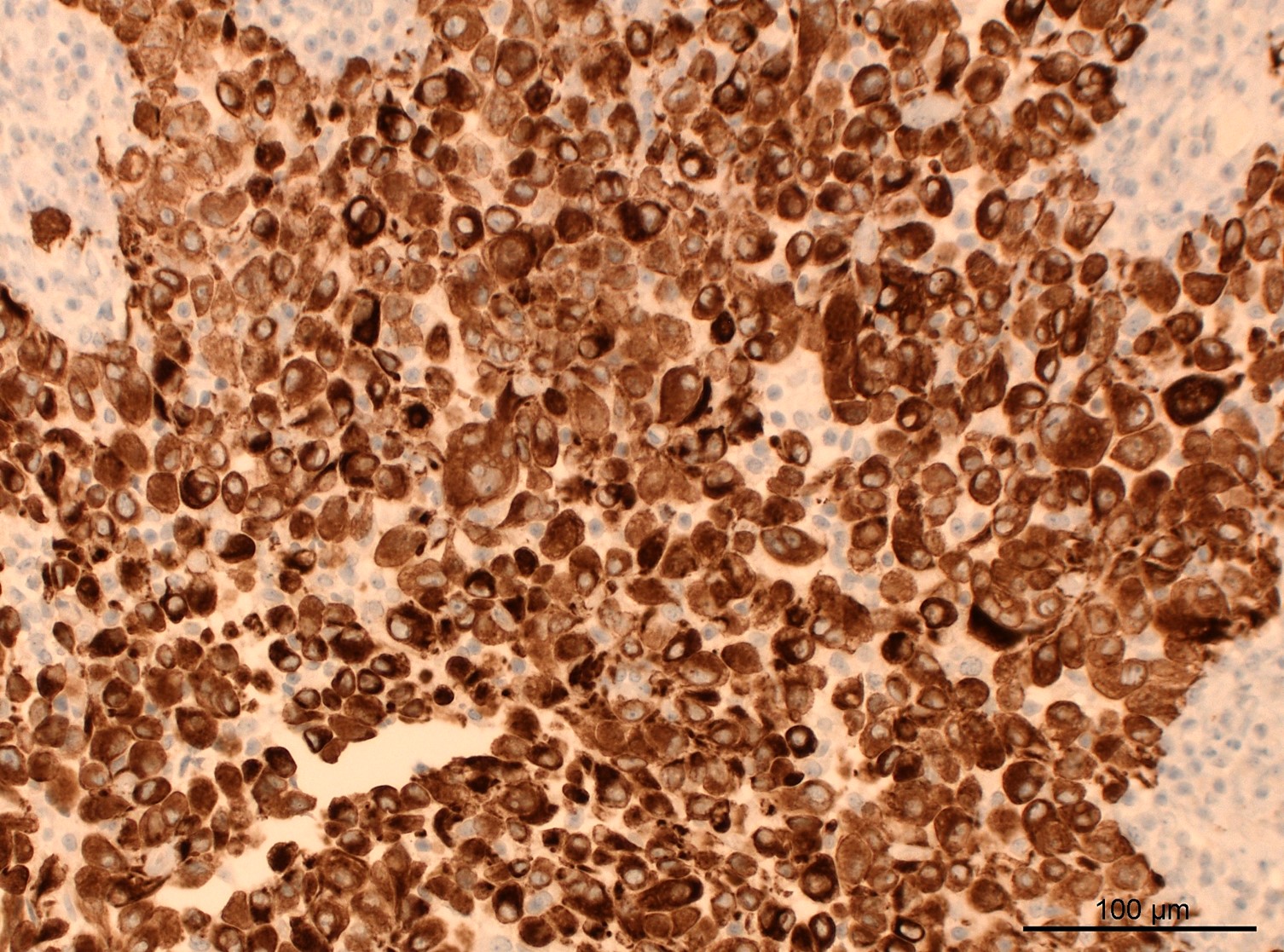
The multiple peritoneal masses from this 27 year old man are histologically comprised of epithelioid cells with eosinophilic cytoplasm and a solid growth pattern. The tumor nodules additionally show a thick fibrous capsule, prominent lymphocytic cuffing and extensive necrosis. With an initial suspicion of a carcinoma, a series of immunohistochemical stainings was performed but showed negativity for all stained markers (TTF1, CDX2, SALL4, synaptophysin, chromogranin A, PAX8, GATA3, p40, calretinin) except AE1/3; this tumor could not be further classified into one of the known malignant epithelial neoplasms. A following next generation sequencing analysis revealed a gene fusion EWSR1::ATF1 (one of the CREB family of transcription factors), so that the entity of this tumor was successfully clarified.
Malignant epithelioid neoplasm with EWSR1/FUS::CREB fusion is a distinctive entity which was recently reported by Argani et al. 2020 (Mod Pathol 2020;33:2233) and Shibayama et al. 2021 (Am J Surg Pathol 2022;46:134). These tumors most commonly show epithelioid cell morphology but can also exhibit round or spindled cell phenotypes. Further morphologic features include a thick fibrous capsule surrounding the tumor and cystic changes filled with secretions or blood and prominent lymphocytic cuffing. Immunohistochemically, this group of tumors demonstrates positivity for at least one of the epithelial markers (AE1/3, EMA, CAM5.2). The predilection sites are mesothelialized cavities, especially the omentum, and the tumor shows overt malignant potential with metastasis to lymph nodes or other organs such as the liver.
A correct diagnosis of these tumors is challenging because they may show common histomorphological features with mesenchymal tumors, which can also harbor EWSR1/FUS::CREB fusions, e.g., angiomatoid fibrous histiocytoma or malignant mesothelioma. Moreover, they cannot be further classified into one of the commonly known epithelial neoplasms by immunohistochemistry alone. Therefore, pathologists should be aware of this entity and its morphologic and immunohistochemical features and how to differentiate it from other mesenchymal and epithelial neoplasms.
Test question answer:
D. This tumor is positive for at least one of the epithelial markers, such as AE1/3
All cases are archived on our website. To view them sorted by case number, diagnosis or category, visit our main Case of the Month page. To subscribe or unsubscribe to Case of the Month or our other email lists, click here.
Thanks to Drs. Hyunkyu Shin and Christian M. Schürch, University Hospital and Comprehensive Cancer Center, Tübingen, Germany, for contributing this case and discussion and to Dr. Borislav Alexiev, Northwestern University Feinberg School of Medicine, Chicago, Illinois, USA for reviewing the discussion.
Advertisement
Case of the Month #516
Clinical history:
A 27 year old man presented with multiple peritoneal masses, up to 3.5 cm in size, with extension into the retroperitoneum and abdominal lymphadenopathy with necrosis.
Histopathology images:
What is your diagnosis?
Click here for diagnosis, test question and discussion:
Diagnosis: Malignant epithelioid neoplasm with EWSR1/FUS::CREB fusion
Test question (answer at the end):
Which is a true feature of malignant epithelioid neoplasm with EWSR1/FUS::CREB fusion?
A. A confirmed gene fusion EWSR1/FUS::CREB is considered pathognomonic for this entity
B. Predilection sites are visceral organs
C. This tumor generally shows excellent prognosis without metastasis
D. This tumor is positive for at least one of the epithelial markers, such as AE1/3
E. This tumor should not exhibit morphologic features of a mesenchymal neoplasm
Stains
AE1/3
p40
SALL4
Discussion:
The multiple peritoneal masses from this 27 year old man are histologically comprised of epithelioid cells with eosinophilic cytoplasm and a solid growth pattern. The tumor nodules additionally show a thick fibrous capsule, prominent lymphocytic cuffing and extensive necrosis. With an initial suspicion of a carcinoma, a series of immunohistochemical stainings was performed but showed negativity for all stained markers (TTF1, CDX2, SALL4, synaptophysin, chromogranin A, PAX8, GATA3, p40, calretinin) except AE1/3; this tumor could not be further classified into one of the known malignant epithelial neoplasms. A following next generation sequencing analysis revealed a gene fusion EWSR1::ATF1 (one of the CREB family of transcription factors), so that the entity of this tumor was successfully clarified.
Malignant epithelioid neoplasm with EWSR1/FUS::CREB fusion is a distinctive entity which was recently reported by Argani et al. 2020 (Mod Pathol 2020;33:2233) and Shibayama et al. 2021 (Am J Surg Pathol 2022;46:134). These tumors most commonly show epithelioid cell morphology but can also exhibit round or spindled cell phenotypes. Further morphologic features include a thick fibrous capsule surrounding the tumor and cystic changes filled with secretions or blood and prominent lymphocytic cuffing. Immunohistochemically, this group of tumors demonstrates positivity for at least one of the epithelial markers (AE1/3, EMA, CAM5.2). The predilection sites are mesothelialized cavities, especially the omentum, and the tumor shows overt malignant potential with metastasis to lymph nodes or other organs such as the liver.
A correct diagnosis of these tumors is challenging because they may show common histomorphological features with mesenchymal tumors, which can also harbor EWSR1/FUS::CREB fusions, e.g., angiomatoid fibrous histiocytoma or malignant mesothelioma. Moreover, they cannot be further classified into one of the commonly known epithelial neoplasms by immunohistochemistry alone. Therefore, pathologists should be aware of this entity and its morphologic and immunohistochemical features and how to differentiate it from other mesenchymal and epithelial neoplasms.
Test question answer:
D. This tumor is positive for at least one of the epithelial markers, such as AE1/3
