
Stains & CD markers
NADH

Copyright: 2021-2024, PathologyOutlines.com, Inc.
PubMed Search: NADH
NADH
Author: Moiz Vora, M.D.
Editorial Board Member: Christian M. Schürch, M.D., Ph.D.
Deputy Editor-in-Chief: Chunyu Cai, M.D., Ph.D.
Last author update: 28 July 2022
Last staff update: 28 July 2022
Copyright: 2021-2024, PathologyOutlines.com, Inc.
PubMed Search: NADH

Table of Contents
Definition / general | Essential features | Terminology | Interpretation | Uses by pathologists | Microscopic (histologic) description | Microscopic (histologic) images | Positive staining - normal | Positive staining - disease | Negative staining | Board review style question #1 | Board review style answer #1Cite this page: Vora M. NADH. PathologyOutlines.com website. https://www.pathologyoutlines.com/topic/NADH.html. Accessed April 19th, 2024.
Definition / general
- Nicotinadmide adenine dinucleotide (NADH) is a coenzyme that facilitates substrate reducing reactions associated with glycolysis, oxidative phosphorylation and fermentation
- NADH-TR stain protocol utilizes enzymatic activity to release hydrogen from NADH, producing a purple-blue formazan pigment that marks the reaction site
Essential features
- NADH enzyme histochemical stain reveals myofibrillar architecture, mitochondria and target fibers; it helps differentiate type I (oxidative) and type II (nonoxidative) fibers
- This technique demonstrates patterns of myofiber injury that are characteristic of congenital and mitochondrial myopathies and specific muscular dystrophies
- This stain protocol requires the use of snap frozen muscle biopsies
Terminology
- Nicotinamide adenine dinucleotide tetrazolium reductase (NADH-TR)
- Nicotinamide adenine dinucleotide diaphorase activity
Interpretation
- Mitochondria within sarcoplasmic network of striated muscle
Uses by pathologists
- Reveals architectural changes in the muscle, e.g., central cores, whorled, lobulated and moth eaten fibers; these changes assist in categorizing patterns of myofibril injury (J Histotechnol 2008;31:101, ScienceDirect: Tetrazolium Reductase [Accessed 2 February 2022])
- NADH diaphorase activity, in combination with myosin ATPase activity at pH 9.4, allows fibers to be classified into 3 categories:
- Slow twitch high oxidative (ST)
- Fast twitch high oxidative (FTH)
- Fast twitch low oxidative (FT)
Microscopic (histologic) description
- Speckled pattern within myofibers (J Histotechnol 2008;31:101)
- Intensity is proportional to number of mitochondria and NADH activity
- Type I oxidative fibers (dark, dense purple appearance)
- Type II nonoxidative fibers (light, scattered purple speckles)
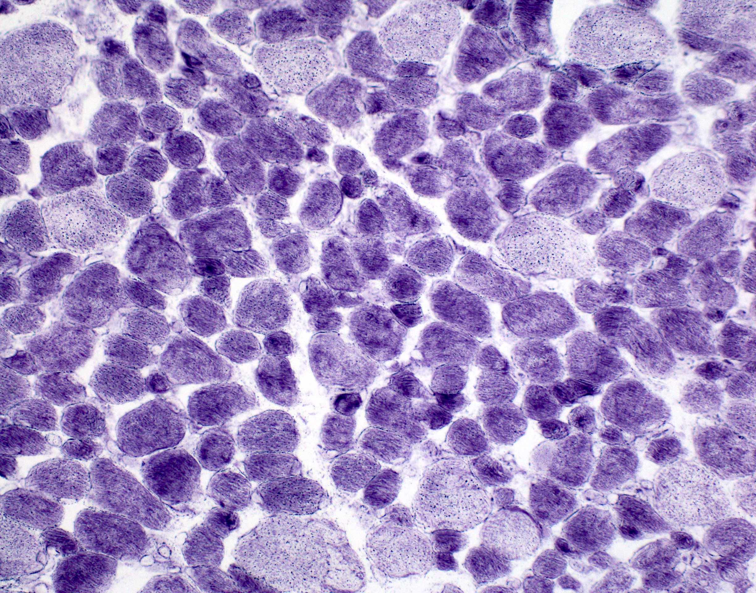
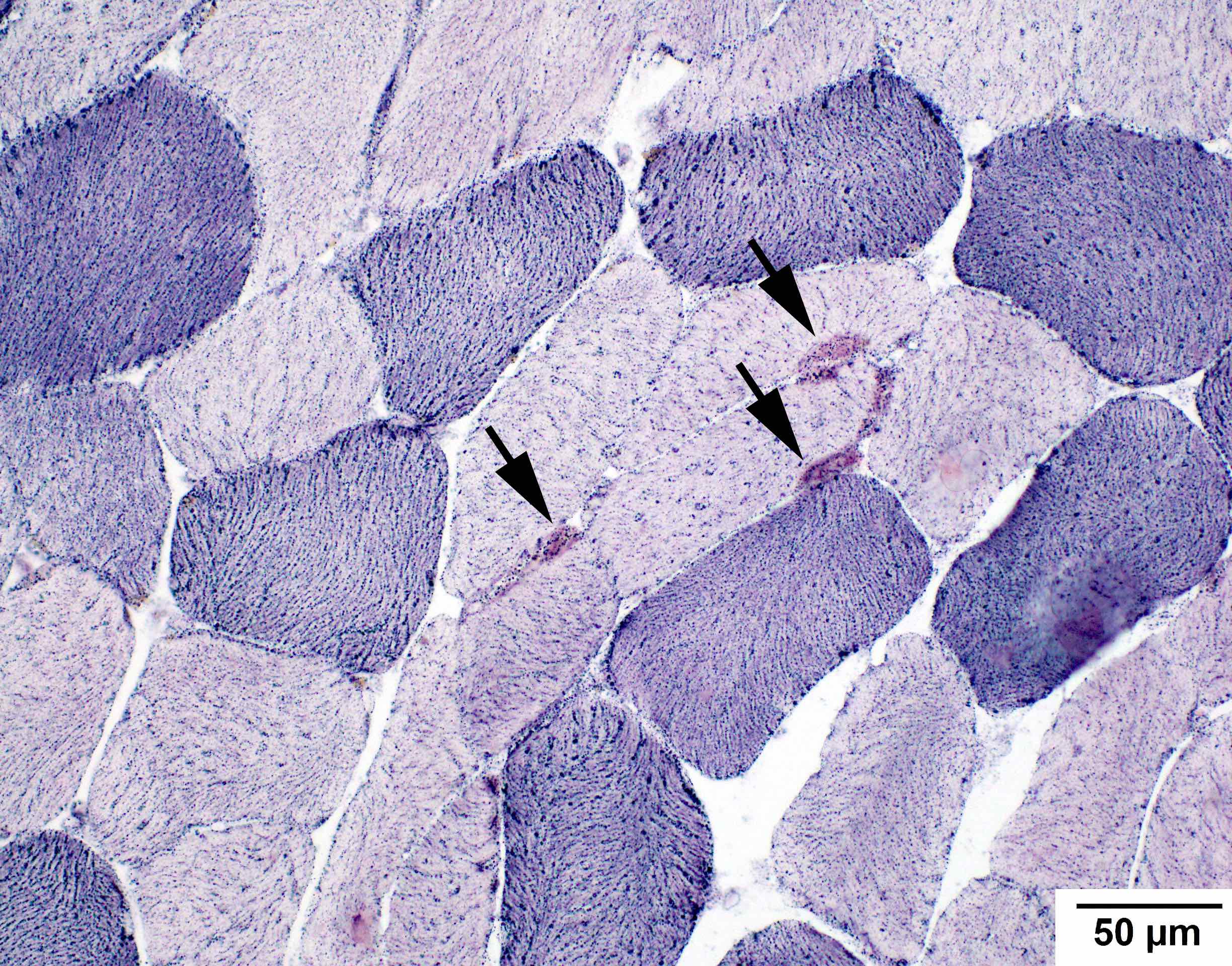
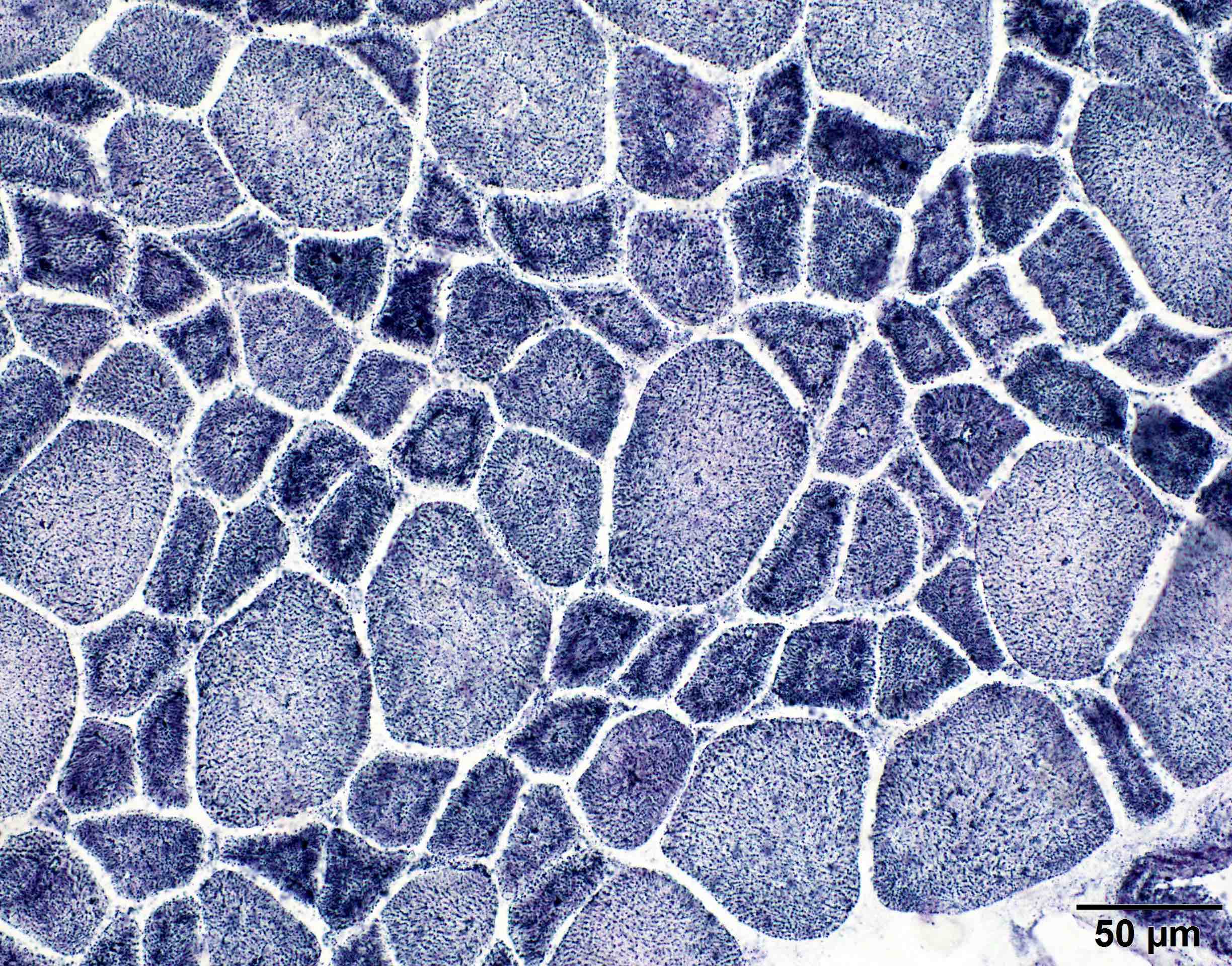
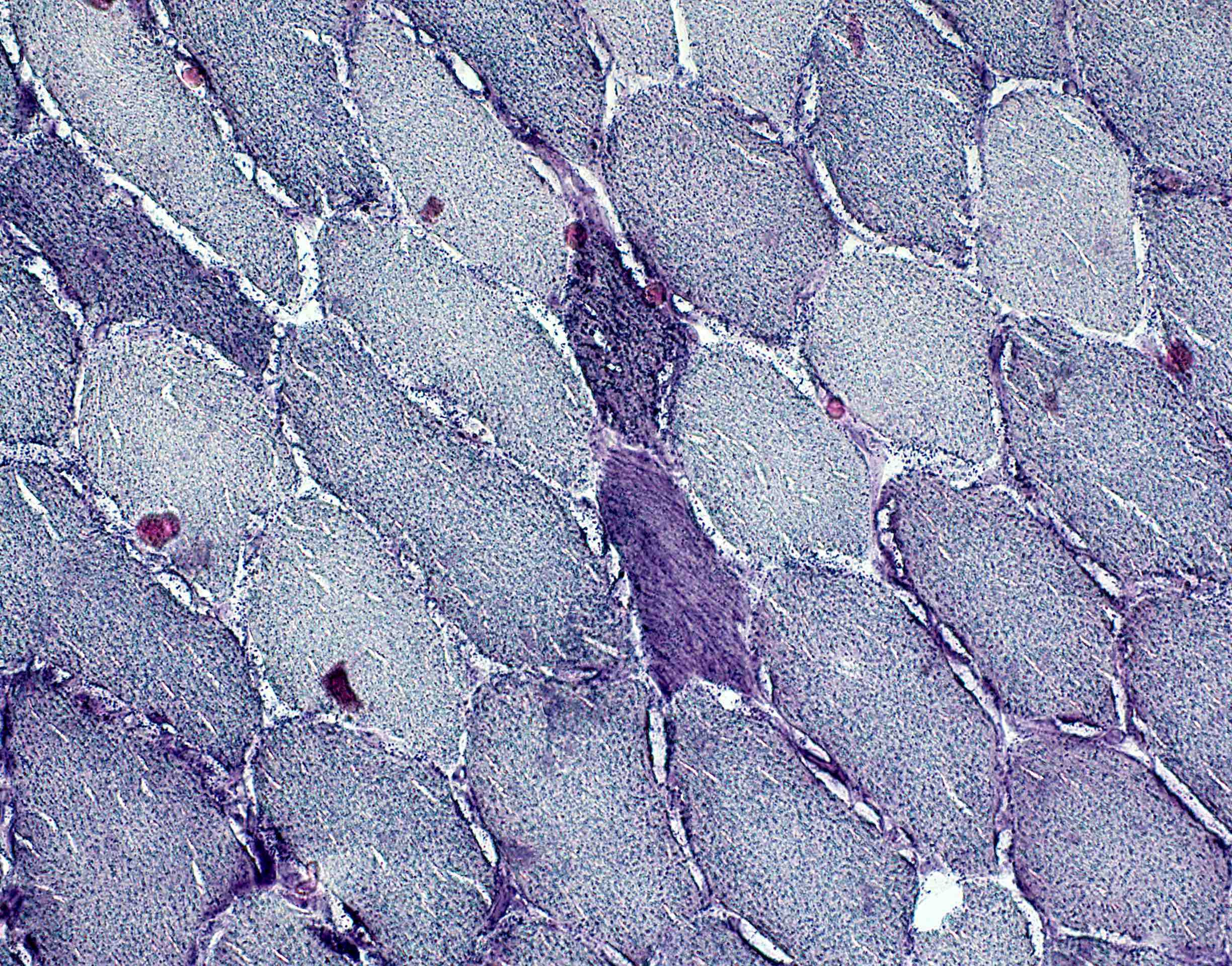
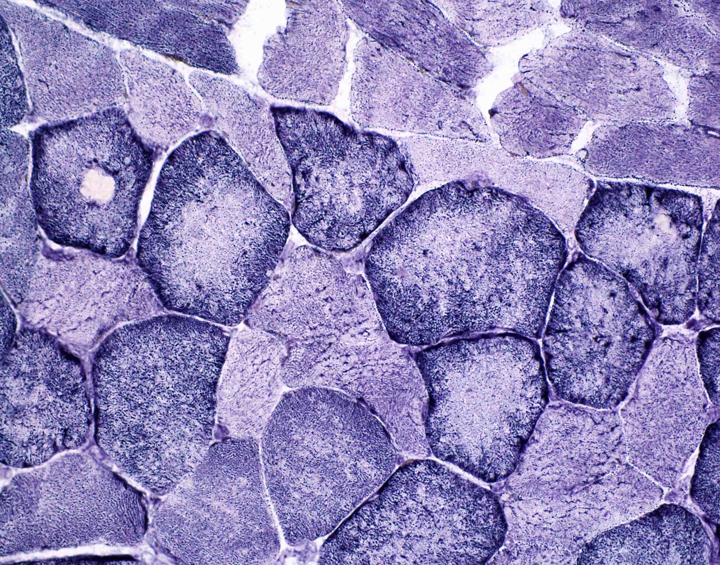
Microscopic (histologic) images
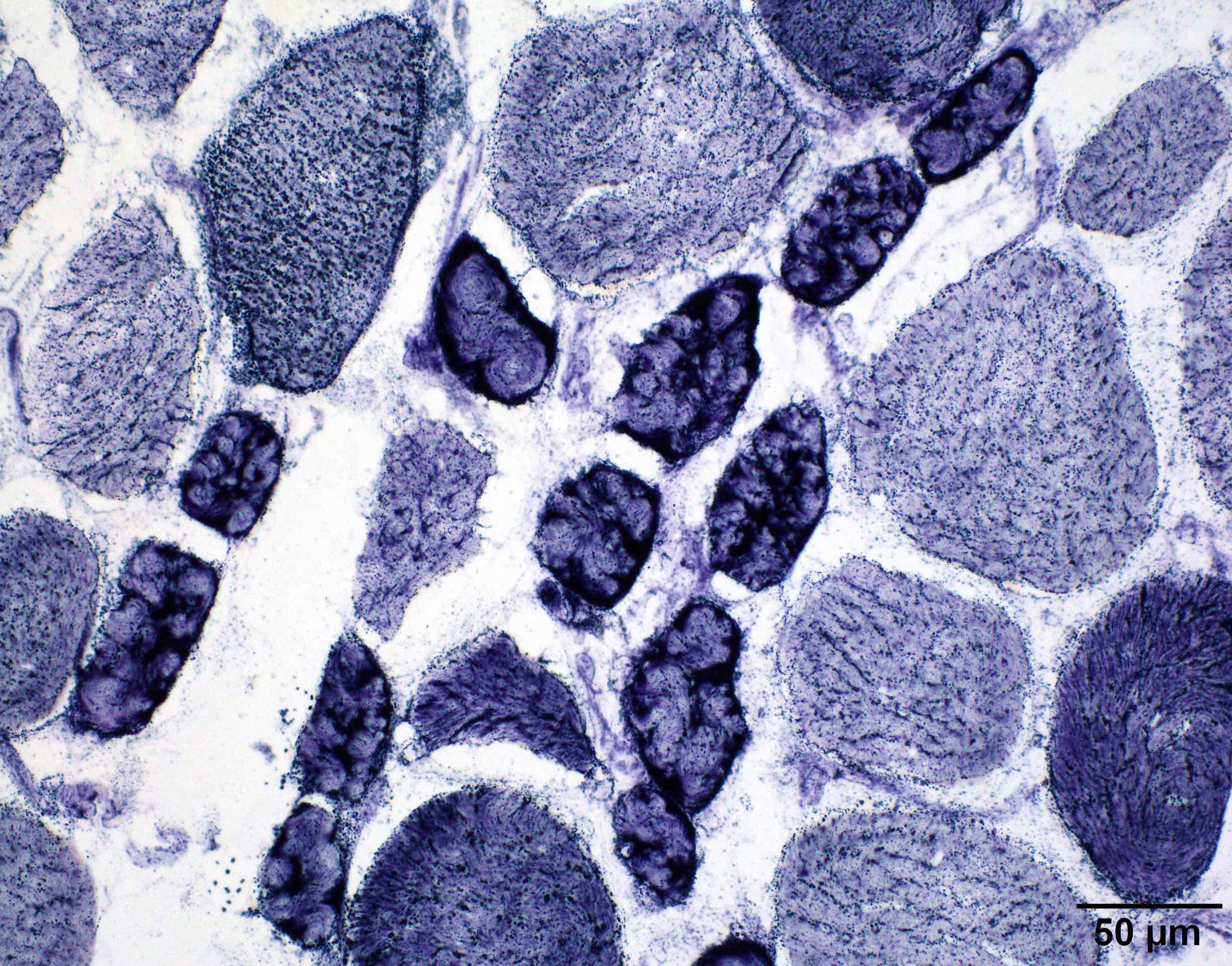
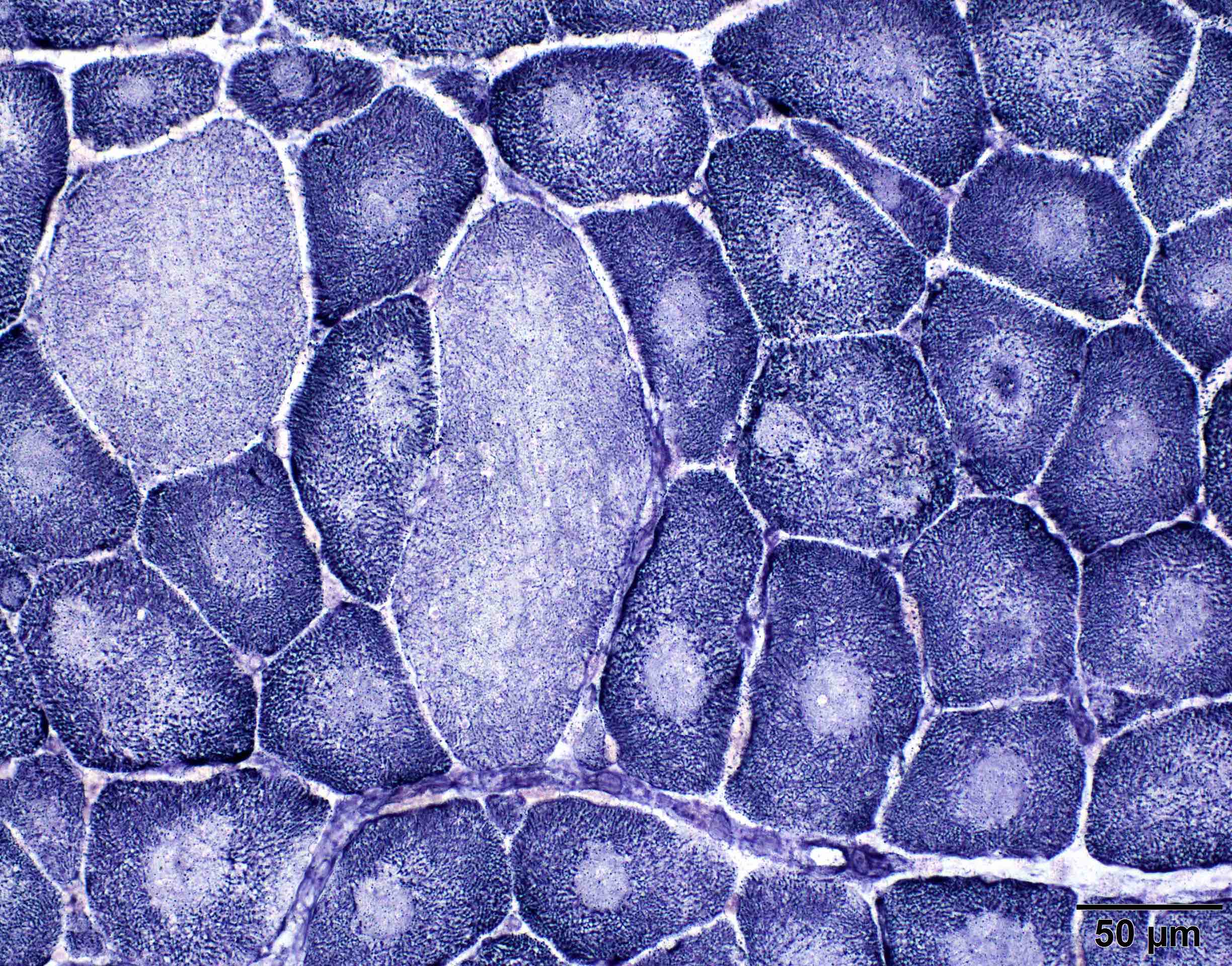
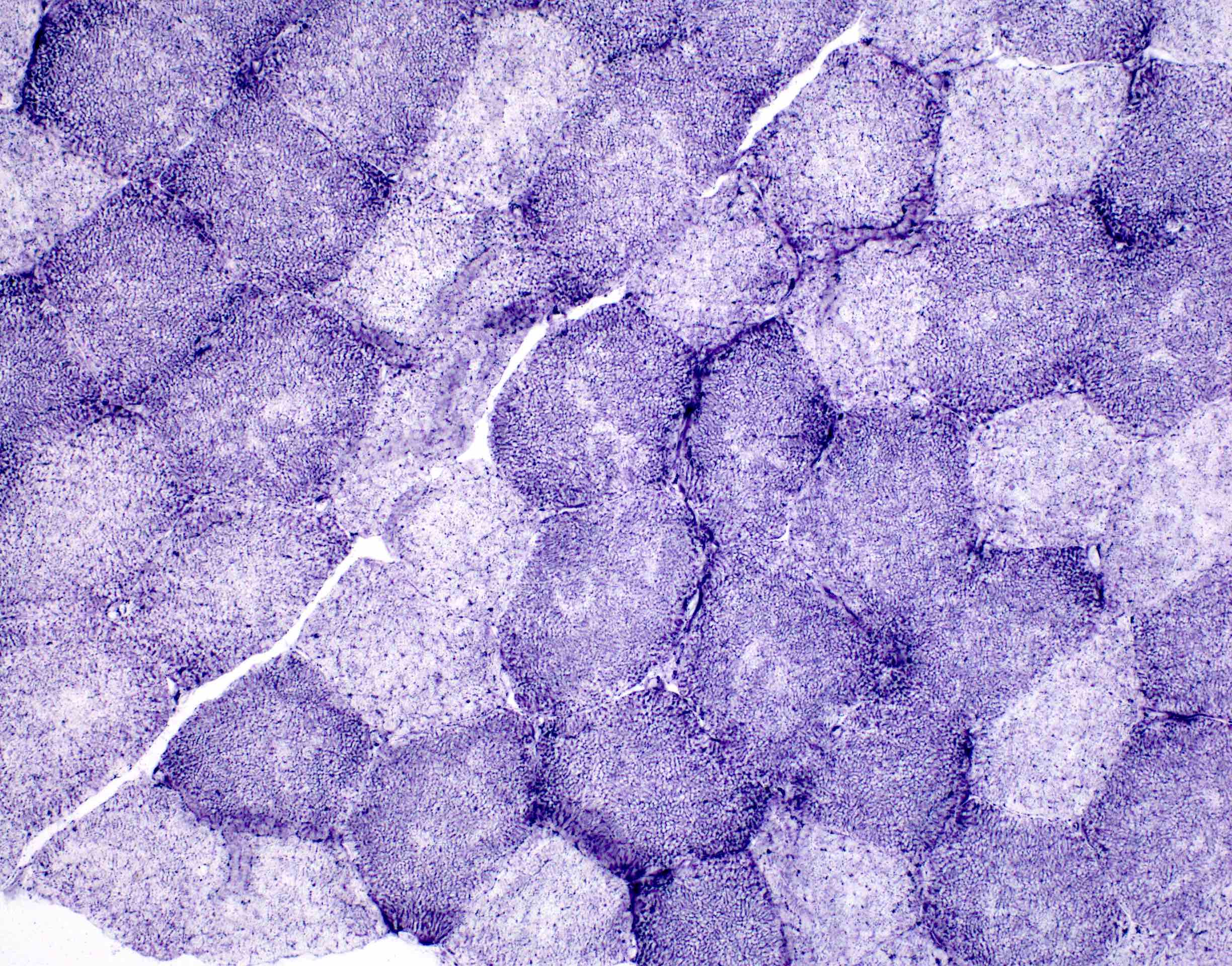
Contributed by Chunyu Cai, M.D., Ph.D.
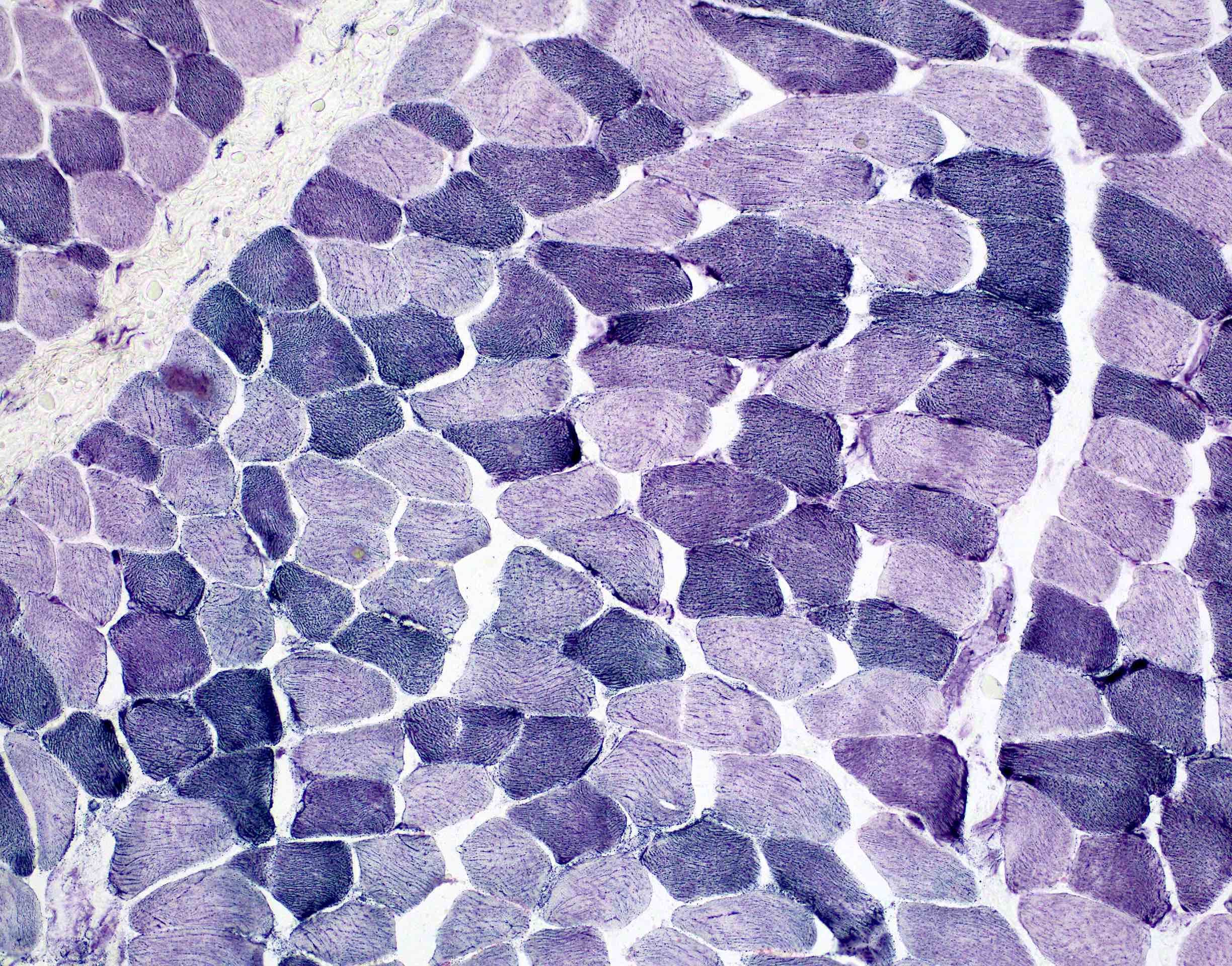
Normal NADH
Myopathy with lobulated fibers
Central core myopathy
Multiminicore myopathy
Nemaline rod myopathy
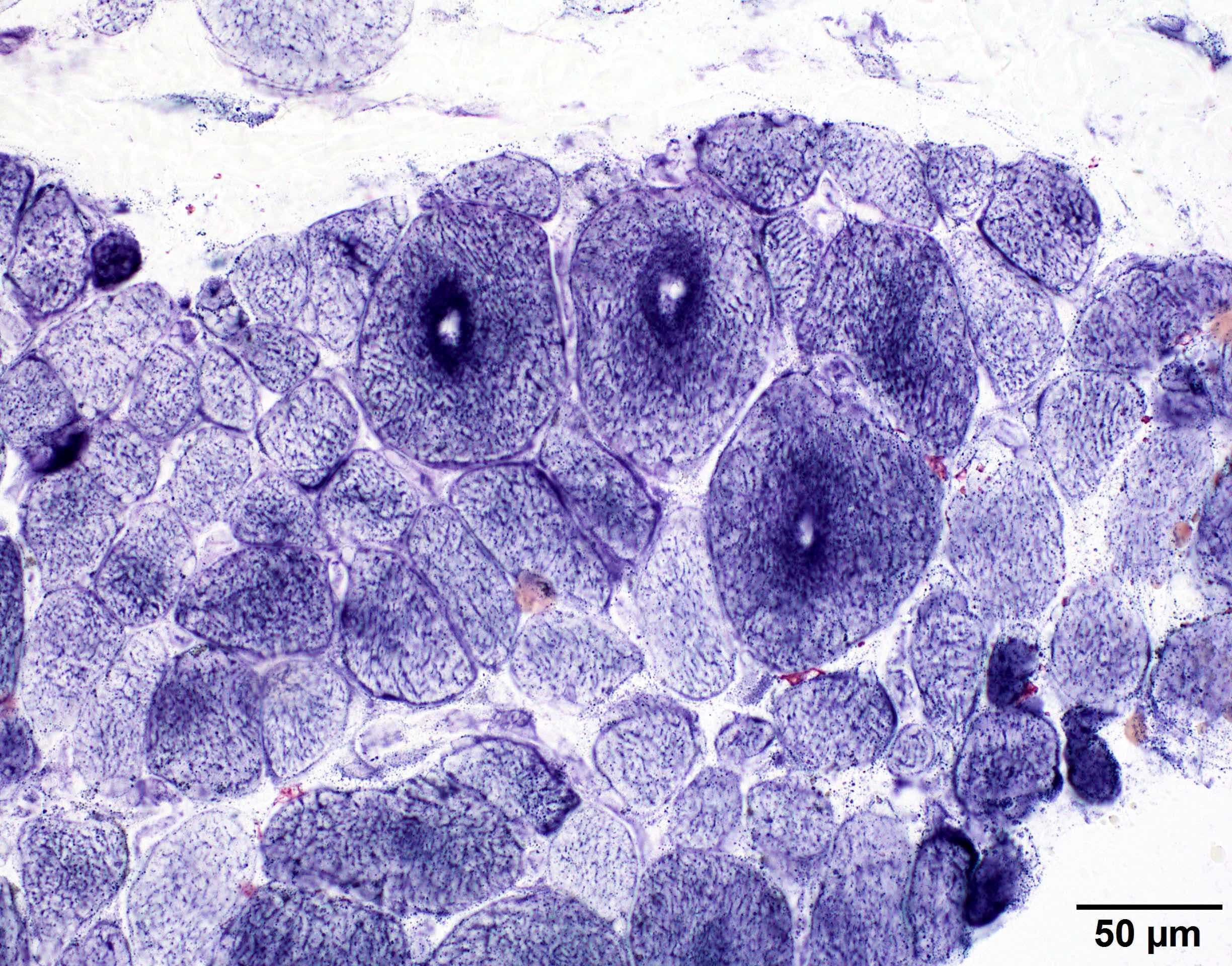
Target fibers
Myopathy with tubular aggregates
Necklace fibers
Ragged blue fibers

Dermatomyositis perifascicular atrophy
Myofibrillar myopathy
Positive staining - normal
- Sarcoplasmic reticulum, T tubules and mitochondria (i.e., the intermyofibrillar network) (Goetz: Textbook of Clinical Neurology, 3rd Edition, 2007, ScienceDirect: Tetrazolium Reductase [Accessed 2 February 2022])
Positive staining - disease
- Nemaline myopathy: type I fiber atrophy and core-like areas devoid of enzyme activity (Mol Med Rep 2014;10:175)
- Mitochondrial myopathies: ragged blue fibers (J Bras Patol Med Lab 2018;54:325)
- Limb girdle muscular dystrophy type 2A: disorganized intermyofibrillar networks showing lobulated appearance (Pediatr Neonatol 2015;56:62)
- Myotonic dystrophy: ring fibers (Phys Med Rehabil Clin N Am 2012;23:609)
- Neurogenic atrophy: target fibers (Phys Med Rehabil Clin N Am 2012;23:609)
- Dermatomyositis: perifascicular atrophy (Phys Med Rehabil Clin N Am 2012;23:609)
- Myofibrillar myopathies: rubbed out myofiber appearance due to diminished or absent staining with NADH (Eur J Neurol 2015;22:1429)
- DNM2 related centronuclear myopathy: radial sarcoplasmic strands (Neuromuscul Disord 2014;24:97)
- Central core disease: type I fibers with central cores (Orphanet J Rare Dis 2007;2:25)
- Tubular aggregate myopathy: tubular aggregates within myofibrils (Int J Mol Sci 2016;17:1952)
Negative staining
- Myopathies related to toxic injury (Phys Med Rehabil Clin N Am 2012;23:609)
- Myopathies related to lipid metabolism (Phys Med Rehabil Clin N Am 2012;23:609)
- Myopathies related to glycogen metabolism except for debrancher enzyme deficiency (Phys Med Rehabil Clin N Am 2012;23:609)
Board review style question #1
In which type of myopathy is this characteristic pattern of injury typically seen by NADH-TR histochemistry?
- Central core disease (type I fibers with central cores)
- Debranching enzyme deficiency (ring fibers)
- Dermatomyositis (perifasicular atrophy)
- Mitochondrial myopathy (ragged blue fibers)
- Tubular aggregate myopathy (tubular aggregates)
Board review style answer #1

