

Adrenal gland & paraganglia
Other nonneoplastic
Ovarian thecal metaplasia
Authors: Natasha Santhanam, B.A., Debra L. Zynger, M.D.
Editorial Board Member: Bonnie Choy, M.D.


Last staff update: 3 April 2024 (update in progress)
Copyright: 2002-2024, PathologyOutlines.com, Inc.
PubMed search: Ovarian thecal metaplasia
Table of Contents
Definition / general | Essential features | Terminology | Epidemiology | Sites | Etiology | Clinical features | Diagnosis | Radiology description | Case reports | Treatment | Gross description | Microscopic (histologic) description | Microscopic (histologic) images | Positive stains | Negative stains | Sample pathology report | Differential diagnosis | Board review style question #1 | Board review style answer #1Cite this page: Santhanam N, Zynger DL. Ovarian thecal metaplasia. PathologyOutlines.com website. https://www.pathologyoutlines.com/topic/adrenalovarianthecal.html. Accessed April 19th, 2024.
Definition / general
- Rare mesenchymal lesion composed of cells that resemble ovarian-like stroma
Essential features
- Fibroblastic spindle cell proliferation
- Often described as wedge shaped lesion
- Continuous with adrenal gland capsule
- Can be multifocal or a solitary lesion
Terminology
- Spindle cell foci of unknown significance has been recommended as a new diagnostic term, as ovarian thecal metaplasia is a misnomer (Endocr Pathol 2011;22:222)
Epidemiology
- Found mainly in postmenopausal women with a mean age range of 39 - 65 years old (Am J Clin Pathol 1977;67:318)
- Autopsy series of women with metastatic breast cancer and a range of 2 - 10 adrenal slides reviewed per case yielded an incidence of 4.3% (12 out of 276 patients) (Am J Clin Pathol 1977;67:318)
- Rarely found in men (Arch Pathol Lab Med 1989;113:1071)
Sites
- In the adrenal cortex adjacent to the adrenal gland capsule
Etiology
- Originally thought to be due to nodular hyperplasia of adrenal cortical blastema (Arch Pathol 1971;92:319)
- Now considered to be due to metaplasia of adrenal capsule mesenchymal cells into spindle cells, which resemble ovarian stroma in response to unopposed pituitary gonadotropin following menopause (Arch Pathol 1971;92:319)
Clinical features
- Asymptomatic and found incidentally (Am J Clin Pathol 1977;67:318)
Diagnosis
- Diagnosed on surgical excision via histologic analysis (World J Radiol 2014;6:919)
Radiology description
- Not seen on imaging (World J Radiol 2014;6:919)
Case reports
- 17 year old girl with Beckwith-Wiedemann syndrome and right adrenal mass lesion (World J Radiol 2014;6:919)
- 49 year old woman with incidental left adrenal mass found on computed tomography (CT) (Arch Pathol Lab Med 2004;128:1294)
- 77 year old man with acquired bilateral testicular atrophy and incidental metaplasia on autopsy (Arch Pathol Lab Med 1989;113:1071)
Treatment
- No treatment is needed (Endocr Pathol 2011;22:222)
Gross description
- Cannot be visualized grossly (Am J Clin Pathol 1977;67:318)
Microscopic (histologic) description
- Single or multiple nodules (Arch Pathol 1971;92:319, Am J Clin Pathol 1977;67:318)
- Size of nodules ranges from 0.4 to 1.7 mm (Arch Pathol 1971;92:319, Am J Clin Pathol 1977;67:318)
- Basophilic spindle cell whorls found beneath the adrenal capsule with variable cellularity (Arch Pathol 1971;92:319)
- Reticular fibers surround individual spindle cells (Arch Pathol 1971;92:319)
- Can contain nest of adrenal cortical cells (Arch Pathol 1971;92:319)
- Lesions can contain patches of dense eosinophilic hyalinized fibers (Arch Pathol 1971;92:319)
- Some can contain areas of central sclerosis (Endocr Pathol 2011;22:222)
- Dystrophic focal calcification can be present (Endocr Pathol 2011;22:222)
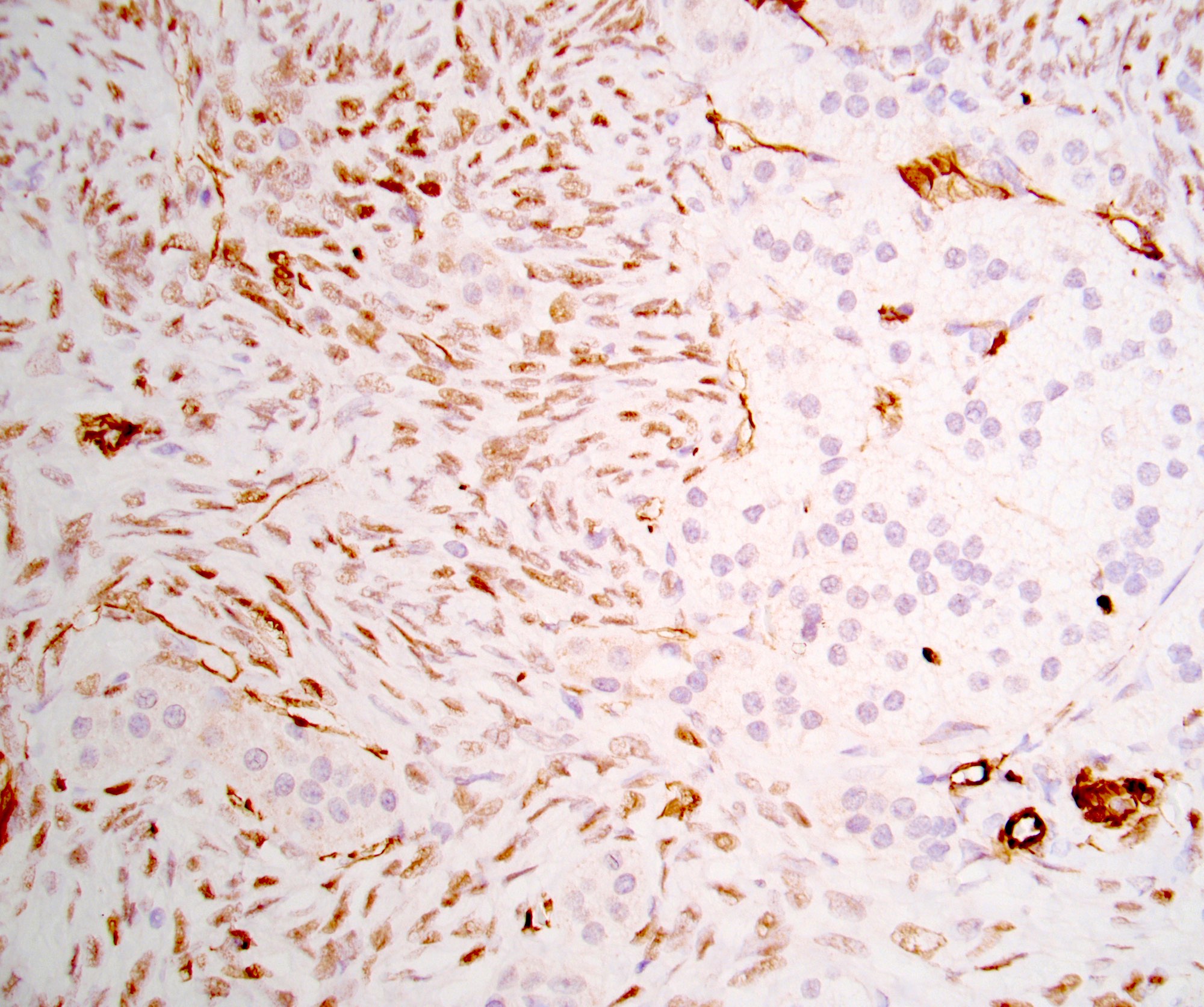
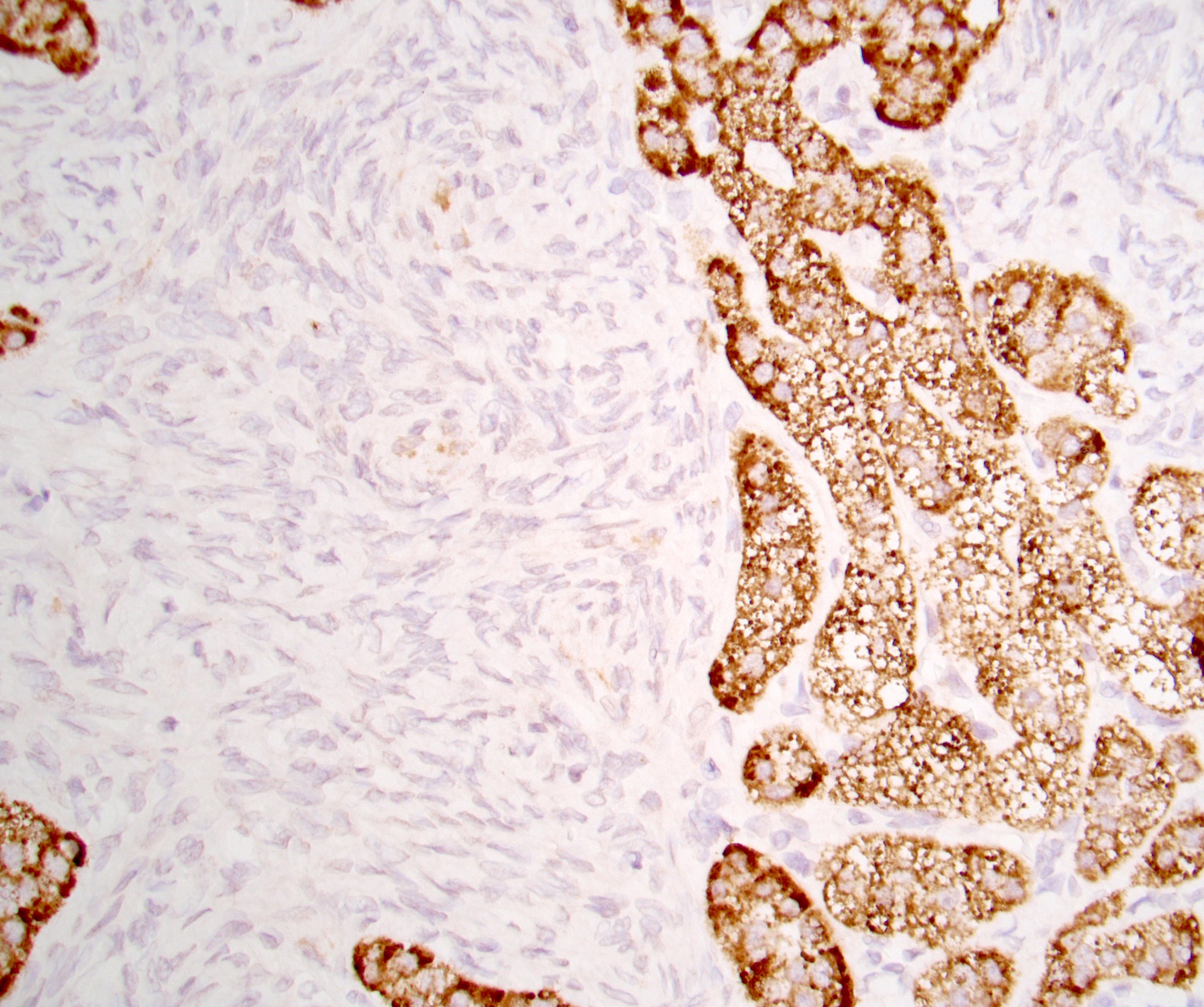
Microscopic (histologic) images
Contributed by Debra L. Zynger, M.D.
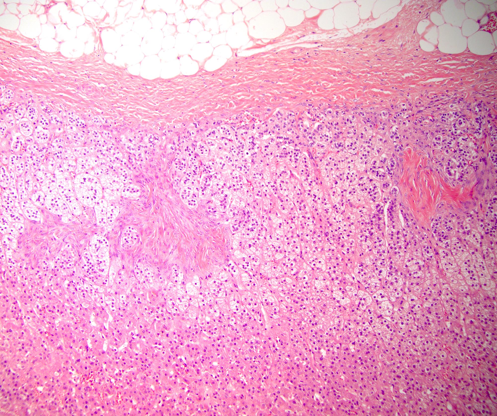

Subcapsular spindle cells

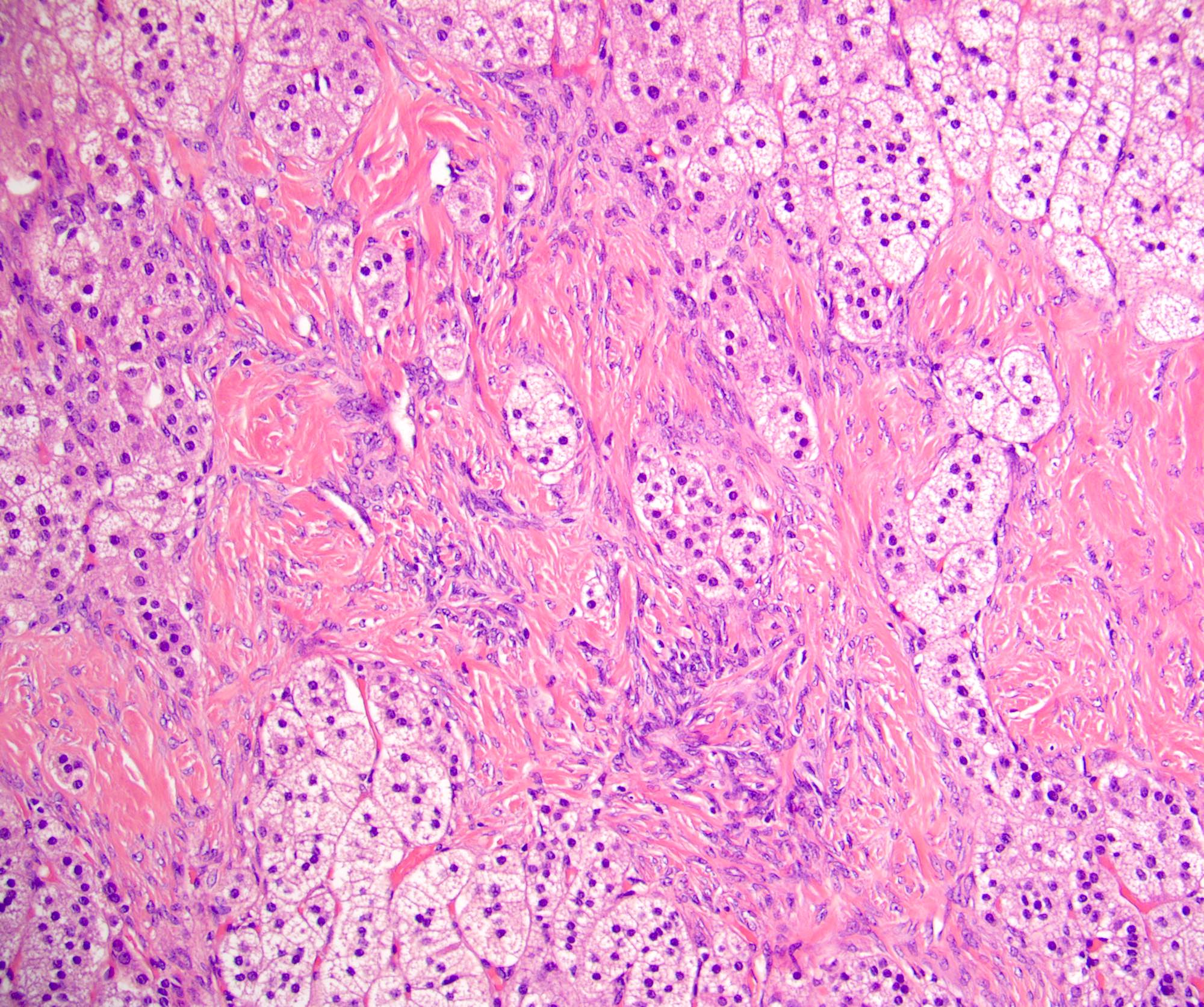
Spindle cells with entrapped adrenal cortical cells


Bland spindle cells


Thick hyalinized fibers
WT1

Desmin
MelanA
Positive stains
Negative stains
- α inhibin (negative or focal), SF1, MelanA, HMB45
- AE1 / AE3
- S100
- CD34
- Estrogen receptor
- Reference: Endocr Pathol 2011;22:222
Sample pathology report
- Left kidney and adrenal, radical nephrectomy:
- Kidney with renal cell carcinoma, clear cell type (see synoptic report)
- Adrenal with spindle cell proliferation (also known as ovarian thecal metaplasia) (0.15 cm) (see comment)
- Comment: A bland micronodule of spindle cells is seen; it expresses desmin, SMA and WT1 and is negative for S100, CD34, inhibin, SF1, AE1 / AE3 and ER, consistent with the incidental lesion referred to as ovarian thecal metaplasia.
Differential diagnosis
- Calcifying fibrous tumor (Endocr Pathol 2011;22:222):
- Present in subcutaneous and deep soft tissue
- Exceptionally rare in the adrenal gland
- Contains sparsely cellular hyalinized fibrotic stroma and few spindle cells
- Negative for SMA and MSA
- Positive for factor XIIIa and CD34
- Inflammatory myofibroblastic tumor, sclerosing type (Endocr Pathol 2011;22:222):
- Neoplasm containing myofibroblastic cells with abundant inflammation
- Lymphocytes form aggregates with germinal centers
- Positive for SMA and MSA and up to 40% positive for ALK (Am J Surg Pathol 2001;25:761, Am J Surg Pathol 2001;25:1364)
- Leiomyoma (Mod Pathol 2014;27:S17):
- Exceptionally rare in the adrenal gland
- Grossly identifiable lesion
- Does not grow in micronodular pattern
Board review style question #1

The photo above shows a spindle cell proliferation in the adrenal cortex that was incidentally identified upon microscopic examination. This lesion is described in the literature as ovarian thecal metaplasia. Based on IHC expression, this lesion is not related to either the ovary or the adrenal gland. Which combination of the following markers is usually expressed in this lesion?
- AE1 / AE3
- Inhibin, SF1, MelanA, HMB45
- S100, CD34
- SMA, desmin, WT1
Board review style answer #1
D. SMA, desmin, WT1. This rare spindle cell proliferation of the adrenal cortex, termed ovarian thecal metaplasia, expresses SMA, desmin and WT1. Answer A is incorrect because this entity does not express keratins such as AE1 / AE3. Answer B is incorrect because this entity does not express adrenal cortical markers. Answer C is incorrect because this entity does not express S100 or CD34.
Comment Here
Reference: Ovarian thecal metaplasia
Comment Here
Reference: Ovarian thecal metaplasia
