Bladder & urothelial tract
Other nonneoplastic
Malakoplakia
Author: Mustafa Goksel, M.D.
Editorial Board Member: Michelle R. Downes, M.D.
Deputy Editor-in-Chief: Maria Tretiakova, M.D., Ph.D.

Last author update: 25 June 2024
Last staff update: 25 June 2024
Copyright: 2003-2025, PathologyOutlines.com, Inc.
PubMed Search: Malakoplakia
Table of Contents
Definition / general | Essential features | Terminology | ICD coding | Epidemiology | Sites | Pathophysiology | Etiology | Clinical features | Diagnosis | Case reports | Treatment | Clinical images | Microscopic (histologic) description | Microscopic (histologic) images | Positive stains | Negative stains | Sample pathology report | Differential diagnosis | Practice question #1 | Practice answer #1 | Practice question #2 | Practice answer #2Cite this page: Goksel M. Malakoplakia. PathologyOutlines.com website. https://www.pathologyoutlines.com/topic/bladdermalakoplakia.html. Accessed September 11th, 2025.
Definition / general
- Chronic inflammatory disorder that affects many organs, most commonly occurs in urinary bladder
- Single or multiple white-yellow soft raised plaques on the mucosal surface, can mimic malignancy (Nephrourol Mon 2014;6:e18522)
Essential features
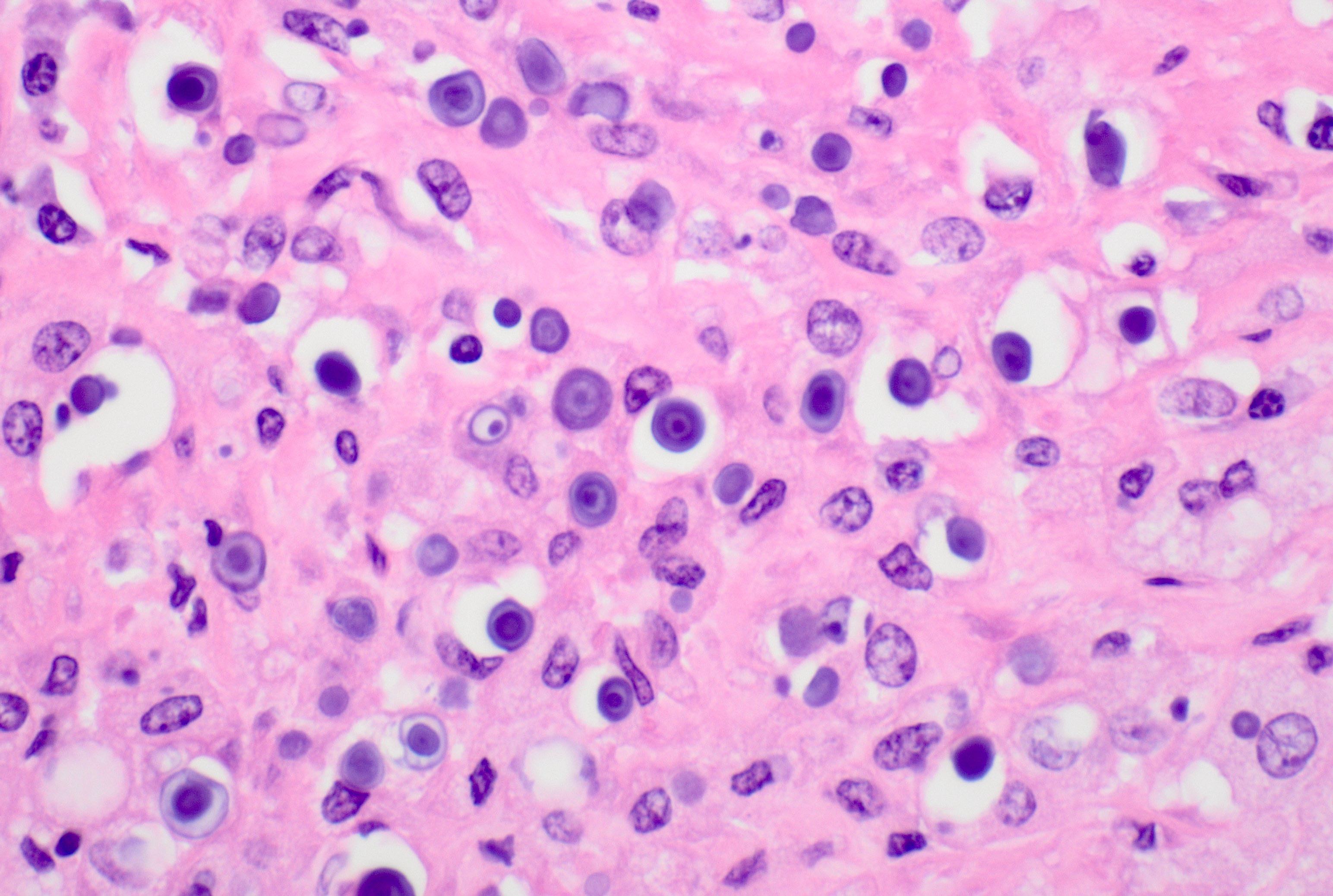
- Histiocytic infiltrate with characteristic cytoplasmic basophilic inclusions
- Round histiocytes (von Hansemann cells) and rounded, concentric, basophilic, cytoplasmic inclusions (Michaelis-Gutmann bodies)
- Mass forming, typically polypoid bladder mass with intact mucosa
Terminology
- First described by Michaelis and Gutmann in 1902 and later named by von Hansseman (Ztschr Kin Med 1902;47:208, Arch Ital Urol Androl 2022;94:350)
- Malakoplakia comes from Greek words malakos (soft) and plakos (plaque)
ICD coding
Epidemiology
- More common in immunocompromised patients, patients with diabetes mellitus, renal transplantation recipients (Arch Ital Urol Androl 2022;94:350)
- Prevalence: 4 times higher in women
- Mostly affects people over 40 years old
Sites
- Mostly occurs at genitourinary tract (most commonly urinary bladder, followed by renal parenchyma, prostate, rarely ureter) (Arch Ital Urol Androl 2022;94:350)
- Can affect many organs including gastrointestinal tract, skin, thyroid, testis, lung, retroperitoneum, adrenal gland, cerebrum, vertebrae, pleura, tonsils, spleen (Proc (Bayl Univ Med Cent) 2020;33:389, N Engl J Med 2019;380:580, Int J Surg Pathol 2015;23:308, Transplant Proc 2022;54:173, J Urol 1981;125:139)
Pathophysiology
- Thought to be a defect in macrophage phagolysosomal response to bacterial infection, resulting in accumulation of macrophages with calcified inclusions composed of undigested bacteria and products (Am J Kidney Dis 2016;68:e27)
Etiology
- Exact etiology unknown
- Recurrent urinary infections (E. coli and other gram negative bacilli) and immunosupression
Clinical features
- Polypoid mass with intact mucosa
- Patients usually present with urinary symptoms and urinary tract infection (72% due to E. coli)
- Hydroureteronephrosis (Arch Ital Urol Androl 2022;94:350)
- Rarely associated with calcified plaques (encrusted cystitis) and renal failure (Ir J Med Sci 2006;175:74, Nat Clin Pract Urol 2008;5:516)
Diagnosis
- Histological by biopsy
Case reports
- 55 year old woman with history of kidney disease presented with general malaise and worsening renal failure and was found to have a bladder mass (Pathol Res Pract 2022;237:153852)
- 61 year old immunocompromised man with symptoms of urinary obstruction was found to have exuberant malakoplakia of prostate presenting as prostatic abscess (J Endourol Case Rep 2020;6:231)
- 82 year old woman with intravesical malakoplakia mimicking an aggressive transitional cell carcinoma (Bladder (San Franc) 2020;7:e44)
Treatment
- Antibiotics or surgical excision
Clinical images
Contributed by Zachary Gordon, M.D.

White light cystoscopy
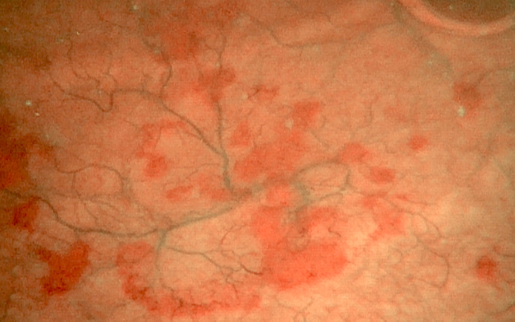
Narrow band cystoscopy
Microscopic (histologic) description
- Sheets of histiocytes with granular eosinophilic cytoplasm (von Hansemann cells) and basophilic intracytoplasmic inclusion (Michaelis-Gutmann bodies)
Microscopic (histologic) images
Contributed by Mustafa Goksel, M.D.
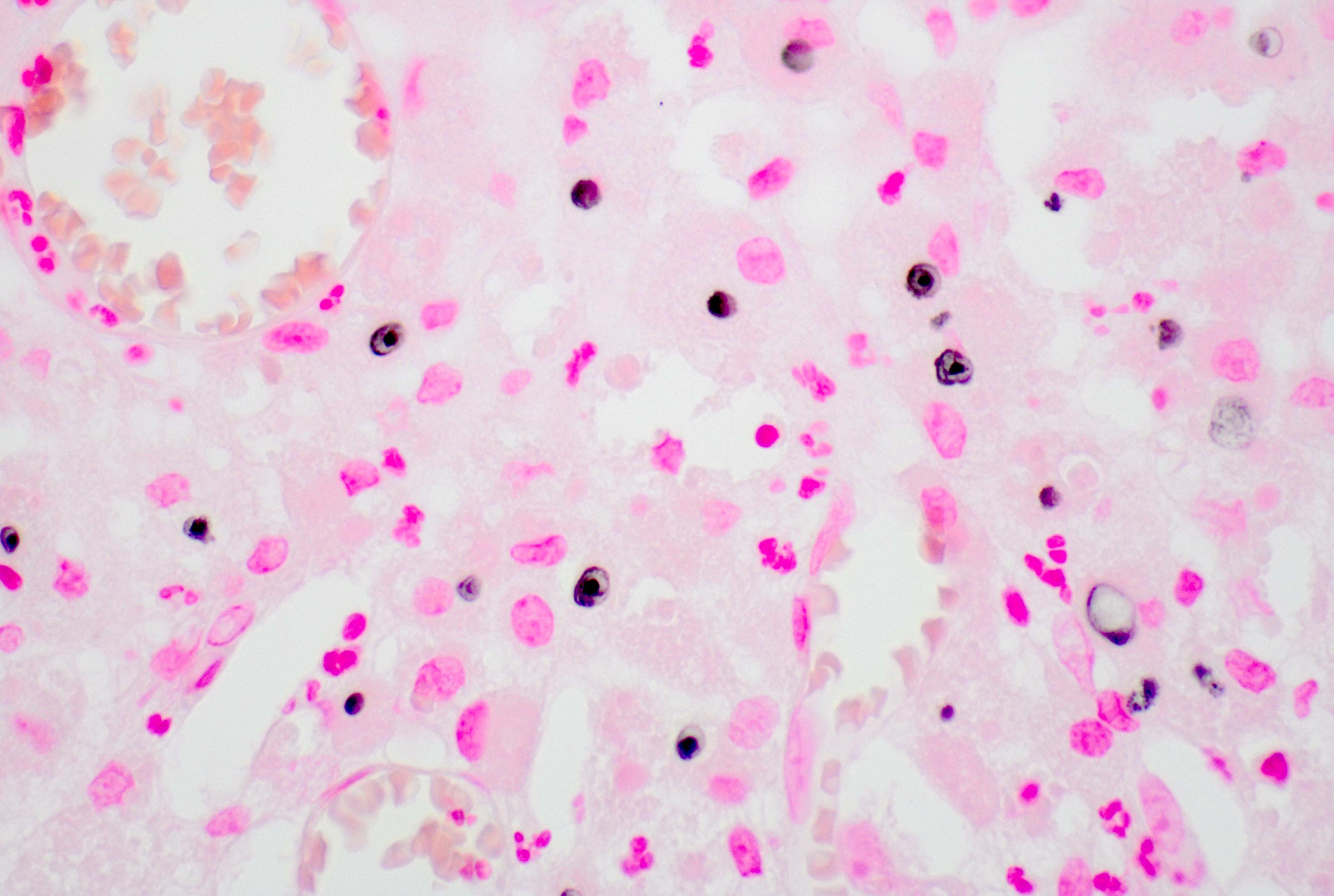
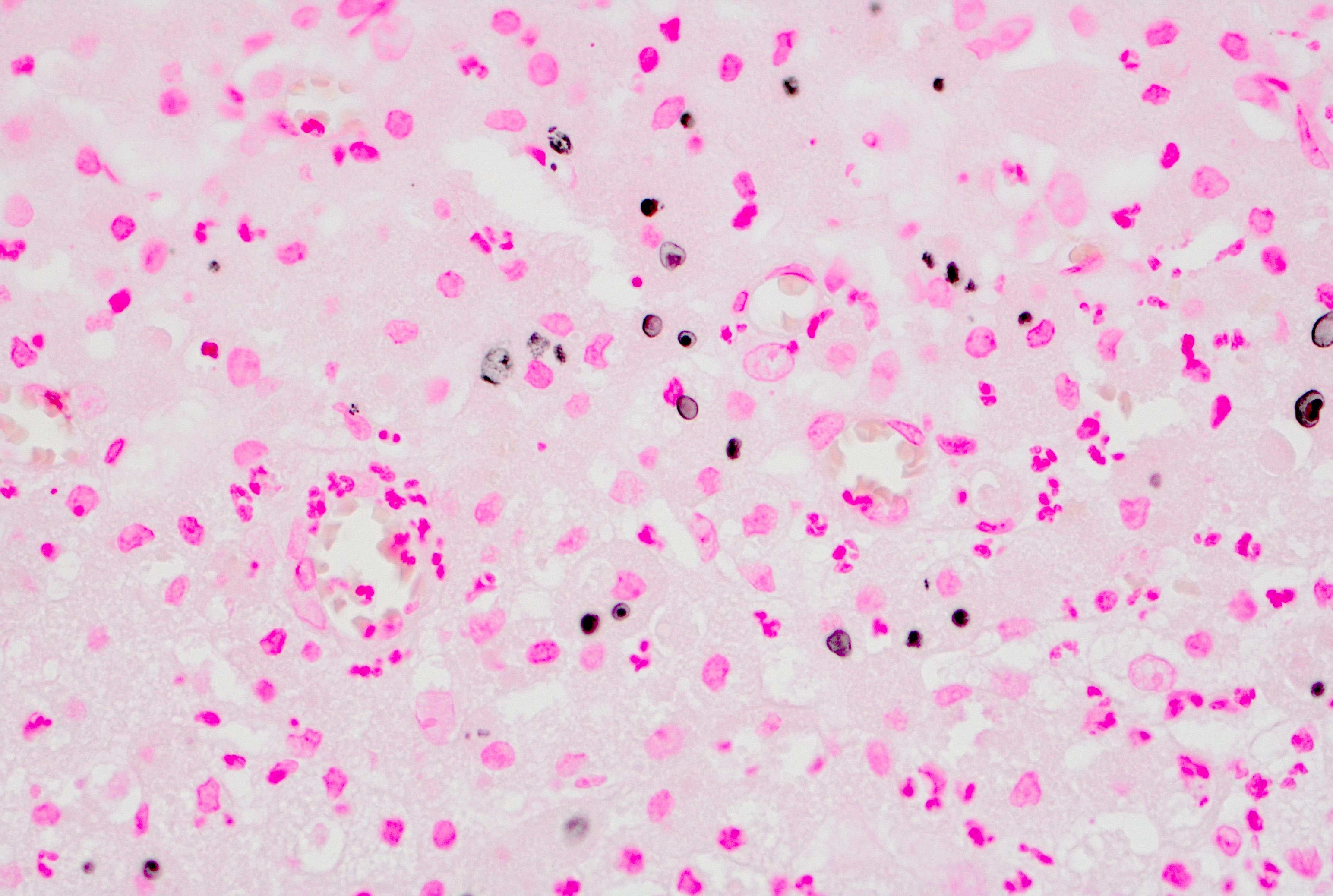
Sheets of eosinophilic histiocytes
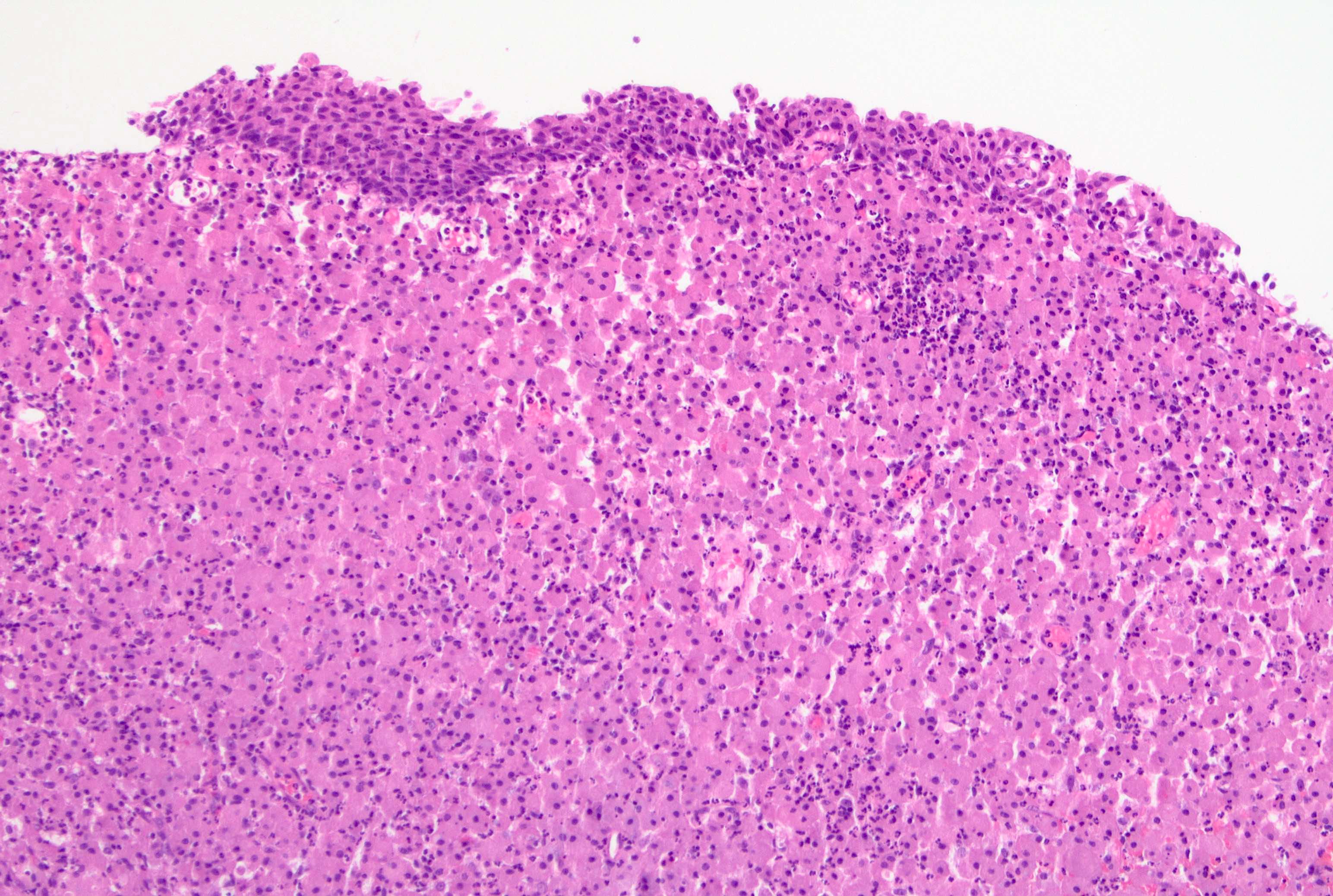
Urinary bladder with histiocytic infiltration
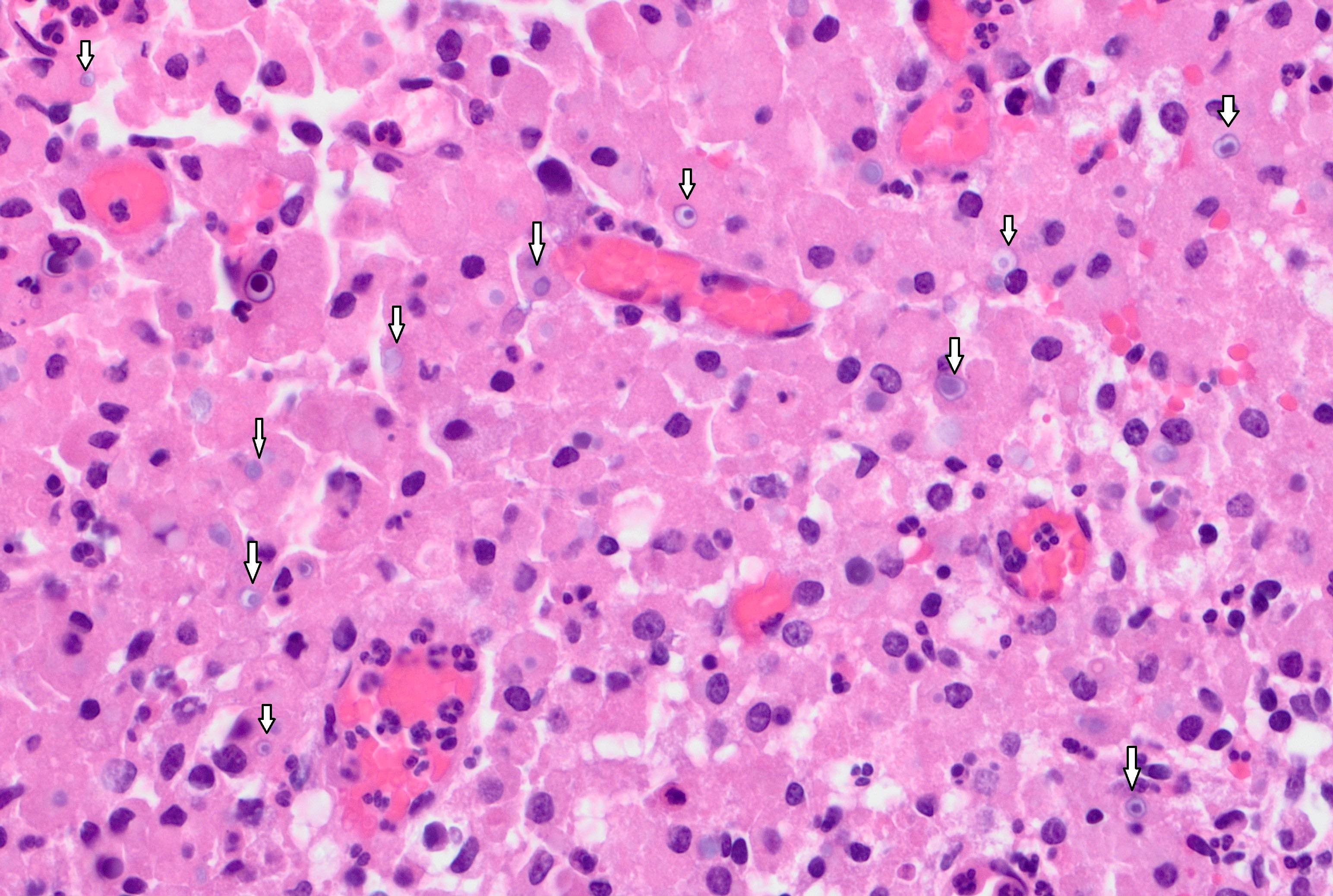
Numerous intracytoplasmic inclusions
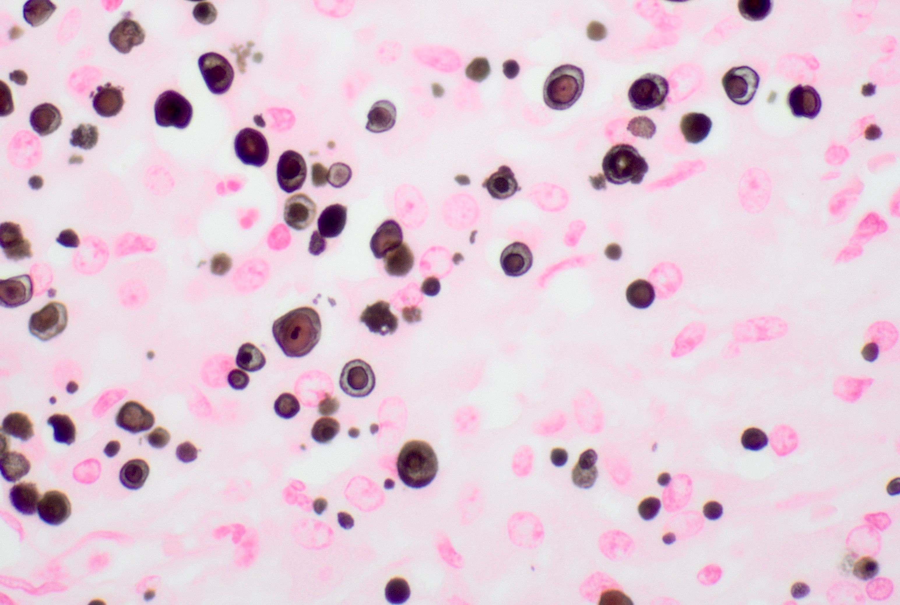
Von Kossa stain
Von Kossa stain
Positive stains
- Von Kossa, Alizarin red
- Prussian blue
- CD68, CD163
- Reference: Histopathology 2022;81:520
Negative stains
Sample pathology report
- Bladder lesions, biopsy:
- Benign urothelial mucosa with acute and chronic inflammation and prominent histiocytic infiltrate, consistent with malakoplakia (see comment)
- Comment: Special stains for von Kossa (calcium) were performed on block A1 and highlight concentrically layered basophilic inclusions (Michaelis-Gutmann bodies), supporting the diagnosis of malakoplakia.
- Bladder lesions, biopsy:
- Malakoplakia
Differential diagnosis
- Urothelial carcinoma (poorly differentiated, plasmacytoid and lymphoepithelioma-like):
- Overlying epithelium may show in situ lesion
- Cytokeratin stains are positive
- Gleason pattern 5 prostatic adenocarcinoma:
- Cytokeratin stains are positive
- NKX3.1 stain is positive
Practice question #1
What is the name of the targetoid bodies that are pathognomonic for malakoplakia?
- Asbestos bodies
- Aschoff bodies
- Howell-Jolly bodies
- Michaelis-Gutmann bodies
- Psammoma bodies
Practice answer #1
D. Michaelis-Gutmann bodies are characteristic inclusions seen in many cases of malakoplakia. They are rounded, concentric, intracytoplasmic, basophilic inclusions, caused by accumulation of undigested bacteria and products that are calcified. They can be highlighted by von Kossa stain. Answer A is incorrect because asbestos bodies are golden brown colored and fusiform to beaded rod shaped structures. Answer B is incorrect because Aschoff bodies are microscopic nodules of cells composed of pleomorphic cells, lymphocytes, plasma cells and fibrinoid necrosis. Answer C is incorrect because Howell-Jolly bodies are spherical intracytoplasmic inclusions within red blood cells. Answer E is incorrect because psammoma bodies are a round collection of calcium, a form of calcification that is mostly extracellular and much bigger than an intracellular inclusion.
Comment Here
Reference: Malakoplakia
Comment Here
Reference: Malakoplakia
Practice question #2
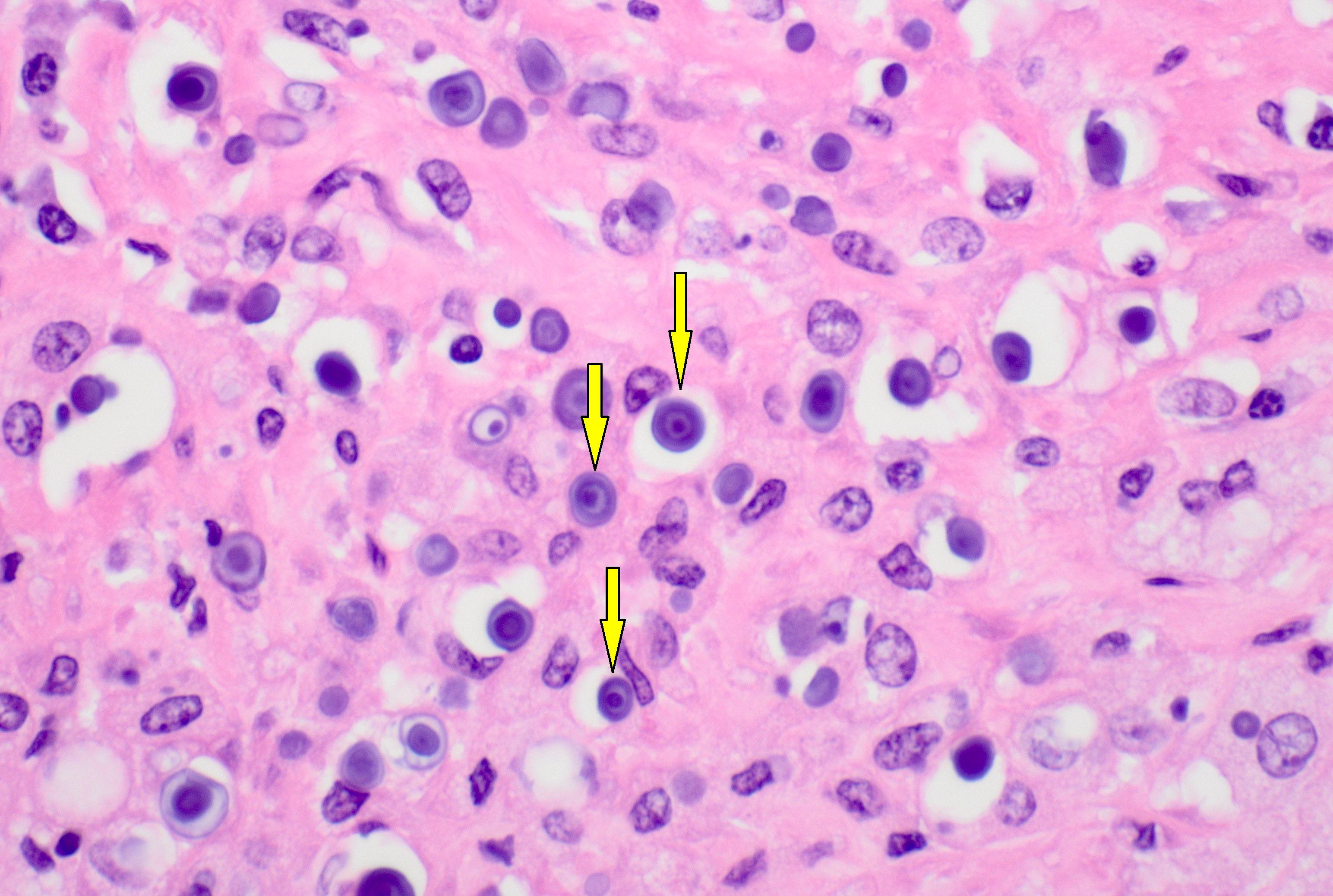
Which of the following stains used in the diagnosis of malakoplakia targets structures shown with yellow arrows in the image above?
- CD4
- CD68
- CD163
- Von Kossa
Practice answer #2
D. Von Kossa. The von Kossa stain highlights the Michaelis-Gutmann bodies in malakoplakia. Answers B and C are incorrect because CD68 and CD163 stains highlight the histiocytes in malakoplakia. Answer A is incorrect because CD4 has no role in the diagnosis of malakoplakia.
Comment Here
Reference: Malakoplakia
Comment Here
Reference: Malakoplakia
