

Bone & joints
Other nonneoplastic
Paget disease
Editorial Board Member: Jose G. Mantilla, M.D.
Deputy Editor-in-Chief: Borislav A. Alexiev, M.D.


Last author update: 8 February 2023
Last staff update: 8 February 2023
Copyright: 2003-2024, PathologyOutlines.com, Inc.
PubMed Search: Paget disease of bone
Table of Contents
Definition / general | Essential features | Terminology | ICD coding | Epidemiology | Sites | Pathophysiology | Etiology | Clinical features | Diagnosis | Laboratory | Radiology description | Radiology images | Prognostic factors | Case reports | Treatment | Frozen section description | Microscopic (histologic) description | Microscopic (histologic) images | Virtual slides | Cytology description | Positive stains | Molecular / cytogenetics description | Videos | Sample pathology report | Differential diagnosis | Board review style question #1 | Board review style answer #1 | Board review style question #2 | Board review style answer #2Cite this page: Enniss BA, Hariri DJ, de la Roza G. Paget disease. PathologyOutlines.com website. https://www.pathologyoutlines.com/topic/bonepagets.html. Accessed April 20th, 2024.
Definition / general
- Chronic disease of bones with episodes of increased bone resorption (osteoclast activity, osteolytic phase) followed by excessive mixed osteoblast and osteoclast activity (mixed phase) leading to disordered, poorly formed bone with increased density (osteosclerotic phase) and increased likelihood of fractures
- Not related to Paget disease of breast or extramammary Paget disease, although both were discovered by Sir James Paget, one of the founders of pathology (Wikipedia: James Paget [Accessed 1 December 2022], Orthop Clin North Am 2015;46:577)
Essential features
- Essentially, accelerated bone turnover making disordered bone
- Multiple phases are present, showing poorly formed bone, increased osteoclast activity (bites in the bone) and osteoblastic rimming
- Diagnosis is usually made radiographically
- Can be monostotic or polyostotic
Terminology
- Also called osteitis deformans
ICD coding
Epidemiology
- Most are over age 55; rare before age 40 (Am J Med 2018;131:1298)
- Fairly common in older people with estimates ranging near 2.3% in that population (J Bone Miner Res 2000;15:461)
- Most common in Western Europe and locations settled by those populations (U.S., Canada, Australia, etc.); rare in Africa and those of African descent, Scandinavia and Asia (Am J Med 2018;131:1298)
- Slight male predominance (4:3)
Sites
- Patients can present as polyostotic (multiple bones) or monostotic (one bone)
- Most common sites:
- Spine and pelvis (30 - 75%)
- Sacrum (30 - 60%)
- Skull (25 - 65%)
- Femur (25 - 25%)
- Rare in hands / feet, ribs, fibula (Wien Med Wochenschr 2017;167:9)
Pathophysiology
- Believed to be a disease of osteoclasts
- Genetic and environmental problems can lead to disruption in osteoclast differentiation and activation:
- Receptor activator of nuclear factor kappa Β ligand (RANKL), osteoclast differentiation
- Macrophage colony stimulating factor (MCSF), macrophage differentiation
- Nuclear factor kappa B (NFκB), apoptosis regulation
- Osteoprotegrin, bone remodeling (Curr Osteoporos Re 2021;19:327)
- Abnormal osteoclast activity leads to exaggerated physiologic response
- Not fully understood
Etiology
- Not fully understood
- May be due to slow virus infection of paramyxovirus, similar to subacute sclerosis leukoencephalitis (virus identified in osteoblasts)
- Several gene mutations have been identified in familial and sporadic forms (Curr Osteoporos Re 2021;19:327)
Clinical features
- Patients are generally asymptomatic or have mild symptoms
- May show bone pain and deformities
- Bone overgrowth or deformity can cause osteoarthritis and nerve impingement
- Initial presentation is commonly after pathologic fracture
- Incidental finding after imaging studies or serum alkaline phosphatase for other reasons
- Also associated with high output congestive heart failure in polyostotic disease due to shunting of blood through highly vascular lesions (Am J Med 2018;131:1298)
- While uncommon, osteitis deformans can progress to sarcoma, most of which are osteosarcoma; it has a very poor prognosis (Orthop Clin North Am 2015;46:577)
Diagnosis
- Diagnosis is primarily radiologic
- Serum alkaline phosphatase (ALP) can be helpful if positive and used to monitor disease progression
- Biopsy is not needed in routine cases but can be useful if radiographically distinct lesions appear in an abnormal population or if there is concern that the lytic lesion is a metastasis; monostotic lesions that are not radiographically characteristic should be biopsied
Laboratory
- Elevated serum ALP
- ALP isoenzyme electrophoresis for bone type
- Most patients with Paget have normal ALP, more so than those with monostotic disease
- Other markers of increased bone turnover: procollagen type I N terminal propeptide (PINP), serum C telopeptide (CTx), urinary N telopeptide (NTx) and urinary hydroxyproline (Am J Med 2018;131:1298)
- Serum calcium and phosphorus are generally normal
Radiology description
- Early: radiolucency
- Well defined lytic lesion is called osteoporosis circumscripta and is fairly specific for Paget disease when occurring in the frontal bone (Wien Med Wochenschr 2017;167:9)
- Late: increased bone density, cortical thickening with marked tunneling, increased prominence of trabeculae, increased microfractures, loss of distinction between cortex and medulla; may have sharp demarcation between normal and affected bone; may extend into soft tissue if florid disease
- Long bone can become bowed
- Bone scintigraphy will show increased uptake in active lesions
Radiology images
Contributed by Jose G. Mantilla, M.D.
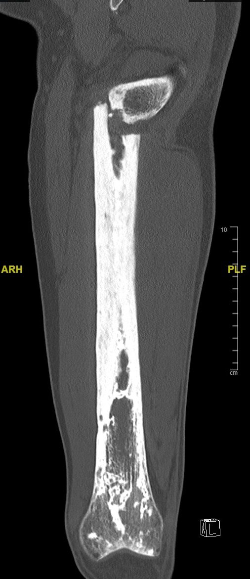
Coronal CT of femur
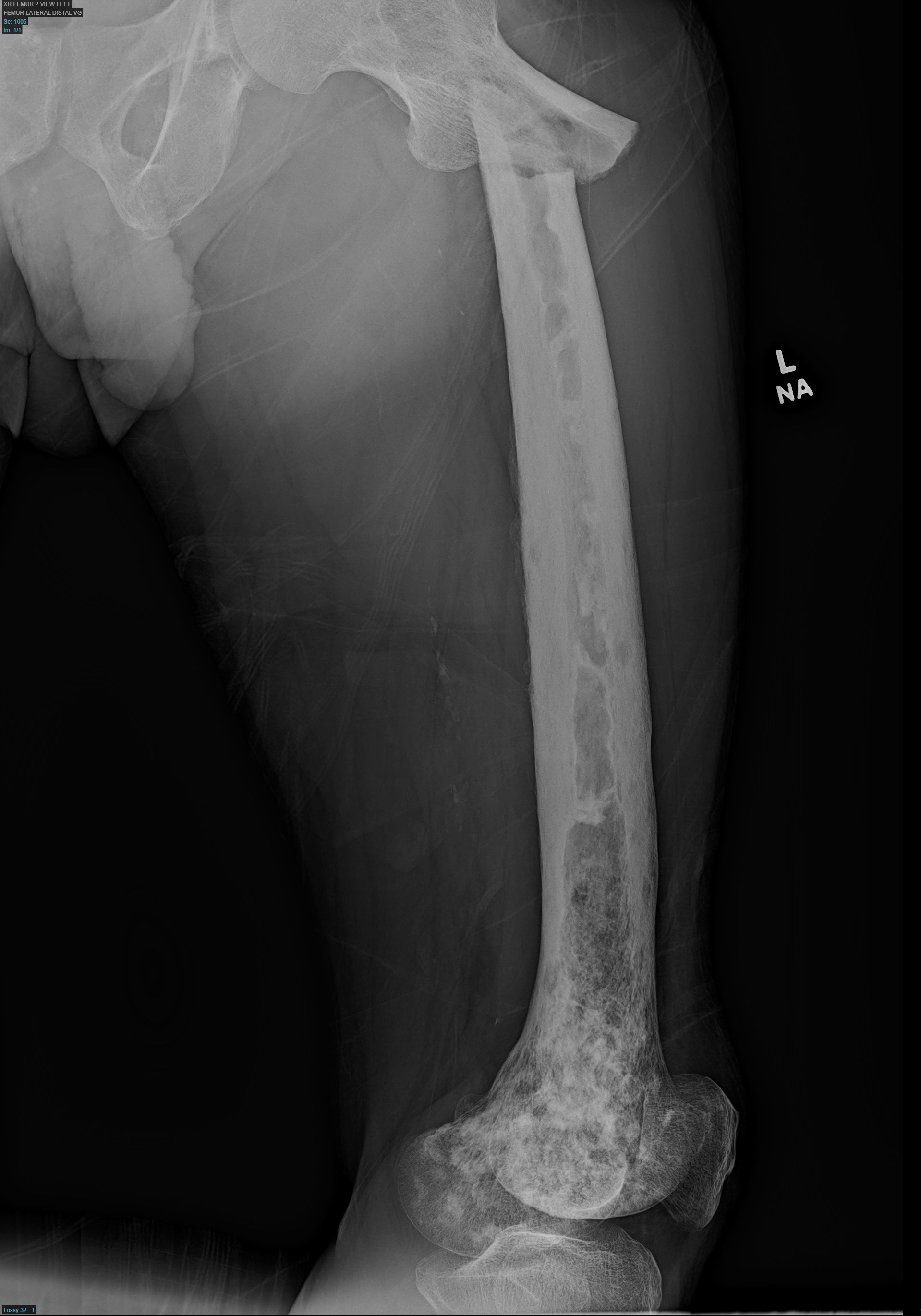
Xray of femur
Prognostic factors
- Progression to sarcoma is a poor prognostic factor
Case reports
- 60 year old man with JPD1 who described hardening of his external ears at age 45 years, after 4 years of treatment with bisphosphonates (Am J Med Genet A 2016;170A:978)
- 74 year old African American man was referred for the investigation of symmetrical polyarthritis, left upper arm joint deformity and low back pain (Am J Case Rep 2019;20:764)
- 75 year old man with a rapidly progressive cervical myelopathy on a background of a 3 year history of neck pain and a severely degenerative cervical spine (Ann R Coll Surg Engl 2019;101:e38)
Treatment
- Bisphosphonates
- Calcitonin
Frozen section description
- Intraoperative evaluations of bone should not be performed
Microscopic (histologic) description
- Major findings are haphazard layers of lamellar bone with woven bone, irregular cement lines between layers of lamellar bone, thickened trabeculae with osteoblastic rimming and areas of bone resorption (bite marks) with abnormal osteoclasts, peritrabecular fibrosis and increased vascularity of marrow
- Early: primarily woven bone
- Focal mosaic pattern of lamellar bone, resembles jigsaw puzzle with prominent irregular cement lines
- Osteoclasts present at surface of bone with bite marks into the bone
- In osteolytic phase, osteoclasts are increased and abnormal with up to 100 nuclei, peritrabecular fibrosis, increased vascularity
- Middle phase: enlarged trabeculae with very irregular shapes and abnormal cement lines; prominent osteoblast activity
- Late: significantly decreased osteoblast and osteoclast activity, thick trabeculae and thicker bones with mosaic pattern of cement lines; fine fibrosis of marrow
- All phases may be identified within a single specimen
Microscopic (histologic) images
Contributed by Dana J. Hariri, M.D.
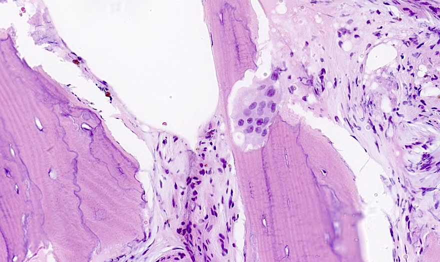
Osteoblastic rimming
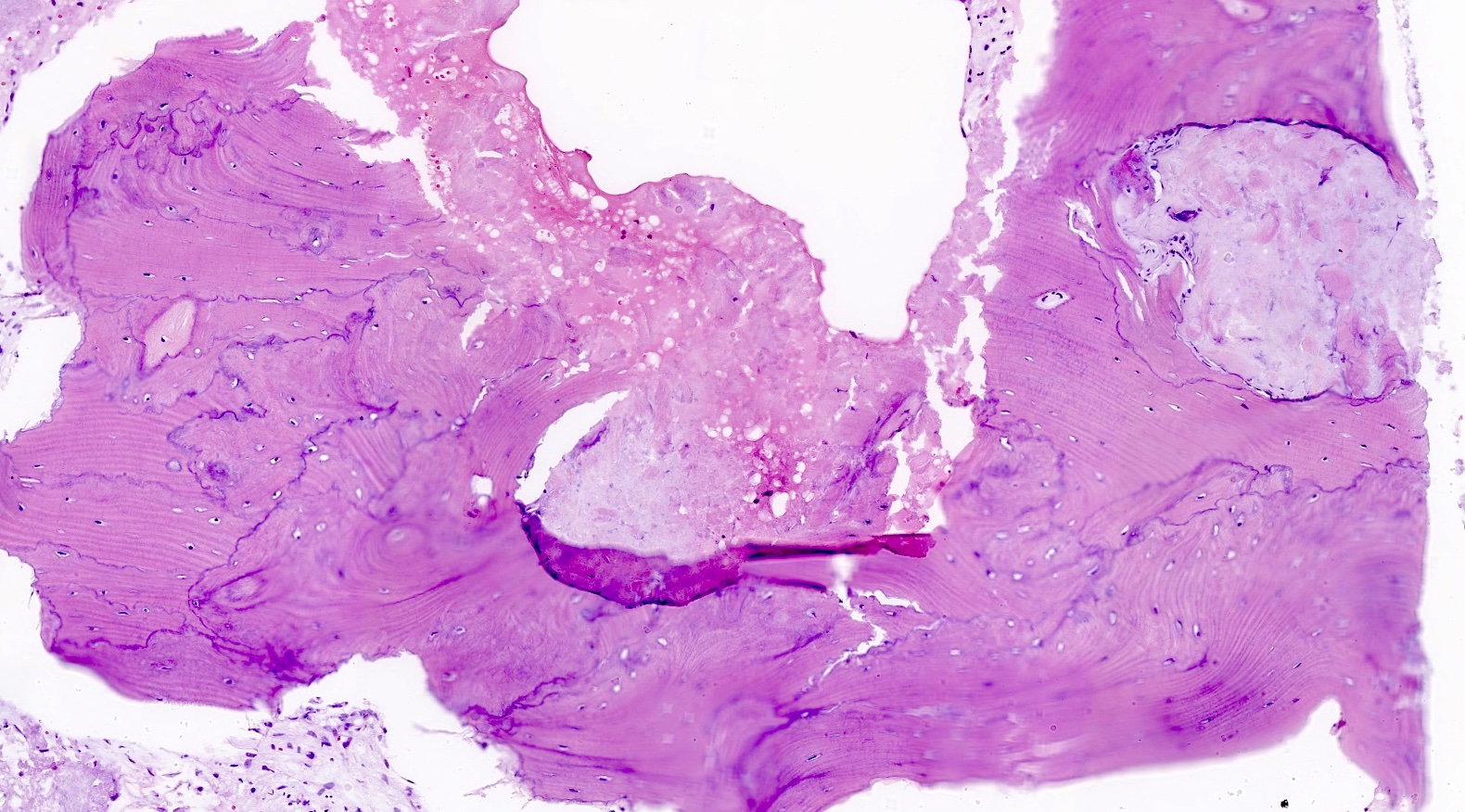
Jigsaw pattern
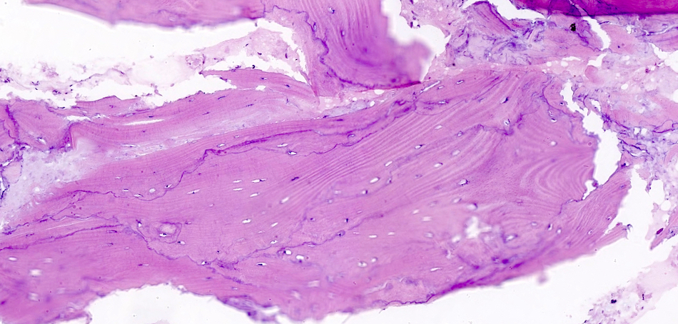
Haphazard lamellar bone
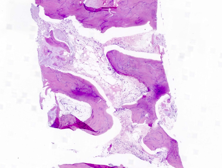
Peritrabecular fibrosis
Virtual slides
Images hosted on other servers:

Prominent mosaic pattern and marrow fibrosis
Cytology description
- There is no utility for cytology
Positive stains
- Reticulin (highlights disorganization of lamellar bone)
- IHC has little utility
Molecular / cytogenetics description
- SQSTM1 mutations are the most common in familial Paget disease of bone (Curr Osteoporos Re 2021;19:327)
- TNFRSF11A gene encoding RANKL leads to increased osteoclast activity
Videos
Overview of diagnosis and management of Paget disease
Sample pathology report
- Bone, distal femur, excision:
- Osteitis deformans, Paget disease of bone
Differential diagnosis
- Reactive bone formation adjacent to neoplasm:
- Cement lines not as prominent
- Clinical setting of adjacent neoplasm which can be seen on imaging
- Chronic osteomyelitis:
- Lack of mosaic pattern of bone formation
- Can have fibrosis with new bone formation
- Fibrosis usually accompanied by chronic inflammatory cells
- Polyostotic fibrous dysplasia:
- Branching and anastomosing irregular trabeculae of woven bone
- Lacks prominent osteoblastic rimming
Board review style question #1
Which of the following is true of the lesion shown in the picture above?
- Heat stable alkaline phosphatase will be elevated
- It has a very poor prognosis and the patient will likely die within a year
- It will likely progress to a low grade sarcoma
- The lesion rarely has more than one site of involvement
- Pathologic fractures are a common initial presentation
Board review style answer #1
E. Pathologic fractures are a common initial presentation
Paget disease of bone most commonly presents with a pathologic fracture but can present as generic bone pain. It very rarely progresses to any form of sarcoma but it is a complication that presages a poor outcome; otherwise, the outcome is more benign. Alkaline phosphatase is sometimes elevated but the bone isoenzyme of ALP is heat labile (bone burns, liver lasts). It is important to note that while temperature stability is often mentioned for alkaline phosphatase isoenzymes, electrophoresis is the preferred way to subtype them. The lesions can be monostotic or polyostotic.
Comment Here
Reference: Paget disease
Board review style question #2
Of the following sites, which is the most likely to show Paget disease?
- Metatarsal of the hand
- Pelvis
- Ribs
- Tibia
Board review style answer #2
B. Pelvis. Common sites of involvement are pelvis, spine, skull, tibia, femur, skull, vertebrae and humerus. Hand, feet and ribs are very rare.
Comment Here
Reference: Paget disease
Comment Here
Reference: Paget disease
