
Breast
Other carcinoma subtypes, not WHO classified
Cystic hypersecretory carcinoma

Copyright: 2001-2024, PathologyOutlines.com, Inc.
PubMed search: cystic hypersecretory [title] carcinoma breast invasive
- Not part of WHO breast classification
- First described in 1984 (Am J Surg Pathol 1984;8:31)
- Very rare (< 100 cases reported)
- DCIS or hyperplasia is more common
- Usually low grade for several years but may metastasize
- 40 year old woman with painful breast mass (Arch Pathol Lab Med 2005;129:e79)
- 43 year old woman (Jpn J Radiol 2011;29:660)
- 45 year old woman (J Korean Med Sci 2004;19:149)
- 48 year old woman with Paget’s disease of nipple (Int J Surg Pathol 2008;16:208)
- 49 year old woman with invasive lobular carcinoma in opposite breast 10 years after diagnosis (Arch Pathol Lab Med 1999;123:1108, free full text)
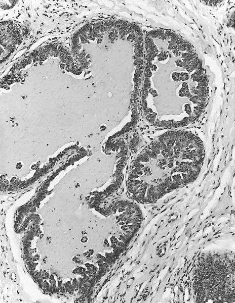
- Numerous cysts with mucoid or gelatinous secretions
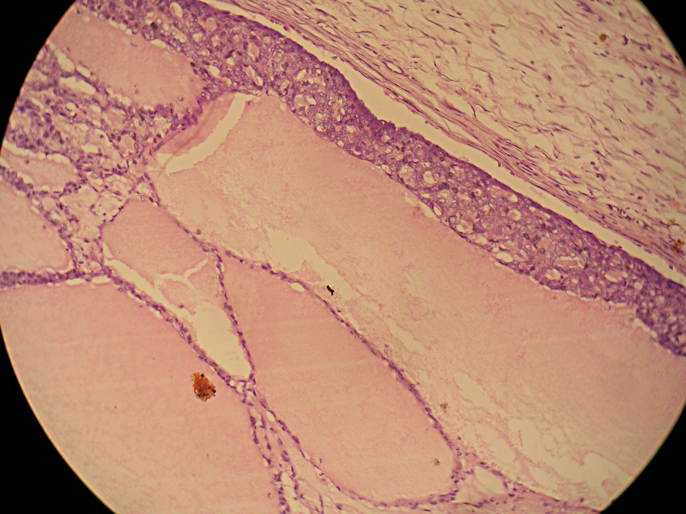
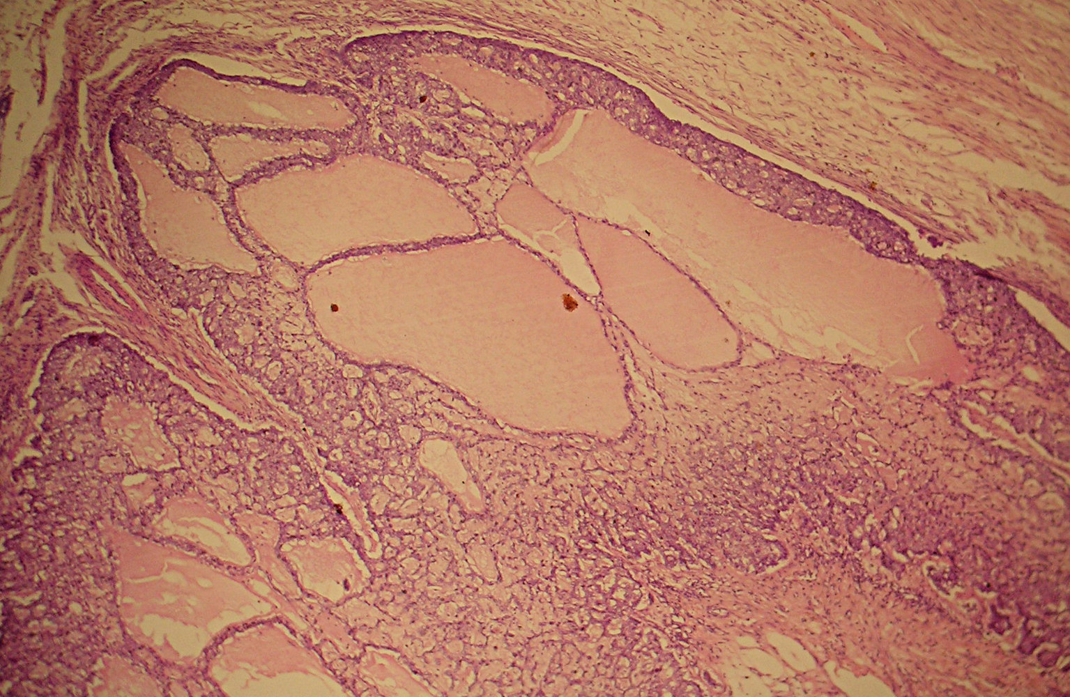
Contributed by Dr. Okechukwu C. Okafor
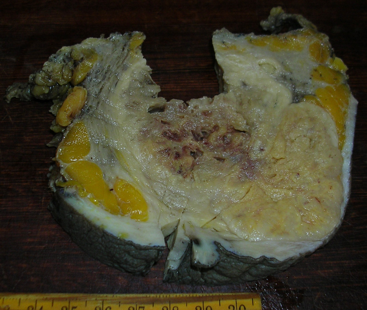
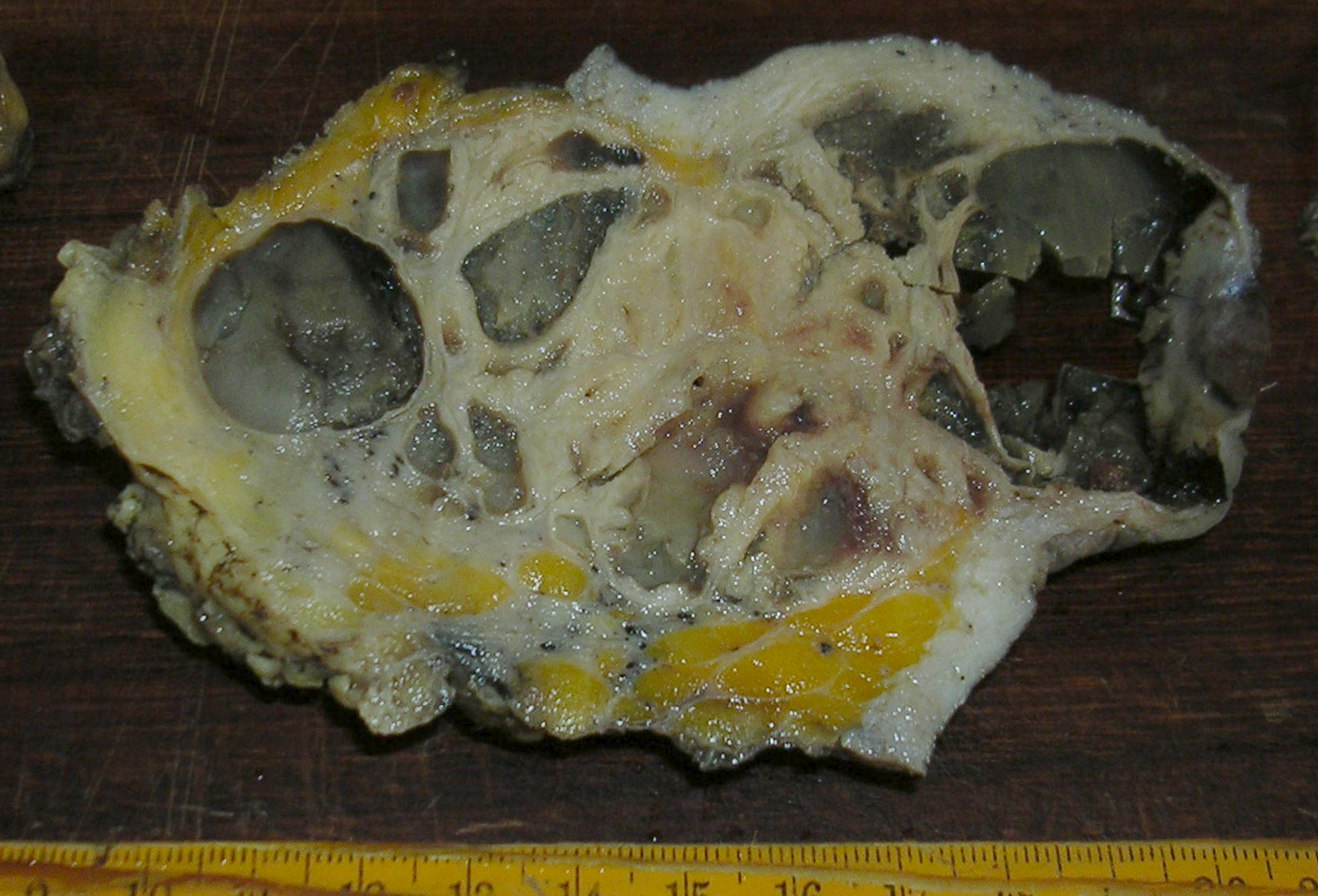
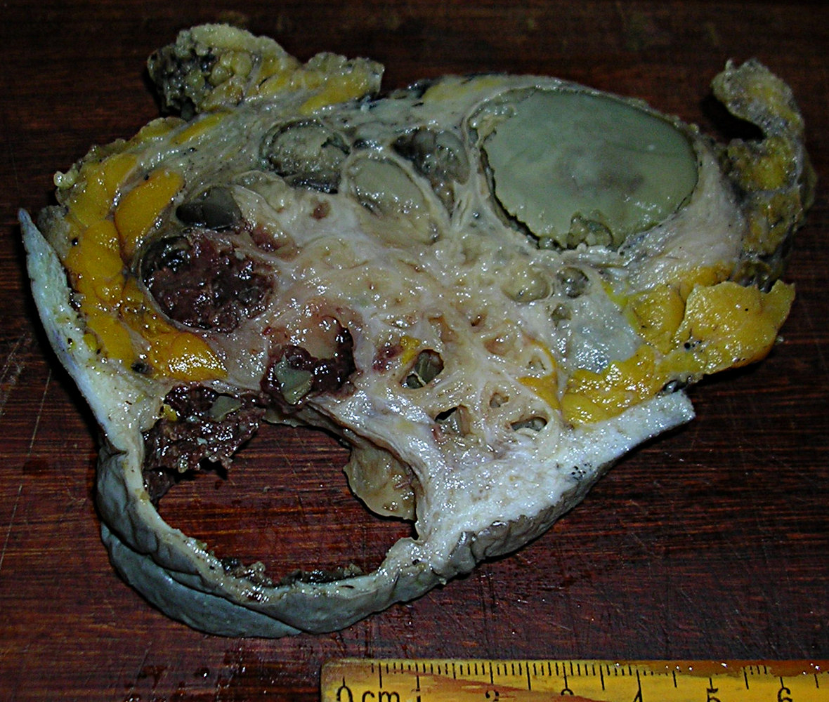
Mastectomy with 12 cm multicystic tumor
Images hosted on other servers:

Numerous cysts with a gelatinous secretion
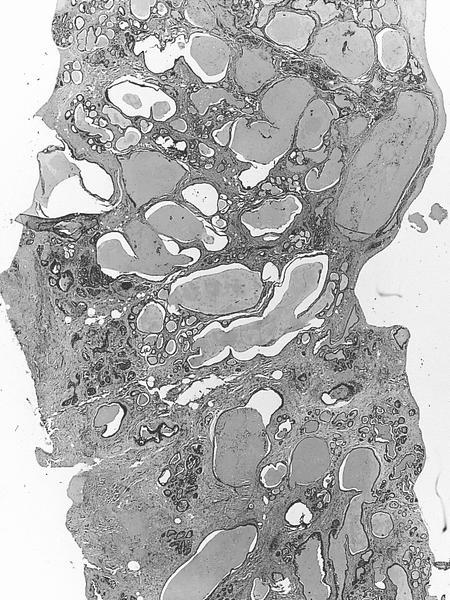
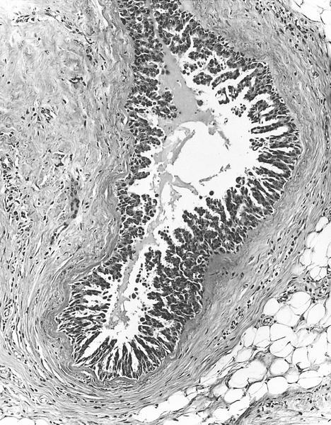
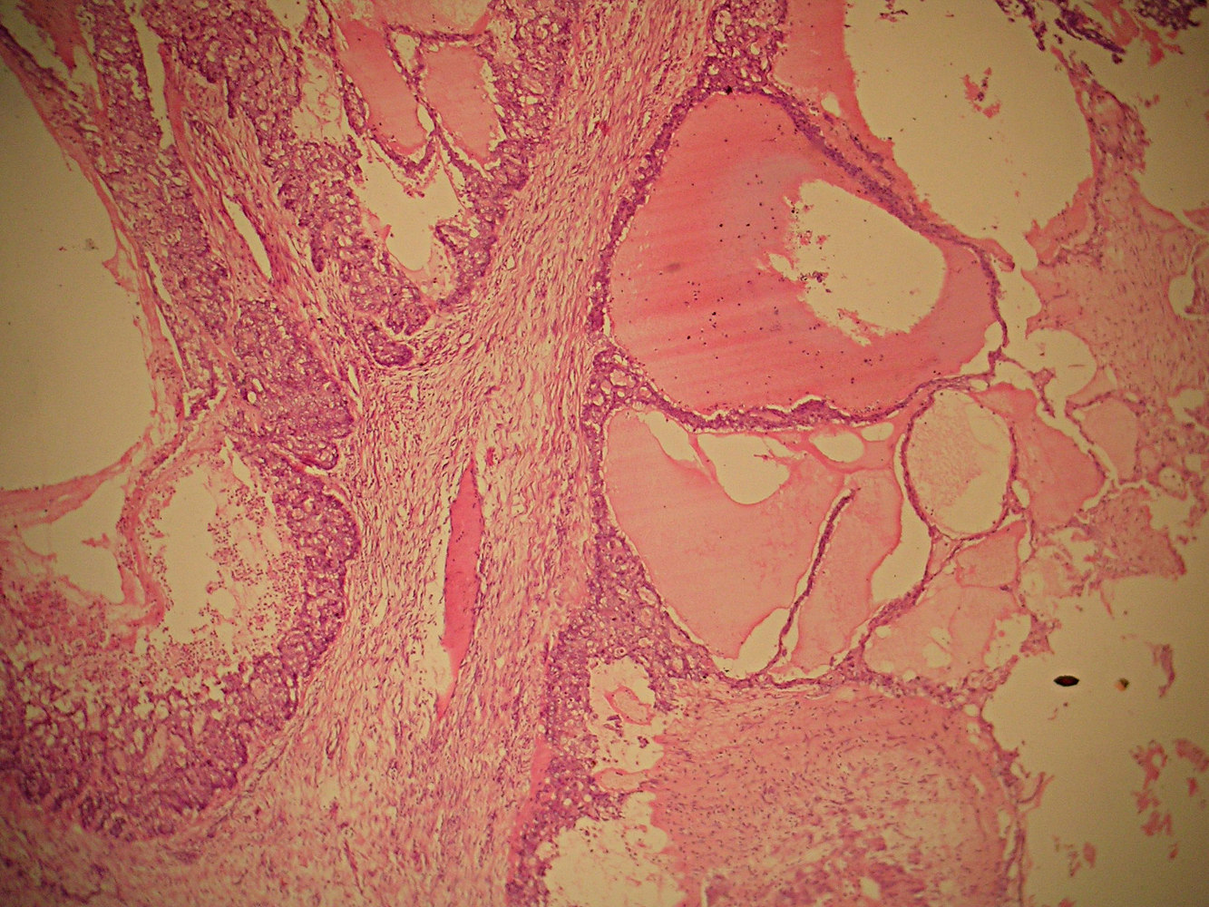
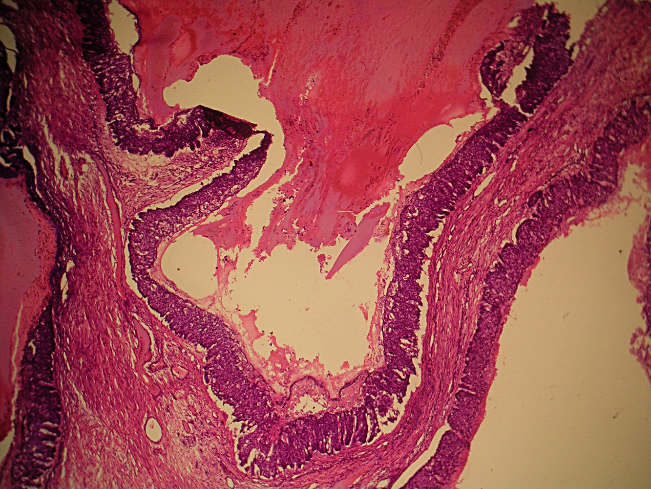
- Cystic dilation of ducts containing colloid-like eosinophilic material that often retracts from epithelium
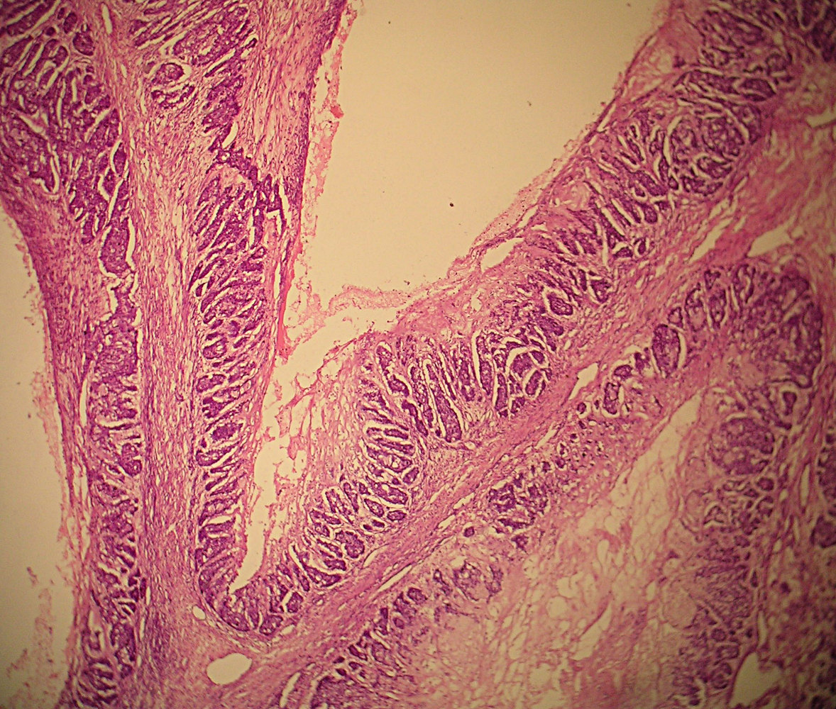
- Epithelium focally has micropapillary DCIS
- Also invasion of surrounding stroma by nests of carcinoma, which may be high grade, usually without hypersecretory characteristics
- Extravasation of cyst material into stroma is not invasion
AFIP images

ated carcinoma
invades stroma
next to cyst
prominent cysts with
no apparent ducts
containing carcinoma
Associated micropapillary DCIS, invasion elsewhere

Associated micropapillary DCIS with no
evident secretion in tumor cells, which have
a hobnail appearance, nuclei are relatively
clear with small, discrete nucleoli
Associated micropap-
illary DCIS with sparse
secretion that is re-
tracted from epithelium

Cysts lined by flat cuboidal epithelium contain
homogeneous secretions, these cysts are
nonspecific - they can be found in cystic
hypersecretory hyperplasia or carcinoma
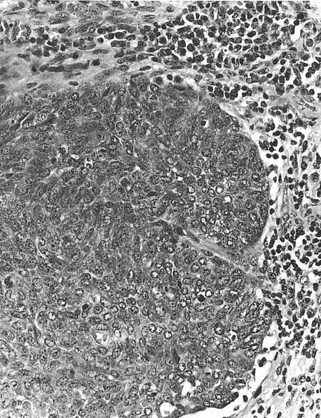
Axillary nodal
metastases, with
some cells exhib-
iting clear nuclei

Note transition in cyst epithelium with plaque of tumor cells
in bottom half, micropapillary pattern is obscured where carcinoma
nearly fills ducts, but traces of retracted secretion remain
(arrows), clear nuclei are also evident, even at this magnification
Contributed by Dr. Okechukwu C. Okafor
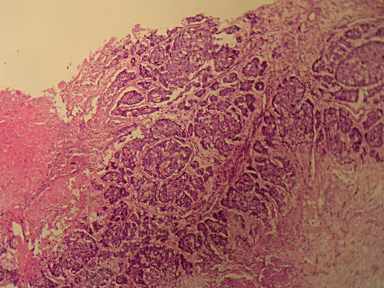
12 cm tumor
Images hosted on other servers:

Various images
- Orange to grayish green colloid-like background with cracking artifact (Pap stain), clusters of malignant cells
- Also histiocytes and apocrine cells (Acta Cytol 1999;43:273, Acta Cytol 1997;41:892)
- Androgen receptors, HER2 (Ceska Gynekol 2005;70:73)
- Variable p53, ER and PR (Histopathology 2005;46:43)
- Secretory carcinoma: predominantly microcysts, t(12;15)(p13;q25) in most cases
- Mucinous / colloid carcinoma: extracellular mucin, not intracystic secretions
- Cystic hypersecretory hyperplasia: no invasion present (Cancer 1988;61:1611)
