
Breast
Other nonneoplastic
Secretory change
Author: Hind Warzecha, M.D.

Last author update: 1 June 2010
Last staff update: 23 April 2024 (update in progress)
Copyright: 2002-2024, PathologyOutlines.com, Inc.
PubMed Search: Pregnancy-like hyperplasia
Table of Contents
Definition / general | Terminology | Epidemiology | Etiology | Clinical features | Treatment | Gross description | Microscopic (histologic) description | Microscopic (histologic) images | Differential diagnosis | Additional referencesCite this page: Warzecha H. Secretory change. PathologyOutlines.com website. https://www.pathologyoutlines.com/topic/breastpseudolactionalhyperplasia.html. Accessed April 25th, 2024.
Definition / general
- Pregnancy-like changes in a nonpregnant, nonlactating patient
Terminology
- See also pregnancy / lactation, lactating adenoma
Epidemiology
- Present in 2 - 3% of breast biopsies
- Identified in needle localization and core biopsies due to calcifications or presence of a mass
- Women, mean age 44 years, range 38 - 52 years
- Not lactating, not pregnant (by definition)
Etiology
- May be associated with phenothiazine or other medications (Am J Clin Pathol 1987;87:23)
Clinical features
- Often multifocal
- Associated with / may merge with cystic hypersecretory hyperplasia
- May have associated ADH or DCIS (Am J Clin Pathol 2004;122:714) but invasive carcinoma is rare (Am J Surg Pathol 2004;28:789)
Treatment
- Recommend excision if atypia found in core biopsy (Am J Surg Pathol 2000;24:1670)
Gross description
- No gross lesion
Microscopic (histologic) description
- Glands and terminal ducts with little or no secretion
- Glandular cells are swollen with abundant pale or clear, finely granular or vacuolated cytoplasm
- Luminal cytoplasmic borders of glandular cells are frayed with small cytoplasmic blebs extending into lumen that may contain nuclei
- Small, uniform, round and darkly stained nuclei
Microscopic (histologic) images
AFIP images
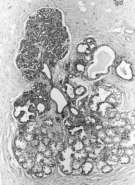
Expanded acinar glands
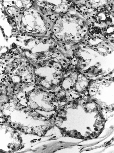
Cells have abundant pale cytoplasm but no secretion
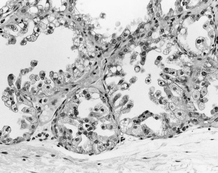
Micropapillary pattern
of columnar cells with
luminal cytoplasmic buds
Images hosted on other servers:


Cells have finely vacuolated cytoplasm
Differential diagnosis
- Apocrine adenosis or apocrine metaplasia: cytoplasm more eosinophilic, vacuoles are usually only focal
- Apocrine DCIS: abundant eosinophilic cytoplasm, architectural or cytologic atypia
- Columnar cell lesion: tightly packed columnar epithelial cells
Additional references
