

CNS & pituitary tumors
Embryonal tumors
Other embryonal tumors
CNS neuroblastoma, FOXR2 activated
Editorial Board Member: P.J. Cimino, M.D., Ph.D.
Deputy Editors-in-Chief: Debra L. Zynger, M.D., Chunyu Cai, M.D., Ph.D.


Last author update: 28 September 2023
Last staff update: 13 October 2023
Copyright: 2002-2024, PathologyOutlines.com, Inc.
PubMed Search: CNS neuroblastoma, FOXR2 activated

Table of Contents
Definition / general | Essential features | Terminology | ICD coding | Epidemiology | Sites | Pathophysiology | Etiology | Clinical features | Diagnosis | Laboratory | Radiology description | Radiology images | Prognostic factors | Case reports | Treatment | Gross description | Microscopic (histologic) description | Microscopic (histologic) images | Cytology description | Positive stains | Negative stains | Electron microscopy description | Molecular / cytogenetics description | Molecular / cytogenetics images | Videos | Sample pathology report | Differential diagnosis | Additional references | Board review style question #1 | Board review style answer #1 | Board review style question #2 | Board review style answer #2Cite this page: Anderson SA, Singh N. CNS neuroblastoma, FOXR2 activated. PathologyOutlines.com website. https://www.pathologyoutlines.com/topic/cnsneuroblastoma.html. Accessed April 16th, 2024.
Definition / general
- CNS neuroblastoma, FOXR2 activated (CNS NB FOXR2) tumors are embryonal neoplasms characterized by poorly differentiated neuroblastic or neuronal differentiation with variable neuropil rich stroma and ganglion cells
- Characterized by the activation of FOXR2 by structural rearrangements
Essential features
- Embryonal tumor with neuronal or neuroblastic differentiation and activation of FOXR2 rearrangement and fusion or a DNA methylation profile aligned with CNS neuroblastoma, FOXR2 activated
Terminology
- CNS neuroblastoma, FOXR2 activated
- Formerly recognized as one of the primitive neuroectodermal tumors of CNS (CNS PNETs)
ICD coding
- ICD-O: 9500/3 - neuroblastoma, NOS
- ICD-10: C71 - malignant neoplasm of brain
- ICD-11: 2A00.1Y & XH85Z0 - other specified embryonal tumors of brain & neuroblastoma, NOS
Epidemiology
- Supratentorial location in all tumors
- Slight female predilection (J Neuroimaging 2023;33:359)
- Range of ages at diagnosis, 1 - 20 years
- Median age at diagnosis is 5 years (Neuro Oncol 2021;23:1597)
Sites
- Supratentorial
Pathophysiology
- Thought to arise from neuroectodermal cells, although the exact cell of origin is not known (J Neuropathol Exp Neurol 2021;80:52)
- Most frequent genetic alterations are on the transcription factor gene FOXR2 (Cell 2016;164:1060)
- Although FOXR2 plays a causative role in the formation of CNS embryonal tumors, the exact underlying mechanisms have yet to be determined (Neuro Oncol 2019;21:993)
Etiology
- Risk factors have not been reported
Clinical features
- Common symptoms: headache, vomiting, seizures and focal neurologic deficits (J Neuroimaging 2023;33:359)
Diagnosis
- Embryonal tumor with neuronal or neuroblastic differentiation and activation of FOXR2 rearrangement and fusion or a DNA methylation profile aligned with CNS neuroblastoma, FOXR2 activated
Laboratory
- No specific laboratory findings for this entity
Radiology description
- Well circumscribed supratentorial mass with a mixed solid and cystic appearance (J Neuropathol Exp Neurol 2021;80:52)
- Solid component may show heterogeneous enhancement
Radiology images
Images hosted on other servers:

T2 weighted and postcontrast T1 weighted axial
Prognostic factors
- Favorable survival for patients with CNS NB FOXR2; higher rate of recurrences in nonirradiated and locally irradiated patients (Neuro Oncol 2021;23:1597)
Case reports
- 3 year old girl with a cystic right frontal lobe tumor (Brain Tumor Pathol 2020;37:100)
- Case series of CNS neuroblastoma, FOXR2 activated (J Neuroimaging 2023;33:359)
Treatment
- Maximal surgical resection followed by radiation to the brain and spinal cord; chemotherapy may also be given (NCI: Childhood Medulloblastoma and Other Central Nervous System Embryonal Tumors Treatment (PDQ®) - Patient Version [Accessed 5 September 2023])
Gross description
- CNS embryonal tumors are often pink, soft, well circumscribed masses; if a prominent desmoplastic component is present, it is often tan-white and firm
Microscopic (histologic) description
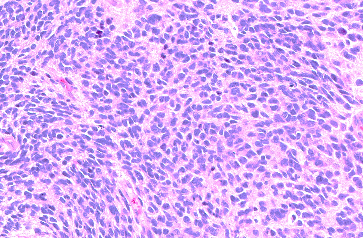
- Poorly differentiated neuroepithelial cells and neurocytic cells (Cell 2016;164:1060)
- Uniform small cells with high N:C ratio, oval to round with hyperchromatic nuclei
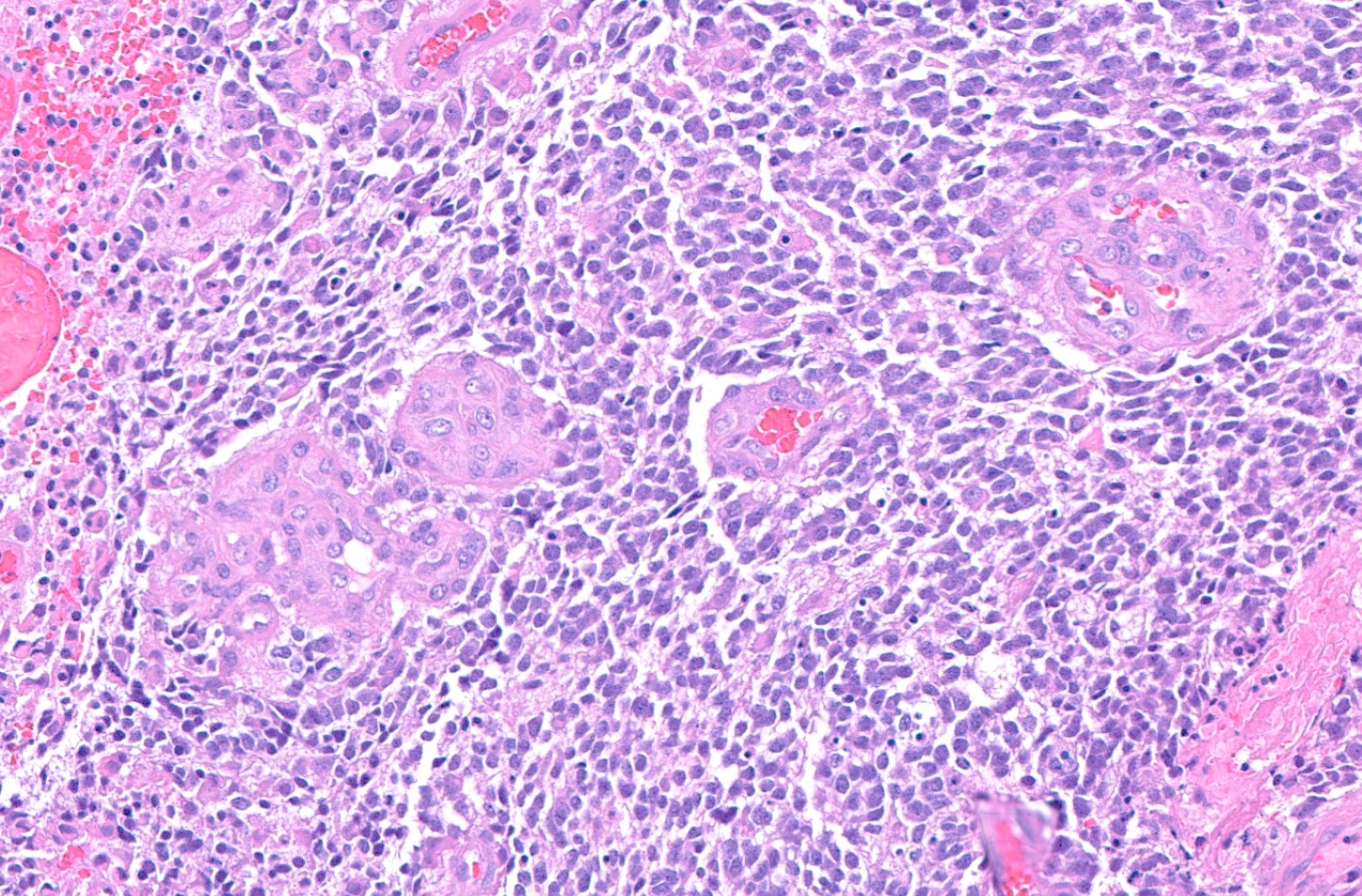
- Cells have little to no apparent cytoplasm in a background of neuropil with or without Homer Wright rosettes (J Neuropathol Exp Neurol 2021;80:52)
- Background of neuropil rich stroma
- Abundant mitotic activity
- Palisading necrosis can be present
- Infiltration of adjacent parenchyma is variable
Microscopic (histologic) images
Contributed by Nirupama Singh, M.D., Ph.D., Carmen Perrino, M.D. and P.J. Cimino, M.D., Ph.D.
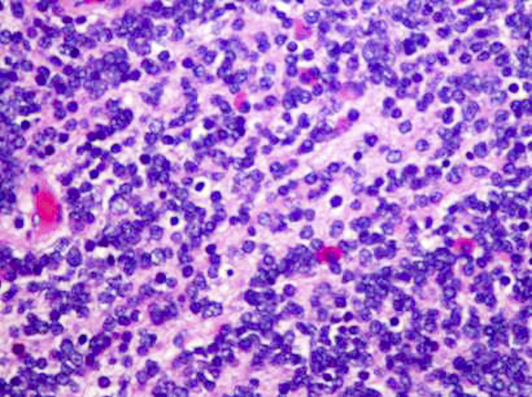
Neuroblasts with no cytoplasm
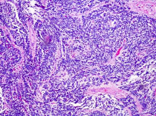
Homer Wright rosettes
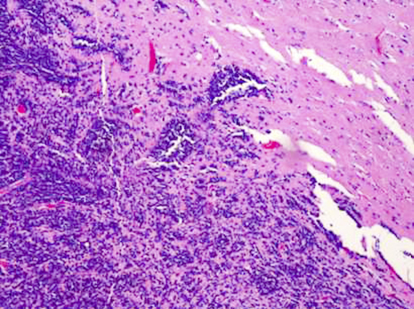
Small tumor cells
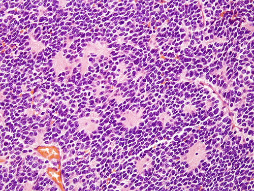
Pseudorosettes
Embryonal cells

Neurocytic cells

Focal ganglion cells
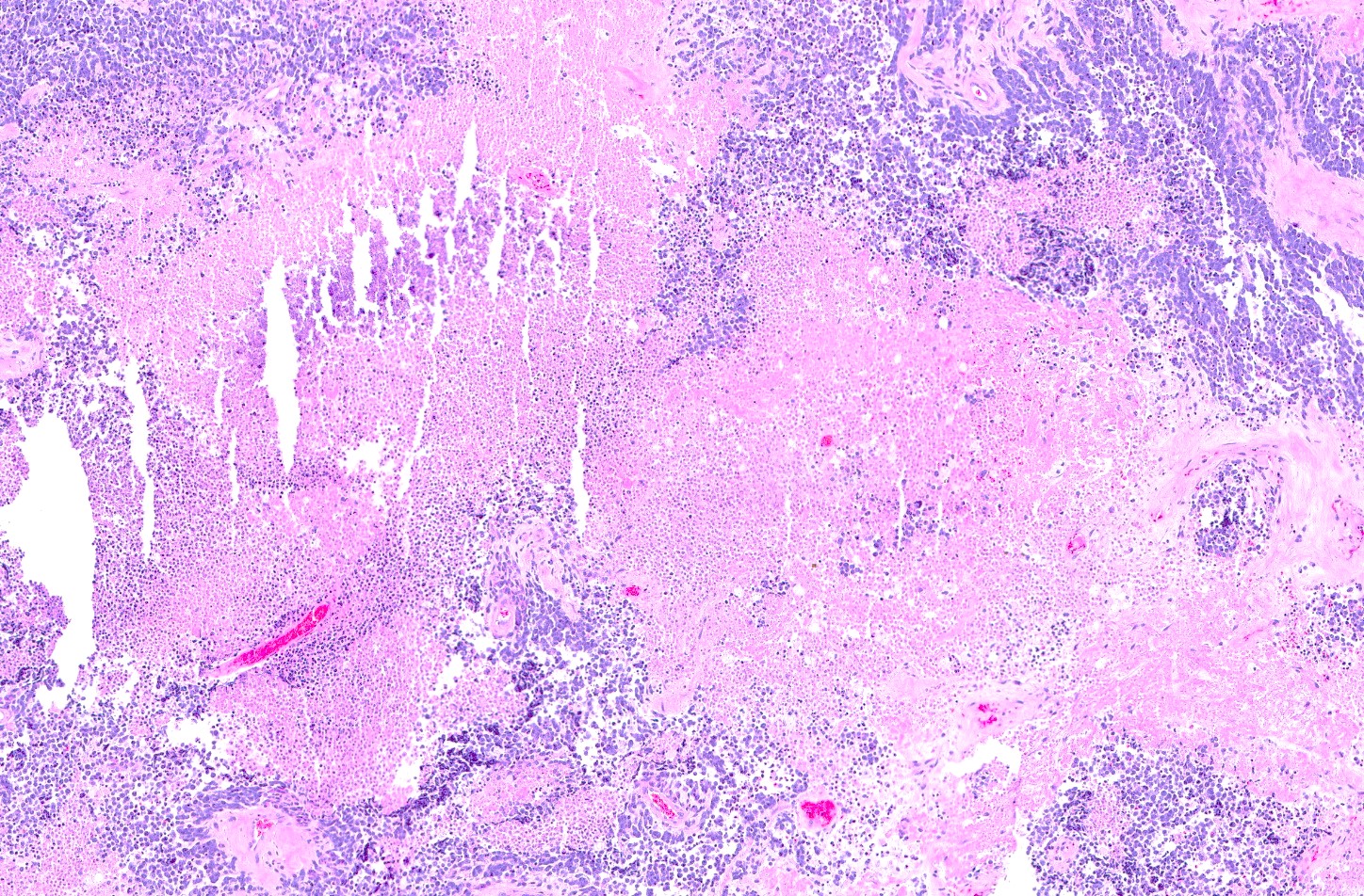
Necrosis
MVP
Cytology description
- Evaluation of cerebrospinal fluid (CSF) for the presence of tumor cells is required for staging (Radiology 1969;93:1351)
Positive stains
- Olig2
- Synaptophysin expression is accentuated in regions of neurocytic or ganglion cell differentiation (J Neuropathol Exp Neurol 2021;80:52)
- Ki67 is high
Electron microscopy description
- Tumor cells are often surrounded by cell processes
- Intermediate filaments and clear vesicles can be found (Brain Tumor Pathol 2020;37:100)
Molecular / cytogenetics description
- Interchromosomal and intrachromosomal rearrangements in forkhead box R2, leading to increased FOXR2 gene expression
- FOXR2 locus on chromosome Xp11.21
- Common fusion partner is JMJD1C locus on chromosome 10q21.3
- Gain of chromosome 1q is present in most cases (Neuro Oncol 2021;23:1597)
- High FOXR2 expression can occur in a subset of high grade gliomas and medulloblastomas; however, DNA methylation profiling can distinguish these entities (Cell 2016;164:1060)
Molecular / cytogenetics images
Images hosted on other servers:

Recurrent molecular alterations
Videos
Case presentation of CNS NB FOXR2
Sample pathology report
- Brain, suprasellar mass, resection:
- CNS neuroblastoma, FOXR2 activated, CNS WHO grade 4 (see comment)
- Comment: Sections of the suprasellar tumor demonstrate small, round, embryonic appearing cells with scant cytoplasm. Necrosis and abundant mitotic activity are appreciated. Immunohistochemical stains for synaptophysin and Ki67 were performed. Synaptophysin is patchy positive. Ki67 proliferation index is ~25%.
- Molecular pathology findings
- FOXR2 fusion
- Gain of chromosome 1q
Differential diagnosis
- CNS embryonal tumors, NEC / NOS:
- Lacks FOXR2 fusion
- Histologically similar
- Embryonal tumor with multilayered rosettes (ETMR):
- Strong and diffuse cytoplasmic LIN28A+
- Cytokeratin, EMA, CD99+ in embryonal cells and rosettes
- Medulloblastoma:
- Larger, more atypical, hyperchromatic nuclei
- Diffuse synaptophysin+
- Atypical teratoid / rhabdoid tumor (AT / RT):
- Presence of rhabdoid cells
- Loss of INI1
- CNS tumor with BCOR internal tandem duplication (ITD):
- Requires ITD in exon 15 of BCOR
- Histologically similar
Additional references
Board review style question #1
A 2 year old boy presents with seizures. MRI reveals a well circumscribed supratentorial mass with a mixed solid and cystic appearance. Which of the following is the most likely diagnosis?
- Ewing sarcoma
- Lymphoma
- Neuroblastoma
- Testicular germ cell tumor
Board review style answer #1
C. Neuroblastoma. In this age group, neuroblastoma is the most common of these tumors. Answers A, B and D are incorrect because these diagnoses are less likely in this age group.
Comment Here
Reference: Neuroblastoma
Comment Here
Reference: Neuroblastoma
Board review style question #2
This supratentorial mass is composed of small, round, embryonic appearing cells and Homer Wright rosettes. Whole genome analysis demonstrated a FOXR2 fusion. What is the most likely diagnosis?
- CNS neuroblastoma, FOXR2 activated
- Embryonal tumors with multilayered rosettes
- Ependymoma
- Medulloblastoma
Board review style answer #2
A. CNS neuroblastoma, FOXR2 activated. This histology along with the molecular findings are consistent with a CNS neuroblastoma, FOXR2 activated. Answers B, C and D are incorrect because although they might be similar morphologically, they will not have a FOXR2 fusion.
Comment Here
Reference: Neuroblastoma
Comment Here
Reference: Neuroblastoma
