
Colon
Infectious colitis
Balantidiasis
Author: Elliot Weisenberg, M.D.

Last author update: 1 June 2015
Last staff update: 12 October 2023
Copyright: 2003-2024, PathologyOutlines.com, Inc.
PubMed Search: Balantidiasis [title] colon


Table of Contents
Definition / general | Epidemiology | Pathophysiology | Diagrams / tables | Clinical features | Diagnosis | Case reports | Treatment | Microscopic (histologic) description | Microscopic (histologic) images | Electron microscopy description | Differential diagnosis | Additional referencesCite this page: Weisenberg E. Balantidiasis. PathologyOutlines.com website. https://www.pathologyoutlines.com/topic/colonbalantidiasis.html. Accessed April 18th, 2024.
Definition / general
- Disease caused by the ciliate protozoan Balantidium coli
Epidemiology
- B. coli is found worldwide, but disease occurs most commonly in parts of the developing world including Latin America, Southeast Asia, Papua New Guinea and parts of the Middle East
- Estimated prevalence is 1% in Aymara children in northern Bolivia (Am J Trop Med Hyg 1998;59:922);and 0.4% in rural northeast Thailand (Korean J Parasitol 2013;51:727)
- Very low prevalence in industrialized countries
- Humans are usually resistant to infection; disease generally occurs in debilitated or poorly nourished patients
- Pigs are the primary reservoir for human infection and most cases occur in people in close proximity to pigs, although rats and other mammals may also transmit disease
- Human to human transmission is also described
- Infection by ingesting fecally contaminated food or water or from ingesting cysts due to other direct contact with pig or rat excrement
Pathophysiology
- Excystation occurs in the small intestine and trophozoites migrate to the colon
- Invasion into the intestinal wall occurs, where they multiply and cyst formation occurs
Diagrams / tables
Images hosted on other servers:


Life cycle
Clinical features
- Most infections are asymptomatic
- Symptomatic patients generally suffer disease similar to amebiasis, with diarrhea, dysentery, abdominal pain and weight loss
- Chronic disease is most common, although fulminant colitis may occur, including perforation leading to peritonitis
- Disease in the lung, urinary bladder and bone has been described (see case reports)
- Similarly to E. histolytica, B. coli causes flask shaped ulcers in the large intestine, most commonly in the cecum and rectosigmoid
- Neobalantidium coli is the largest protozoan parasite and only pathogenic ciliate to infect humans (Pritt: Creepy Dreadful Wonderful Parasites Blog - Answer to Case 532 [Accessed 23 August 2019])
Diagnosis
- Diagnosis is usually made by identification of mobile trophozoites in fresh stool or scraped from an ulcer seen during endoscopy
- Rarely the diagnosis is made by examination of urine or bronchioalveolar lavage fluid, or by identification in biopsy or resection specimens
Case reports
- 20 year old man with pulmonary hemorrhage (S Afr Med J 2010;100:534)
- 28 year old man with dysentery (New Microbiol 2013;36:203)
- 47 year old woman with dysentery and non-Hodgkin lymphoma (World J Gastroenterol 2004;10:458)
- 60 year old man with vertebral osteomyelitis and myelopathy (J Neurosurg Spine 2013;18:310)
- 68 year old man with urinary balantidiasis (Trop Parasitol 2014;4:47)
- 72 year old woman with Balantidium coli in urine sediment (J Parasit Dis 2013;37:283)
- Causing severe peritonitis (Eur J Clin Microbiol Infect Dis 2004;23:393)
Treatment
- Tetracycline is the drug of choice
- Alternative treatments include metronidazole, ampicillin, iodoquinol and nitazoxanide
- Longer treatment is necessary if immunosuppressed
Microscopic (histologic) description
- The trophozoite is oval and large, sometimes large enough to be seen without magnification (generally 30-150 μm in length x 20-120 μm in width, rarely up to 200 μm in length), contains circumferential cilia (B. coli is the only ciliated parasite that infects humans)
- It is rapidly mobile
- In tissue sections, the trophozoites are large with kidney-bean shaped nuclei and visible cilia
- Cytostome (oral groove) can be seen on trophozoite (Pritt: Creepy Dreadful Wonderful Parasites Blog - Answer to Case 532 [Accessed 23 August 2019])
- N. coli cysts may be mistaken for helminth eggs (Pritt: Creepy Dreadful Wonderful Parasites Blog - Answer to Case 532 [Accessed 23 August 2019])
Microscopic (histologic) images
Contributed by Bobbi Pritt, M.D., Richard Bradbury, Ph.D. and Sarah Sapp, Ph.D.

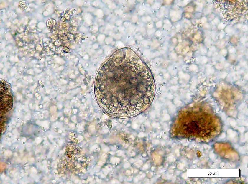

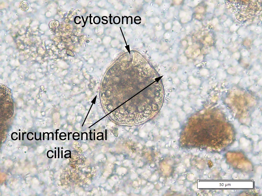
Cyst and trophozoite of Neobalantidium coli (also known as Balantidioides) obtained from West African baboon
Images hosted on other servers:


Various images

Trophic stage (EM)

Intestinal lumen

Encysted ciliate
Electron microscopy description
- Flattened oval organism covered with cilia with gullet at anterior end
Differential diagnosis
