Colon
Other nonneoplastic
Infarcted epiploic appendages
Author: Raul S. Gonzalez, M.D.

Last author update: 3 March 2021
Last staff update: 10 March 2021
Copyright: 2003-2025, PathologyOutlines.com, Inc.
PubMed Search: Infarcted epiploic appendages

Table of Contents
Definition / general | Essential features | Terminology | Sites | Etiology | Clinical features | Diagnosis | Radiology description | Radiology images | Case reports | Gross description | Gross images | Microscopic (histologic) description | Microscopic (histologic) images | Sample pathology report | Differential diagnosis | Additional references | Practice question #1 | Practice answer #1Cite this page: Gonzalez RS. Infarcted epiploic appendages. PathologyOutlines.com website. https://www.pathologyoutlines.com/topic/coloninfarcted.html. Accessed September 7th, 2025.
Definition / general
- Infarction and subsequent fat necrosis of epiploic appendages (fat containing pouches of colonic peritoneum) that may remain attached or autoamputate and lie loose in the peritoneum
Essential features
- Fat necrosis of epiploic appendage, which may detach and lie loose in the peritoneum
- May cause pain or be discovered as an incidental curiosity
Terminology
- Epiploic appendagitis: inflammation but not infarction of appendages
- Unattached infarcted appendages are known as peritoneal loose bodies or peritoneal mice (J Clin Gastroenterol 2006;40:427)
Sites
- Epiploic appendages are chiefly on transverse and sigmoid colon
- Can occur on appendix (S D Med 2006;59:511)
Etiology
- Due to twisting and torsion of normally thick pedicles of epiploic appendages
- May be associated with obstruction and abscess (J Am Assoc Gynecol Laparosc 1996;3:325)
Clinical features
- Abdominal pain, typically in left lower quadrant
- Rarely causes death (South Med J 1986;79:374)
Diagnosis
- Laparoscopy
Radiology description
- Localized pericolic inflammatory changes (Abdom Imaging 1994;19:449)
Radiology images
Images hosted on other servers:

Central calcified oval mass in the pelvis

Pelvic mass
Case reports
- 64 year old man with a 7 cm giant peritoneal loose body (J Med Case Rep 2011;5:297)
Gross description
- Firm, gray-white nodules that may resemble metastatic tumor
- Loose bodies can resemble an egg
Gross images
Images hosted on other servers:

Egg shaped mass

Giant loose peritoneal body

Giant loose body attached to omentum

Round pelvic mass

Central calcifications and a distinct fat plane

Macrograph of giant loose body
Microscopic (histologic) description
- Central infarcted adipose tissue with peripheral fat necrosis and calcification, surrounded by thick, inflamed fibrotic tissue
Microscopic (histologic) images
Contributed by Raul S. Gonzalez, M.D.
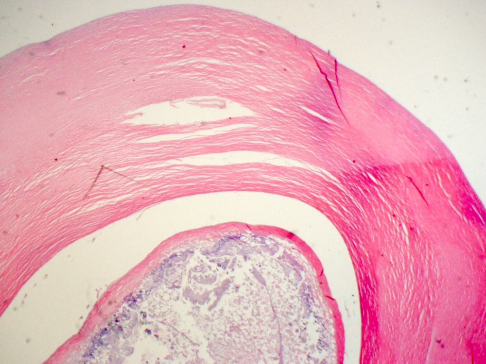
Rounded contour
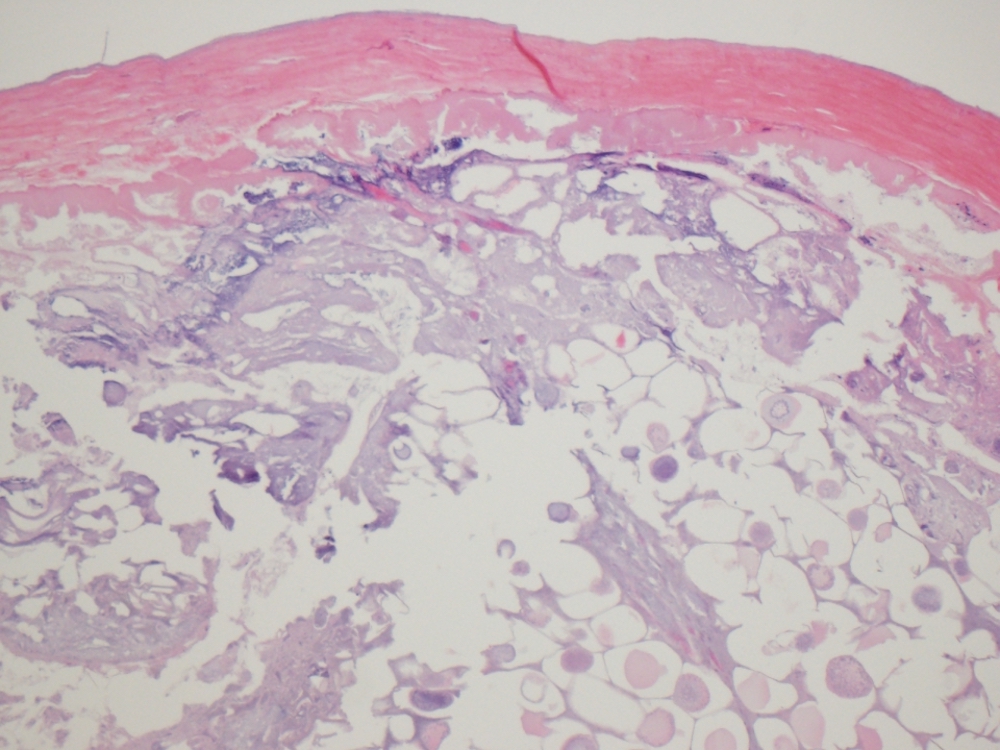
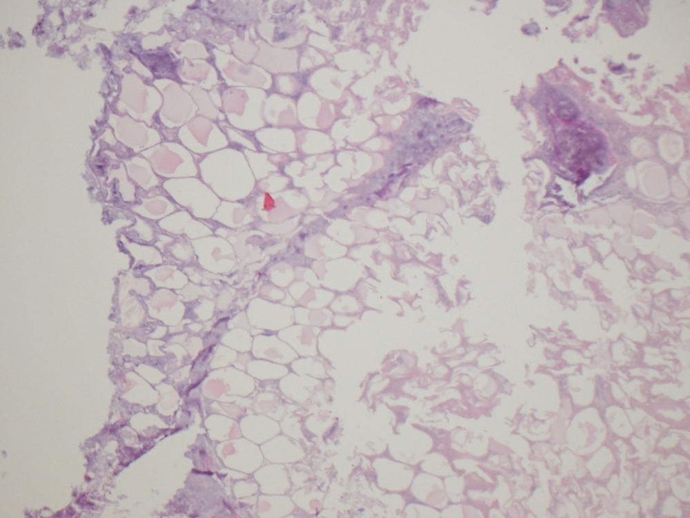
Central fat necrosis
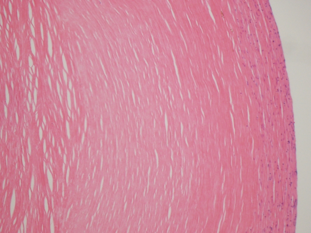
Circumferential fibrous tissue
Sample pathology report
- Abdominal cavity, loose body, removal:
- Fat necrosis with fibrotic rim, consistent with infarcted epiploic appendage
Differential diagnosis
- Fat necrosis from other cause:
- Not rounded / encapsulated or free floating, may show more inflammation
Additional references
Practice question #1
A loose egg-like object is found in a patient's abdomen during planned abdominal surgery. It has a microscopic appearance as shown above. What is the best diagnosis?
- Calcifying fibrous pseudotumor
- Fecal material
- Infarcted epiploic appendage
- Well differentiated liposarcoma
Practice answer #1
