
Colon
Other nonneoplastic
Pulse granuloma
Author: Raul S. Gonzalez, M.D.

Last author update: 4 February 2021
Last staff update: 8 September 2022
Copyright: 2003-2024, PathologyOutlines.com, Inc.
PubMed Search: Pulse granuloma


Table of Contents
Definition / general | Essential features | Terminology | Epidemiology | Sites | Pathophysiology | Etiology | Clinical features | Case reports | Microscopic (histologic) description | Microscopic (histologic) images | Negative stains | Sample pathology report | Differential diagnosis | Board review style question #1 | Board review style answer #1Cite this page: Gonzalez RS. Pulse granuloma. PathologyOutlines.com website. https://www.pathologyoutlines.com/topic/colonpulsegranuloma.html. Accessed April 19th, 2024.
Definition / general
- Granulomatous reaction to legume material that breaches the colonic mucosa
Essential features
- Reactive process arising from foreign body (food material)
- Usually clinically silent and incidental but may form a mass lesion
- Various microscopic appearances but hyaline rings of pulse material must be present
Terminology
- Pulse is the seed material of legumes (beans, peas, peanuts, etc.)
- It has also been described as hyaline rings
Epidemiology
- Can be seen in up to 10% of intestine segments that have undergone injury (Am J Surg Pathol 2016;40:137)
Sites
- Can occur anywhere in gastrointestinal tract but may occur most commonly in colon
- More commonly recognized in oral cavity, mandible / maxilla and lung
- If colon perforates, pulse can access the abdominal cavity and deposit anywhere within (e.g. ovary)
Pathophysiology
- Granulomatous reaction to foreign material
Etiology
- Most often seen in colon damaged by perforation, diverticulitis, inflammatory bowel disease or malignancy
Clinical features
- Generally an incidental finding but can may manifest as a mass lesion clinically
- Associated with use of tobacco and NSAIDs (Am J Surg Pathol 2015;39:84)
Case reports
- 53 year old man with hard, flat rectal lesion (Histopathology 2016;68:938)
Microscopic (histologic) description
- Hyaline predominant
- More than half of lesion consists of pink hyaline rings / ribbons; other food present; inflammation and fibrosis minimal
- Cellular predominant
- Acute and chronic inflammation and fibrosis
- Relatively less pulse and food material
- Sclerosing mesenteritis-like
- Rarest variant
- Arises in mesentery and resembles sclerosing mesenteritis (inflammation, fibrosis) but pulse focally visible
- Average size is 1.0 cm
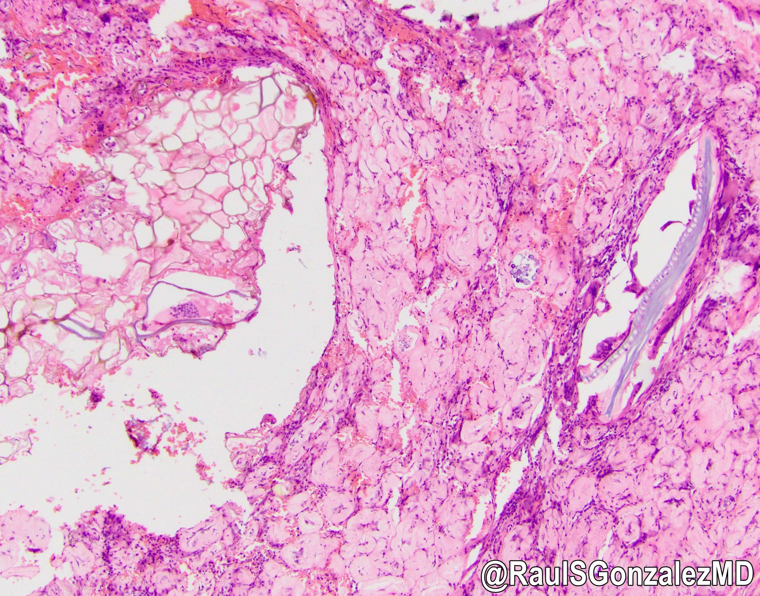
Microscopic (histologic) images
Contributed by Raul S. Gonzalez, M.D.

Serosal hyaline
predominant
pulse granulomas
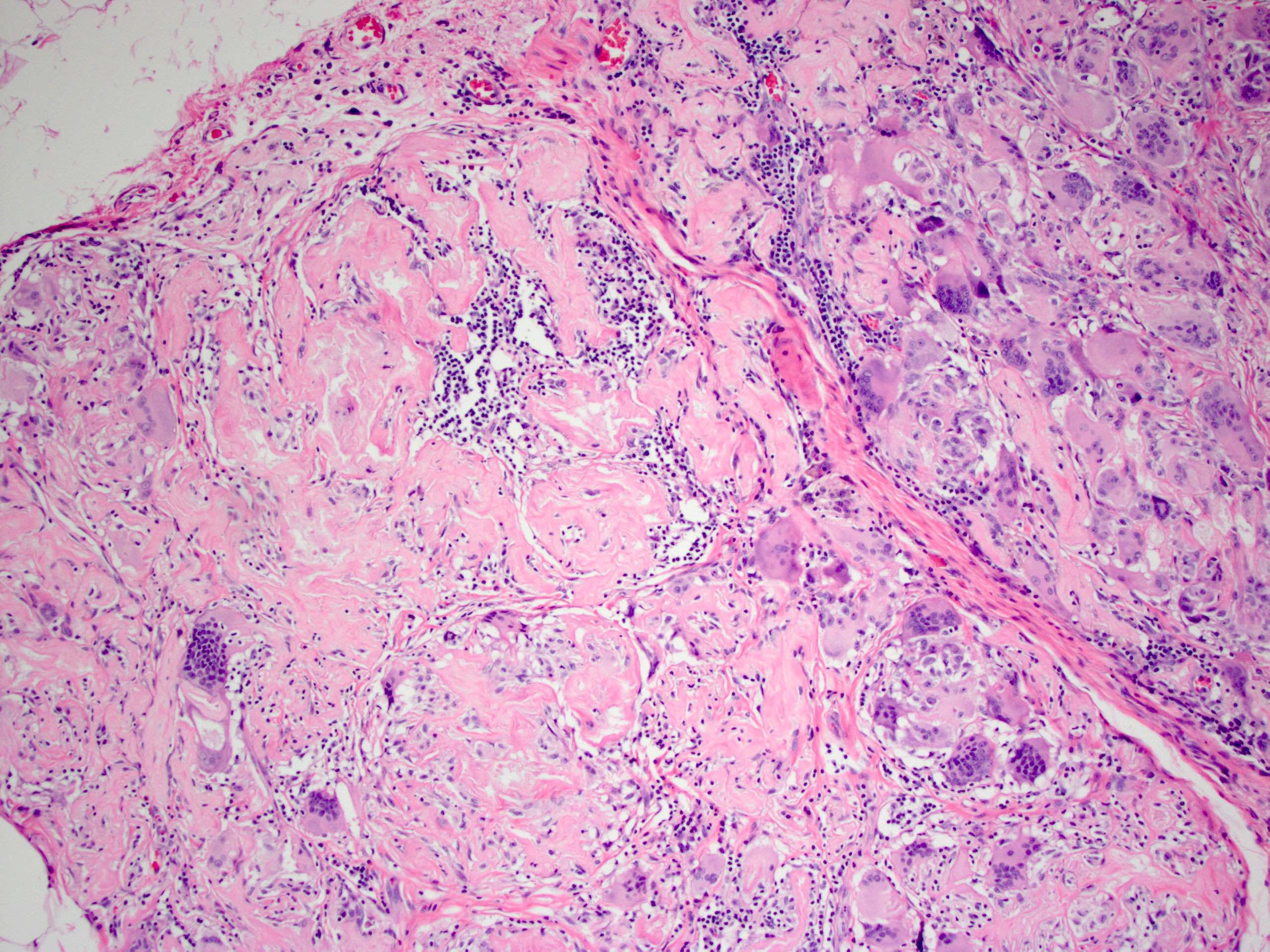
Pulse material
with numerous
foreign body
giant cells
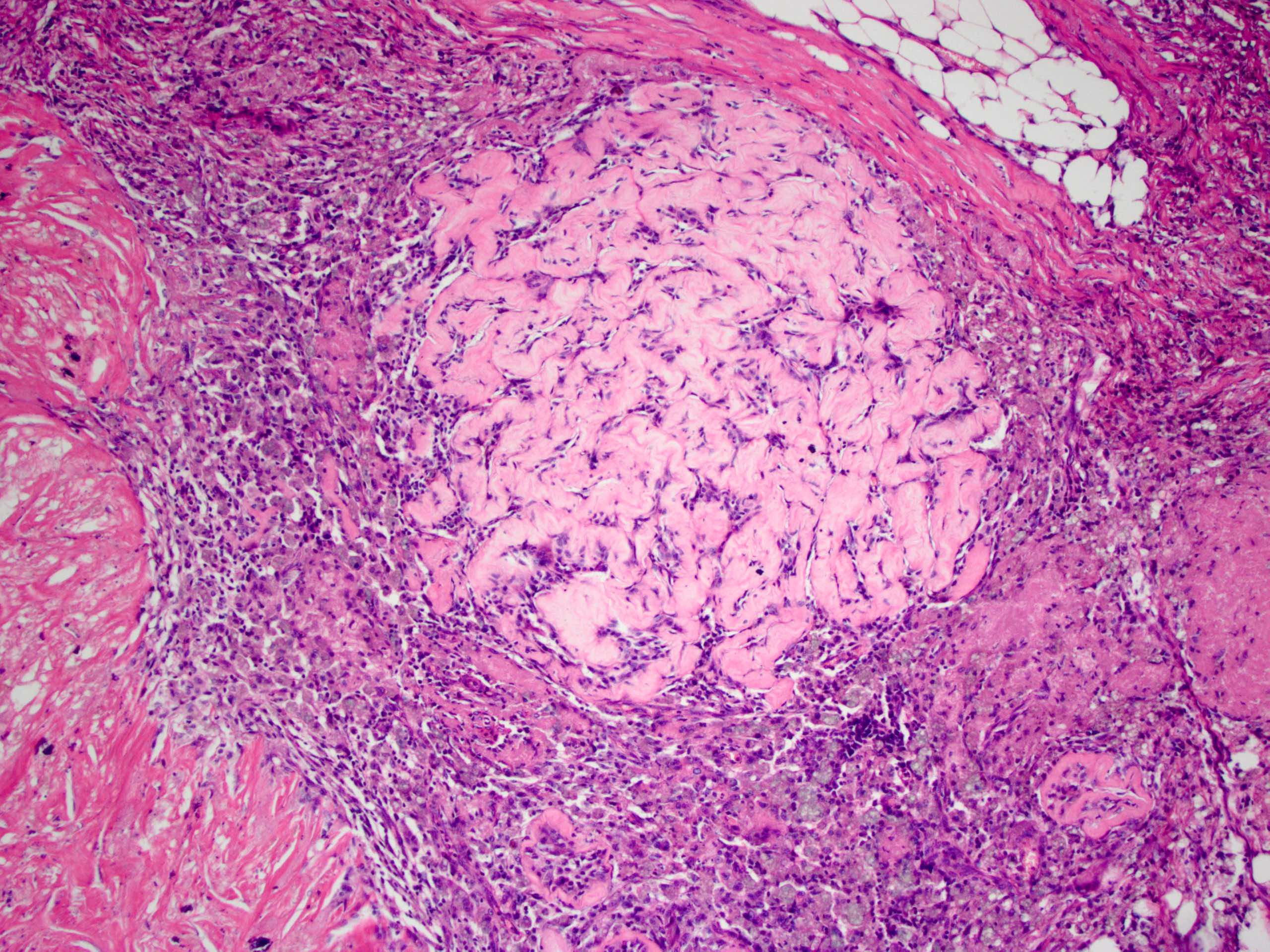
Cellular predominant pulse granuloma
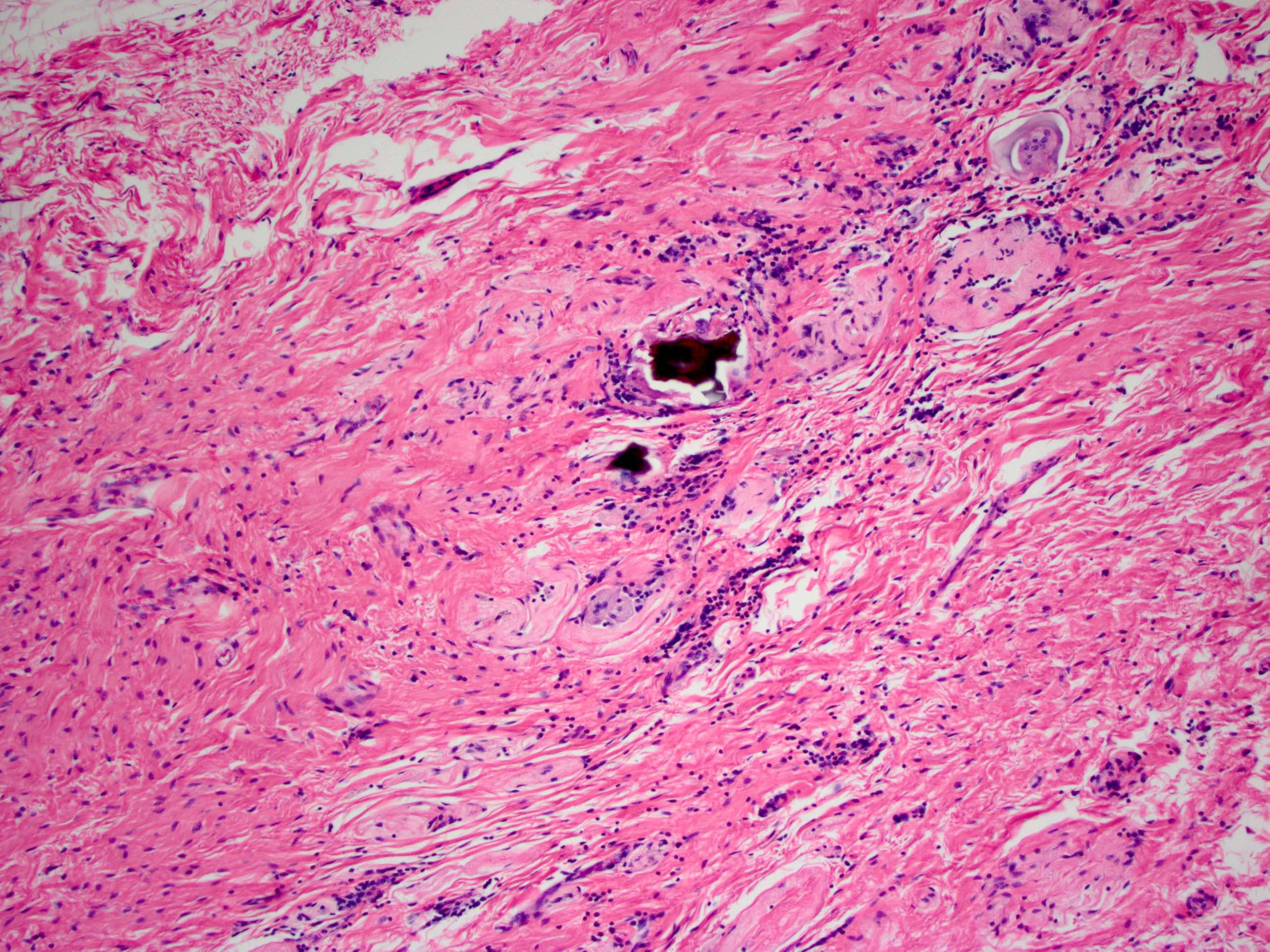
Scant pulse material embedded in colon wall
Contributed by @RaulSGonzalezMD on Twitter
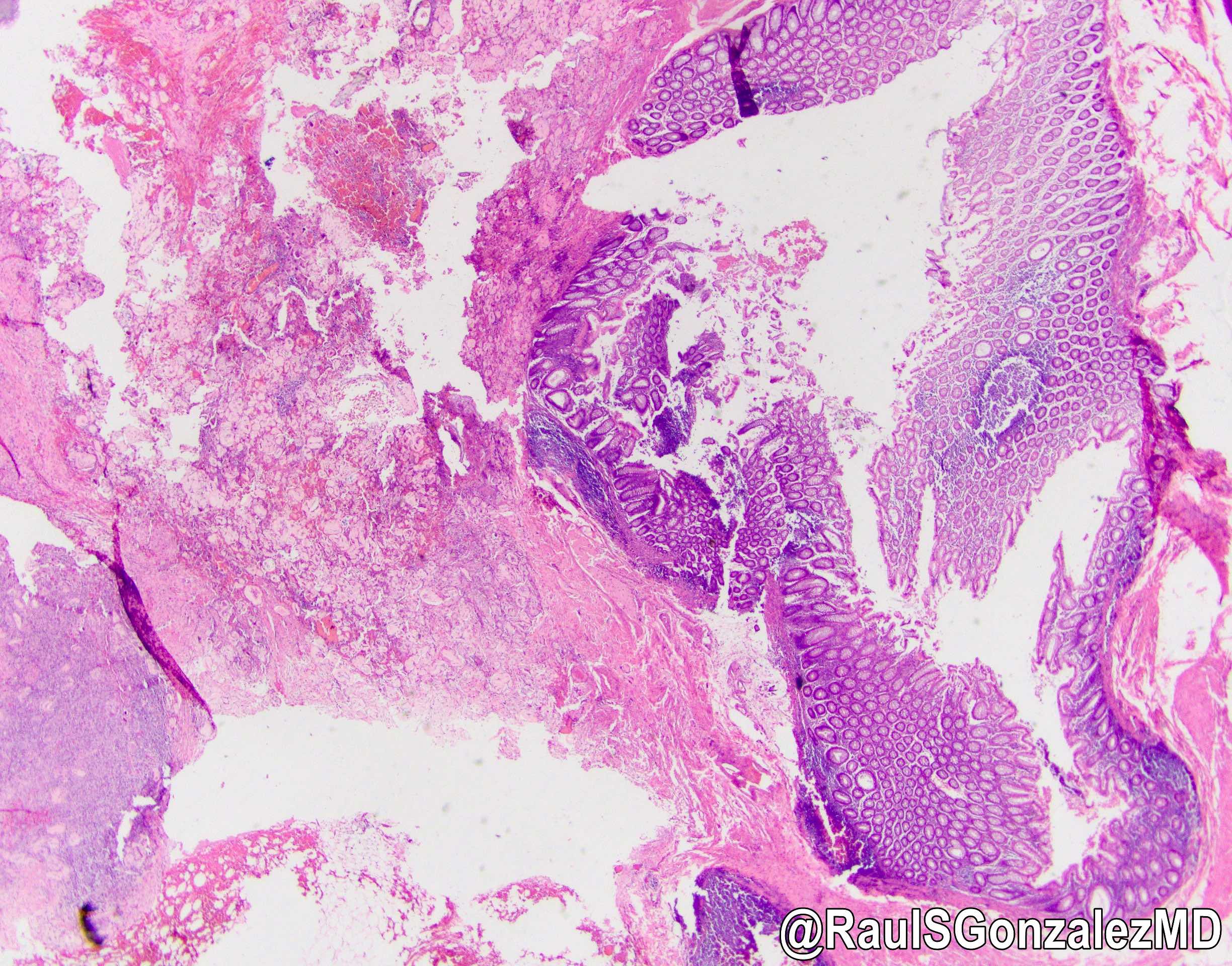
Pulse granuloma

Pulse granuloma
Pulse granuloma
Negative stains
Sample pathology report
- Sigmoid colon, resection:
- Diverticulosis with rupture, mural abscess formation and focal pulse granulomas.
Differential diagnosis
- Amyloidosis:
- Positive with Congo red stain
- Inflammation generally not present
- Lifting agent granuloma:
- Hyaline rings not present
- History of polypectomy at site
- Sclerosing mesenteritis:
- For lesions in the mesentery
- Hyaline rings not present
Board review style question #1

Which of the following is true about pulse granuloma in the colon?
- It always forms a mass lesion
- It has a few described histologic subtypes / forms
- It shows apple green birefringence on Congo red staining
- Pulse refers to any sort of vegetable material
Board review style answer #1
