
Colon
Other nonneoplastic
Vascular ectasia
Author: Raul S. Gonzalez, M.D.

Last author update: 16 February 2021
Last staff update: 17 February 2021
Copyright: 2003-2024, PathologyOutlines.com, Inc.
PubMed search: Vascular ectasia [title] colon


Table of Contents
Definition / general | Essential features | Terminology | Epidemiology | Sites | Etiology | Clinical features | Diagnosis | Case reports | Treatment | Clinical images | Gross description | Microscopic (histologic) description | Microscopic (histologic) images | Sample pathology report | Differential diagnosis | Board review style question #1 | Board review style answer #1Cite this page: Gonzalez RS. Vascular ectasia. PathologyOutlines.com website. https://www.pathologyoutlines.com/topic/colonvascularectasia.html. Accessed April 19th, 2024.
Definition / general
- Abnormally dilated blood vessels in colonic mucosa or submucosa (eMedicine: Angiodysplasia of the Colon [Accessed 17 February 2021])
Essential features
- Cause of lower GI tract bleeding
- More common in right colon and in older patients
- Can be subtle and focal on histology
Terminology
- Also called angiodysplasia, arteriovenous malformation
Epidemiology
- < 1% prevalence but accounts for 20% of patients with lower GI bleeding (#2 most common cause, after diverticulitis)
- Incidence increases with age (J Clin Pathol 1982;35:824)
Sites
- Usually right colon but can occur anywhere in small intestine or colon (Aliment Pharmacol Ther 2014;39:15)
Etiology
- Acquired changes in colonic extracellular matrix which distort veins and capillaries, disposing them to bleed
- Changes may be secondary to chronic vascular obstruction
Clinical features
- Rectal bleeding, often in elderly
- Bleeding episodes typically cease spontaneously but recur
- May be associated with aortic stenosis or von Willebrand disease
Diagnosis
- Colonoscopy, angiography
Case reports
- 22 year old woman with right sided colonic angiodysplasia (Indian J Pathol Microbiol 2006;49:34)
- 74 year old man with myelofibrosis and colonic angiodysplasia (J Clin Pathol 2004;57:999)
Treatment
- Electrocoagulation, surgery (Gastrointest Endosc 2006;64:424)
Clinical images
Images hosted on other servers:

Friable telangiectatic mucosal lesions
Gross description
- Tortuous dilation of multiple small submucosal and mucosal blood vessels
- Easier to identify by angiography than in a surgical specimen unless injected with silicone rubber and cleared with methyl salicylate
Microscopic (histologic) description
- Dilated and thin walled vessels (arteries, veins and capillaries) in mucosa and submucosa, often clustered
- Overlying mucosa may be eroded
- Changes can be subtle and focal
Microscopic (histologic) images
Contributed by Raul S. Gonzalez, M.D.

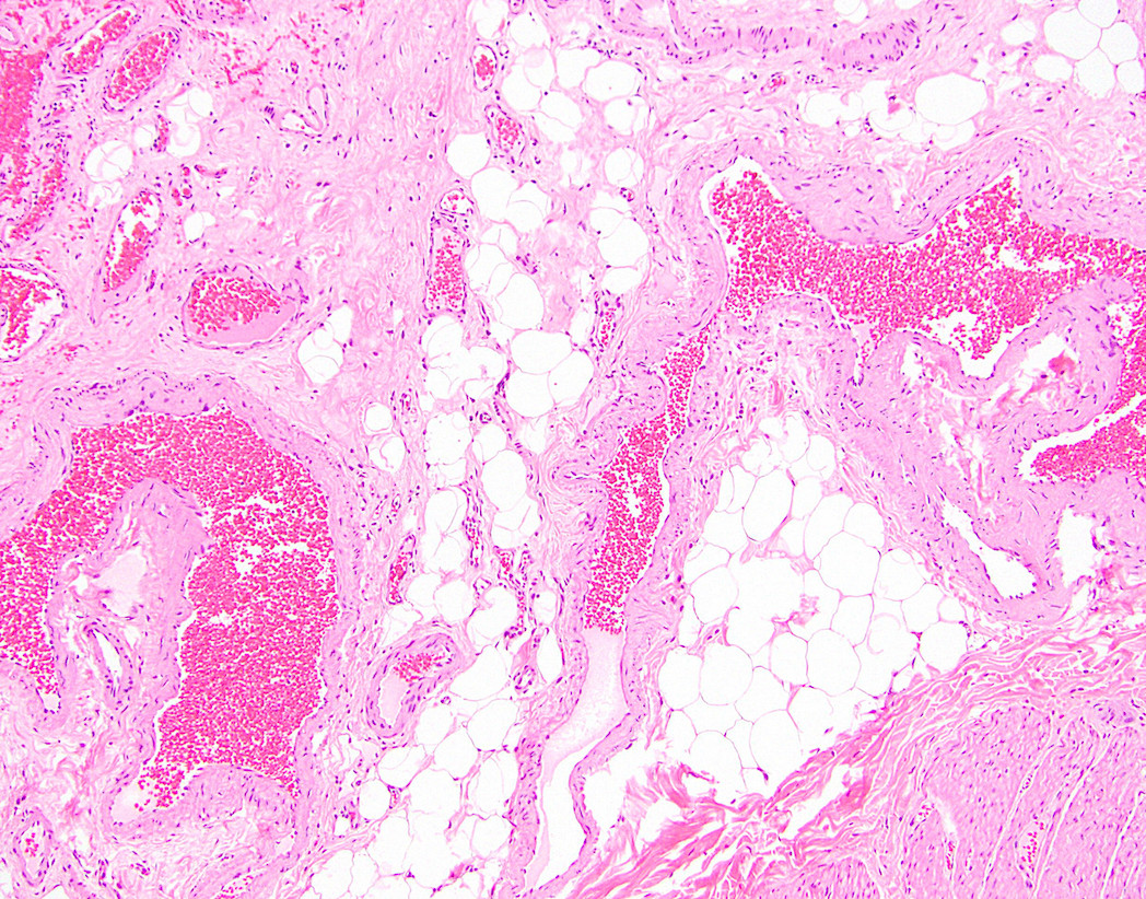
Vascular ectasia
Sample pathology report
- Ascending colon, resection:
- Segment of colon with submucosal angiodysplasia and focal overlying mucosal erosions
- Margins of resection unremarkable.
- Two benign lymph nodes.
Differential diagnosis
- Colonic or anal varices:
- Due to portal hypertension
- Hemangioma:
- Discrete lesion
Board review style question #1
- Which of the following is true about vascular ectasia (angiodysplasia) of the colon?
- Can be found in roughly 20% of resected colons
- Indistinguishable from hemangioma
- More prevalent in younger patients
- Usually found in the right colon
Board review style answer #1
