
Esophagus
Carcinoma
Neuroendocrine carcinoma
Authors: Feriyl Bhaijee, M.D., Israh Akhtar, M.D.


Last author update: 1 July 2013
Last staff update: 12 March 2024 (update in progress)
Copyright: 2003-2024, PathologyOutlines.com, Inc.
PubMed Search: Neuroendocrine (tumor OR carcinoma) esophagus
Table of Contents
Definition / general | Terminology | Epidemiology | Sites | Clinical features | Grading | Prognostic factors | Case reports | Treatment | Gross description | Gross images | Microscopic (histologic) description | Microscopic (histologic) images | Cytology description | Cytology images | Positive stains | Negative stains | Electron microscopy images | Differential diagnosisCite this page: Bhaijee F, Akhtar I. Neuroendocrine carcinoma. PathologyOutlines.com website. https://www.pathologyoutlines.com/topic/esophagusneuroendocrine.html. Accessed April 25th, 2024.
Definition / general
- Epithelial neoplasm with prominent neuroendocrine differentiation
- Pathologic spectrum from low grade neuroendocrine tumors (carcinoid, atypical carcinoid) to high grade neuroendocrine carcinoma (small cell or large cell neuroendocrine carcinoma)
Small cell neuroendocrine carcinoma:
- Small cell neuroendocrine carcinoma is analogous to pulmonary small cell carcinoma
- WHO definition
- Malignant epithelial tumor consisting or small cells with scant cytoplasm, ill defined cell borders, finely granular nuclear chromatin and absent or inconspicuous nucleoi
- Cells are round, oval and spindle shaped
- Nuclear molding is prominent
- Necrosis is typically extensive and the mitotic count is high
Terminology
- Synonyms
- Neuroendocrine tumor (low grade) = carcinoid tumor, atypical carcinoid tumor
- Neuroendocrine carcinoma (high grade) = small cell carcinoma, large cell carcinoma
Epidemiology
- Extremely rare in esophagus: about 100 reported cases (mostly high grade / small cell carcinomas)
- M:F ratio = 3:1 (Am J Surg Pathol 2013;37:467)
- Mean age: 62 years
Sites
- Middle or lower esophagus (Am J Surg Pathol 2013;37:467, Int J Clin Exp Pathol 2013;6:485)
Clinical features
- Usually incidental / unexpected finding on radiologic studies or upper GI endoscopy
- Dysphagia, weight loss, chest pain with high grade carcinoma (Int J Clin Exp Pathol 2013;6:485)
- Typically diagnosed via biopsy or (less commonly) surgical resection
Small cell neuroendocrine carcinoma:
- Very aggressive (Dis Esophagus 2014;27:152, Hepatogastroenterology 2005;52:1738)
- Median survival 18 months (BMC Cancer 2007;7:38)
- 5 year survival 8% or less (Chin Med J (Engl) 2007;120:355, Ann Surg Oncol 2013;20:4239, Hum Pathol 1999;30:216)
Grading
- Grade 1: < 2 mitoses/10 HPF or < 2% Ki67 index
- Grade 2: 2 - 20 mitoses/10 HPF or 3 - 20% Ki67 index
- Grade 3: > 20 mitoses/10 HPF or > 20% Ki67 index (UpToDate: Classification of Neuroendocrine Neoplasms GI Tract [Accessed 27 February 2019], Pancreas 2010;39:707)
Prognostic factors
- Mitotic rate and Ki67 index determine grade
- Low grade lesions have favorable prognosis
- High grade carcinomas are very aggressive, as in other body sites
Case reports
- 51 year old man with collision tumor between papillary adenocarcinoma and large cell neuroendocrine carcinoma (Arch Pathol Lab Med 2000;124:411)
- 54 year old man with atypical carcinoid of the esophagus (Ann Thorac Cardiovasc Surg 2002;8:302)
Small cell neuroendocrine carcinoma:
- 63 year old woman with coexisting paraneoplastic neurological syndrome (Jpn J Clin Oncol 2006;36:109)
- 66 year old man with rare collision tumor of squamous carcinoma and small cell carcinoma in esophagus in separate lymph nodes (J Thorac Dis 2013;5:E203)
- 79 year old man (BMJ Case Rep 2013 Sep 3;2013)
Treatment
- Surgical resection
- Chemoradiation for high grade carcinoma (Rare Tumors 2013;5:e6)
Gross description
- Polypoid or ulcerated mass on upper endoscopy
Gross images
AFIP images

Bulky, ulcerated, infiltrative lesion
Images hosted on other servers:

Small cell
carcinoma (upper),
squamous cell
carcinoma (lower)
Microscopic (histologic) description
- Well differentiated (low grade) tumors
- Uniform, small, bland tumor cells in solid, trabecular, gyriform or glandular patterns
- May have Paneth cell differentiation
- Poorly differentiated (high grade) carcinomas
- Large cell type: nests of pleomorphic, large cells with prominent nucleoli and a moderate amount of cytoplasm
- Small cell type: sheets and nests of small cells with hyperchromatic nuclei and a minimal to moderate amount of cytoplasm; prominent crush artifact and Azzopardi phenomenon, as in small cell carcinomas at other sites
- Necrosis
- Increased mitotic activity
- Angiolymphatic invasion common
- Solid to cribriform growth
- Usually in lamina propria
- May be associated with heterotopic oxyntic mucosa or Barrett esophagus (large cell carcinoma)
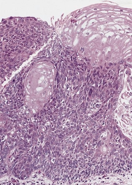
- Neuroendocrine carcinoma may have small component(s) of adenocarcinoma or squamous cell carcinoma differentiation
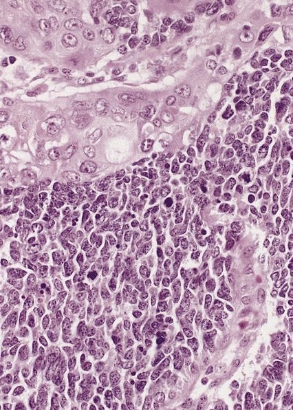
Microscopic (histologic) images
Contributed by Mark R. Wick, M.D. and AFIP images

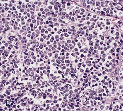
Small cell carcinoma of esophagus
In muscularis
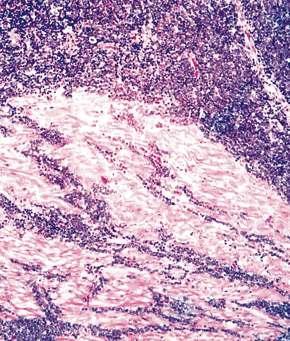
Small cell neuroendocrine carcinoma:

Diffusely infiltrating sheets of small cells
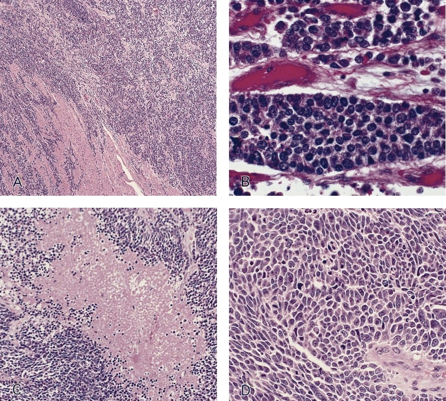
In situ component
Squamous cell differentiation
Cytology description
- Low grade tumors
- Flat sheets or loosely cohesive groups / cords of monotonously uniform plasmacytoid cells
- Eccentric nuclei, coarsely stippled (salt and pepper) chromatin, finely granular cytoplasm
- High grade carcinomas
- Obvious pleomorphism, marked nuclear molding, hyperchromatic nuclei, inconspicuous nucleoli
- Numerous mitoses, crush artifact, necrosis
- Apoptotic figures, blue bodies
Cytology images
Contributed by Mark R. Wick, M.D.
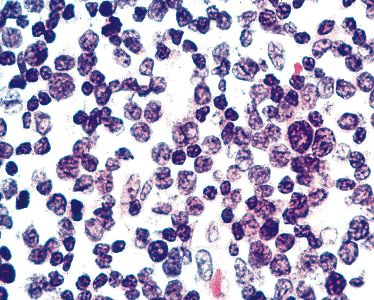
Neuroendocrine tumor
Images hosted on other servers:

Carcinoid tumor

Small cell carcinoma
Positive stains
- Keratins (e.g., CAM 5.2, AE1 / AE3)
- Neuroendocrine markers: synaptophysin, chromogranin, CD56, NSE
- Ki67 (essential for tumor grading see above)
- TTF1 positive in 71% of esophageal neuroendocrine carcinoma (Am J Surg Pathol 2013;37:467)
Negative stains
Electron microscopy images
Images hosted on other servers:

Neurosecretory granules
Differential diagnosis
- Low grade tumors: glomus tumor, paraganglioma
- High grade carcinomas:
- Metastatic pulmonary neuroendocrine carcinoma: exclude clinically, lymphoma, melanoma, basaloid squamous cell carcinoma
