
Eye
Conjunctiva
Epithelial tumors
Squamous cell carcinoma-conjunctiva
Author: Nat Pernick, M.D.

Last author update: 1 September 2013
Last staff update: 4 April 2024
Copyright: 2004-2024, PathologyOutlines.com, Inc.
PubMed Search: Squamous cell carcinoma conjunctiva
Table of Contents
Definition / general | Clinical features | Case reports | Treatment | Clinical images | Gross description | Gross images | Microscopic (histologic) description | Microscopic (histologic) images | Positive stains | Molecular / cytogenetics description | Differential diagnosis | Additional referencesCite this page: Pernick N. Squamous cell carcinoma-conjunctiva. PathologyOutlines.com website. https://www.pathologyoutlines.com/topic/eyeconjSCC.html. Accessed April 16th, 2024.
Definition / general
- See also mucoepidermoid carcinoma
- Rare but more common than basal cell carcinoma at this site
- In U.S., precancerous lesions are excised, so invasive carcinoma is uncommon
- Rates: 0.03 per 100,000 in U.S., 3.5 per 100,000 in Uganda
- Mainly adults
- In U.S., commonly 60+ years, 55 - 70% men (Can J Ophthalmol 2002;37:14)
- Associated with sunlight exposure, actinic keratosis; also xeroderma pigmentosum, albinism, toxins, HPV 16/18 (55%), possibly atopic eczema (Cornea 2003;22:135)
- May invade anterior chamber of globe or orbit but only rarely metastasizes or causes death
Clinical features
- HIV / AIDS patients
- Rising incidence with 8% prevalence in Kenya (East Afr Med J 2006;83:267)
- Recommended to screen HIV / AIDS patients for conjunctival lesions
- Mean age is 35 years
- Usually affects women
- Patients present late with advanced disease
- More aggressive with high recurrence rates
Case reports
- 6 year old boy with 2 conjunctival tumors, atypical fibroxanthoma and xeroderma pigmentosum (Pediatr Dev Pathol 2007;10:149)
- 38 year old woman with AIDS with multifocal disease and intraocular penetration (Cornea 2006;25:745)
- 65 year old man with prosthetic eye (J Postgrad Med 2006;52:234)
- 71 year old man with bony metastases (Klin Monbl Augenheilkd 2002;219:813)
Treatment
- Complete excision of superficial tumors
- Radical surgery for deeply invasive tumors
- 6% recur
- Rarely metastasize to lymph nodes (more common if large or multiple recurrences)
Clinical images
Images hosted on other servers:

Nodular tumor with prominent vascularity at limbus
Gross description
- Papillary or exophytic gray-white mass, often at limbus
- Occasionally jet black resembling melanoma (in heavily pigmented individuals)
- Surrounded by inflamed conjunctiva
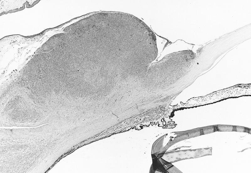
Gross images
AFIP images
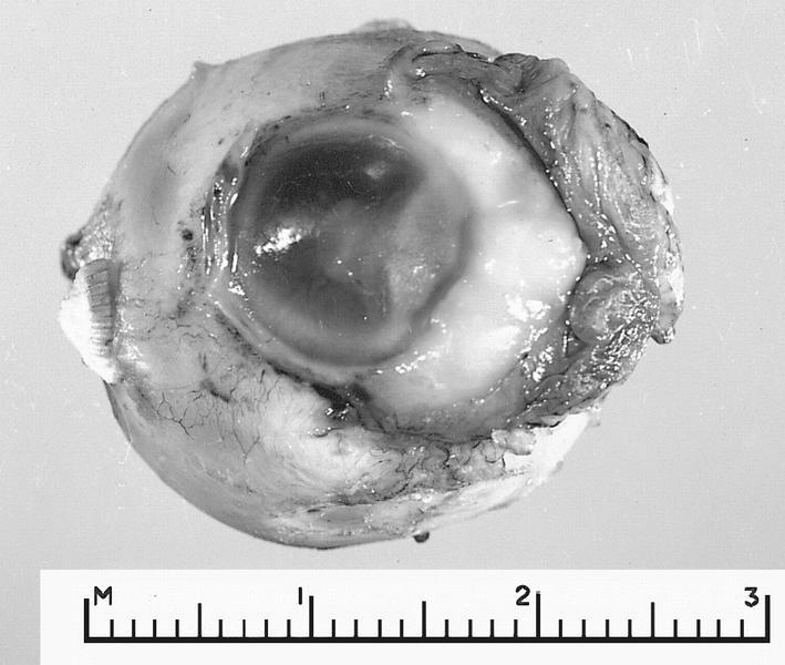
Large limbal tumor invades anterior chamber
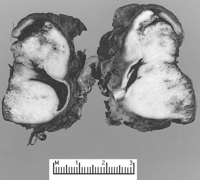
Tumor has destroyed eye and invaded orbit
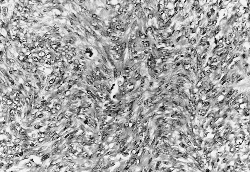
Microscopic (histologic) description
- Atypia throughout full thickness of epithelium (conjunctival intraepithelial neoplasia) with individual tumor cells or nests extending into underlying stroma
- Dense sclera usually limits deepest margins
- Epithelium may be keratinized
- Cells have eosinophilic or clear cytoplasm, intercellular bridges, dyskeratosis, coarse chromatin, prominent nucleoli
- May have pigment within benign and malignant cells in heavily pigmented patients (Ophthalmic Surg Lasers Imaging 2003;34:406)
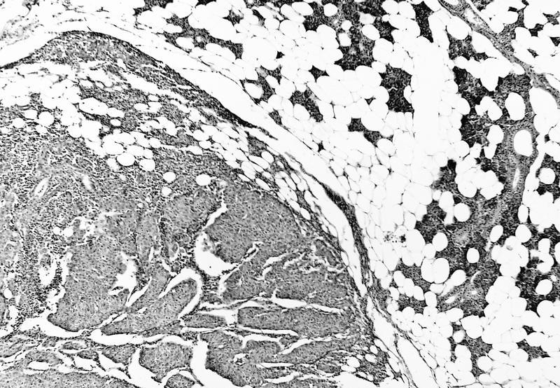
Microscopic (histologic) images
AFIP images
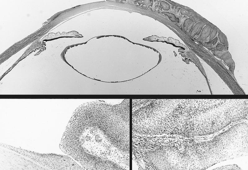
Early invasion with corneal involvement
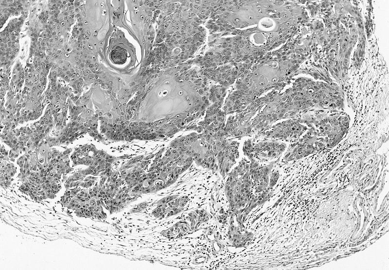
Well differentiated tumor with deep invasion
Metastasis to preauricular node and parotid gland
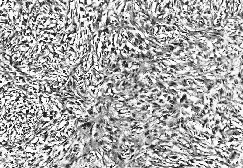
Sarcomatous pattern
Deep scleral and corneal invasion
Images hosted on other servers:

Thick layer of parakeratosis and microinvasion

Lobules of invasive keratinizing carcinoma

Papillomatous pattern
Positive stains
- High molecular weight keratin, EMA, EGFR (tumor and normal) (Ophthal Plast Reconstr Surg 2006;22:113)
Molecular / cytogenetics description
- Usually aneuploid
Differential diagnosis
Additional references
