
Eye
Orbit & optic nerve
IgG4 related disease
Author: Nat Pernick, M.D.

Last author update: 1 February 2014
Last staff update: 8 April 2024
Copyright: 2004-2024, PathologyOutlines.com, Inc.
PubMed Search: Inflammatory pseudotumor orbit
Cite this page: Pernick N. IgG4 related disease. PathologyOutlines.com website. https://www.pathologyoutlines.com/topic/eyeorbitinflammatorypseudo.html. Accessed April 19th, 2024.
Inflammatory pseudotumor
Definition / general
Case reports
Treatment
Gross description
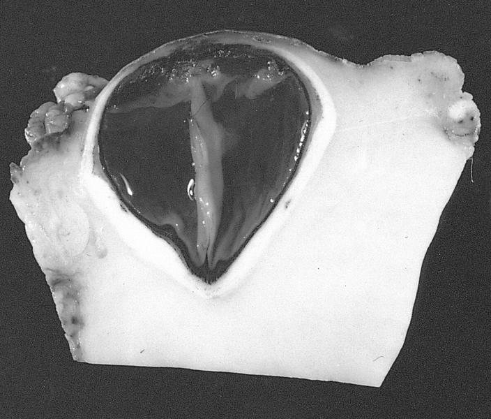
Gross images
AFIP images
Microscopic (histologic) description
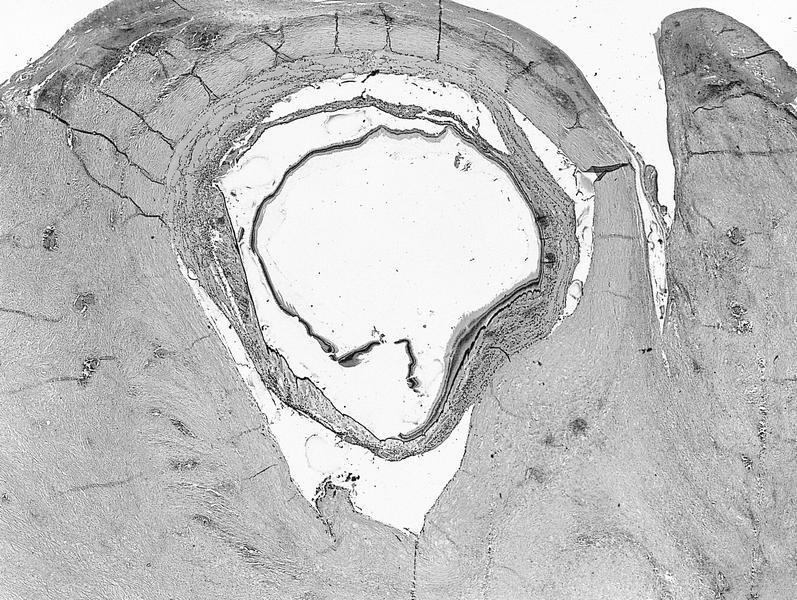
Whole mount images
AFIP images
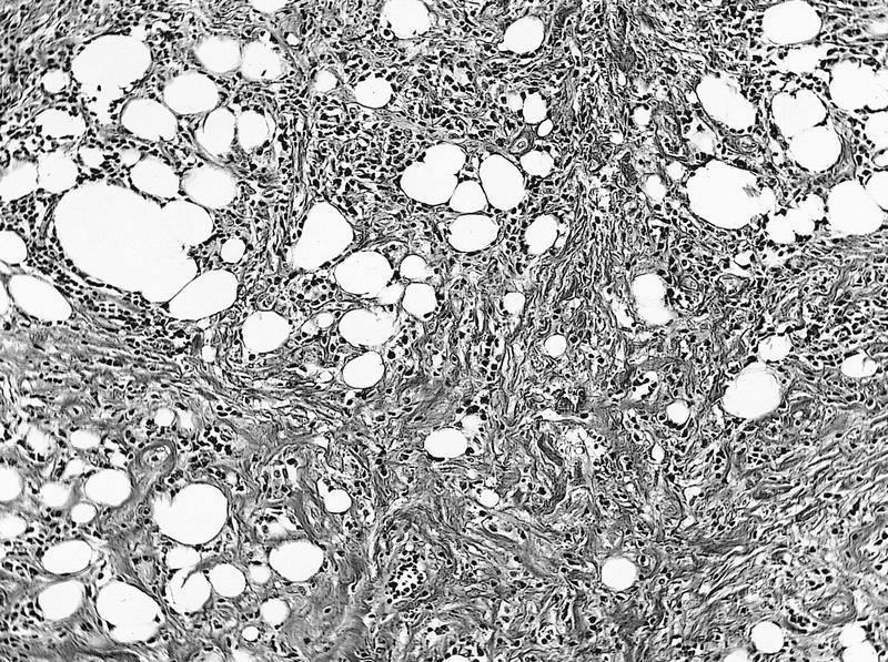
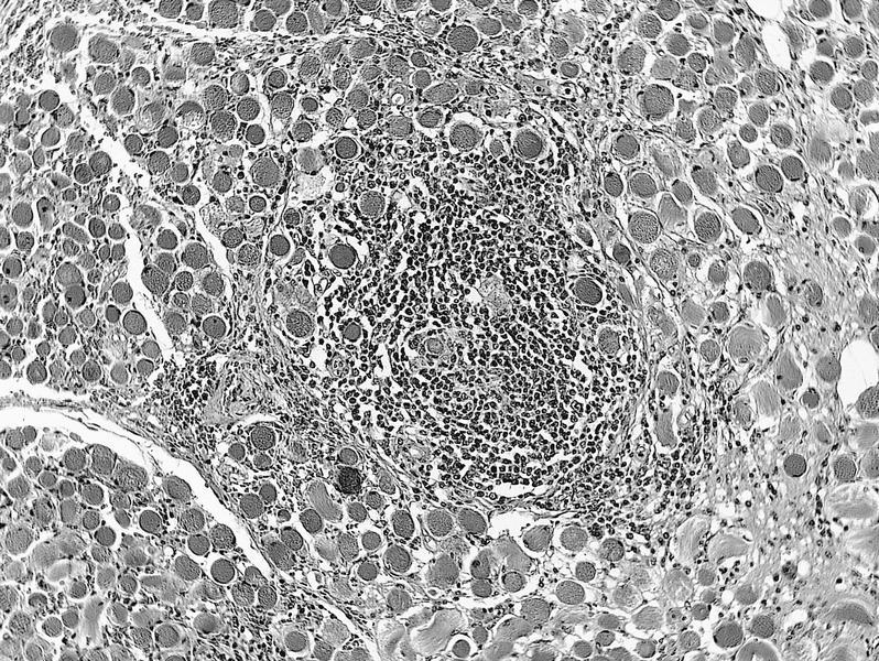
Microscopic (histologic) images
AFIP images
Positive stains
Differential diagnosis
- May not be a specific disease process, but due to various causes (paranasal sinus tumors, Rosai-Dorfman disease, inflammatory fibrosclerosis, dysthyroid ophthalmopathy, cholesterol or keratin granulomas, traumatic fat necrosis, prior hemorrhage or abscess)
- More common than infectious granulomas
- Usually ages 20 - 49 years with good health and sudden onset of exophthalmos with variable lid or conjunctival edema
Case reports
- 50 year old man with intraocular inflammatory myofibroblastic tumor with ALK overexpression (Arch Pathol Lab Med 2004;128:e5)
Treatment
- Steroids (alleviate signs and symptoms)
- Excision
Gross description
- Indurated orbital mass, often surrounding optic nerve and enveloping extraocular muscles
Gross images
AFIP images
Fibrotic mass surrounds the eye
Microscopic (histologic) description
- General:
- Edematous tissue with excessive production of ground substance, chronic inflammatory cells, vascular proliferation and hyperplastic connective tissue
- May have periphlebitis with tissue eosinophilia
- Inflammatory myofibroblastic tumor:
- Combinations of fibroblasts and myofibroblasts in background of plasma cells and other inflammatory cells
- Rosai-Dorfman related:
- Large histiocytes, some with lymphocytophagocytosis, lymphocytes and plasma cells, often with prominent fibrosis
Whole mount images
AFIP images
Dense fibrotic tissue
Microscopic (histologic) images
AFIP images
Inflammation and fibrosis
Chronic inflammatory cells
Positive stains
- Inflammatory myofibroblastic tumor: smooth muscle actin, variable ALK
Differential diagnosis
Idiopathic sclerosing inflammation
Definition / general
Microscopic (histologic) description
- Insidious, chronic and progressive fibrosing process
- Damages orbital structures by entrapment and mass effect
- May have cell mediated pathogenesis, similar to retroperitoneal fibrosis (Mod Pathol 1993;6:581)
Microscopic (histologic) description
- Desmoplasia, sparse lymphocytes (usually T cells), histiocytes, plasma cells, neutrophils, eosinophils
Other
[Pending]
