

Fallopian tubes & broad ligament
Fallopian tube tumor-like lesions
Salpingitis isthmica nodosa
Authors: Laura J. Vidis, D.O., Jessica L. Bentz, M.D.
Editorial Board Member: Kyle Devins, M.D.
Deputy Editor-in-Chief: Gulisa Turashvili, M.D., Ph.D.


Last author update: 31 July 2023
Last staff update: 31 July 2023
Copyright: 2002-2024, PathologyOutlines.com, Inc.
PubMed Search: Salpingitis isthmica nodosa


Table of Contents
Definition / general | Essential features | Terminology | Epidemiology | Sites | Pathophysiology | Etiology | Clinical features | Diagnosis | Radiology description | Radiology images | Prognostic factors | Case reports | Treatment | Gross description | Gross images | Microscopic (histologic) description | Microscopic (histologic) images | Sample pathology report | Differential diagnosis | Additional references | Board review style question #1 | Board review style answer #1 | Board review style question #2 | Board review style answer #2Cite this page: Vidis LJ, Bentz JL. Salpingitis isthmica nodosa. PathologyOutlines.com website. https://www.pathologyoutlines.com/topic/fallopiantubessin.html. Accessed April 19th, 2024.
Definition / general
- Salpingitis isthmica nodosa (SIN) is an outpouching of the fallopian tube epithelium into the smooth muscle wall (adenomyosis of the tube)
- Multiple discrete lumina are enveloped in smooth muscle and in severe cases, the tubal lumen may be completely obstructed
- SIN can lead to infertility or ectopic pregnancy
Essential features
- Adenomyosis of the fallopian tube
- Nodular swelling of the isthmic segment of the fallopian tube
- Multiple glands lined by bland tubal epithelium within the tubal wall, separated and surrounded by smooth muscle
- Associated with infertility and ectopic pregnancy
Terminology
- Diverticulosis of the fallopian tube
- Tubal adenomyosis
Epidemiology
- 0.6 - 11% of healthy fertile women (StatPearls: Salpingitis Isthmica Nodosa [Accessed 22 June 2023], Fertil Steril 1993;60:599)
- Incidence is ~1% in the white female population (Fertil Steril 1978;29:164)
- Primarily affects young women of reproductive age (mean: 26 years)
- 10% incidence in cases of ectopic tubular pregnancies (Gynecol Obstet Invest 1992;34:202)
Sites
- Fallopian tubes, predominantly the isthmus
Pathophysiology
- Invasion of the epithelium into the smooth muscle may lead to muscular hypertrophy, generating the nodular appearance of the lesion
Etiology
- Unclear but early theories include inflammatory or congenital causes (Radiology 1985;154:597)
- Other controversial hypotheses include
- Origination in Wolffian rests or mesonephric rests due to SIN occurring at the location in which these ducts cross during embryologic development (Clin Radiol 1987;38:207)
- Acquired etiology with some studies suggesting diethylstilbestrol exposure; chronic tubal spasm due to increased intramural pressure / weakness in the wall at the site of vasculature (Clin Obstet Gynecol 1987;30:181)
- Infectious etiology due to chronic infection or inflammation
Clinical features
- More common in the background of ectopic pregnancy and infertility
- Associated with infertility in 50% of patients but its presence may not actually affect the number of births (Hum Reprod 1991;6:828)
- Incidence in healthy, fertile women ranges from 0.6 to 11% (Fertil Steril 1993;60:599)
- May be unilateral or bilateral
Diagnosis
- Incidental finding during evaluation for pelvic pain or infertility (hysterosalpingogram [HSG], laparotomy, laparoscopy)
- Tubal evaluation for primary infertility may reveal gross lesion
- HSG can demonstrate the nodular swellings (Fertil Steril 1993;60:599)
- No pathognomonic clinical symptomatology
Radiology description
- Hysterosalpingography (HSG)
- Globular 2 mm diameter collections of contrast medium in isthmus
- Tubal occlusion and hydrosalpinx may be observed
Radiology images
Images hosted on other servers:

Nodular swelling of the isthmus

Multiple nodular diveriticula

Small outpouching or diverticula
Prognostic factors
- Smooth muscle hypertrophy can cause near complete occlusion of the tubal lumen and increase the risk of secondary infection, infertility or ectopic pregnancy
Case reports
- 29 year old woman with subfertility (Eur J Obstet Gynecol Reprod Biol 2015;184:73)
- 33 year old woman with ruptured salpingitis isthmica nodosum (BMJ Case Rep 2021;14:e237860)
- 42 year old woman with abdominal pain and amenorrhea (J Clin Diagn Res 2013;7:2581)
Treatment
- Recanalization using interventional radiology (Fertil Steril 1995;63:715)
- Microsurgical tubocornual anastomosis (Fertil Steril 1993;60:599)
- Assisted reproductive technology (ART) for infertility (Eur J Obstet Gynecol Reprod Biol 2015;184:73)
Gross description
- Firm, nodular swellings of the fallopian tube, ranging in size up to 2 cm in greatest dimension (Br J Radiol 2021;94:20201386)
- May be unilateral or bilateral
- Smooth, intact serosal covering
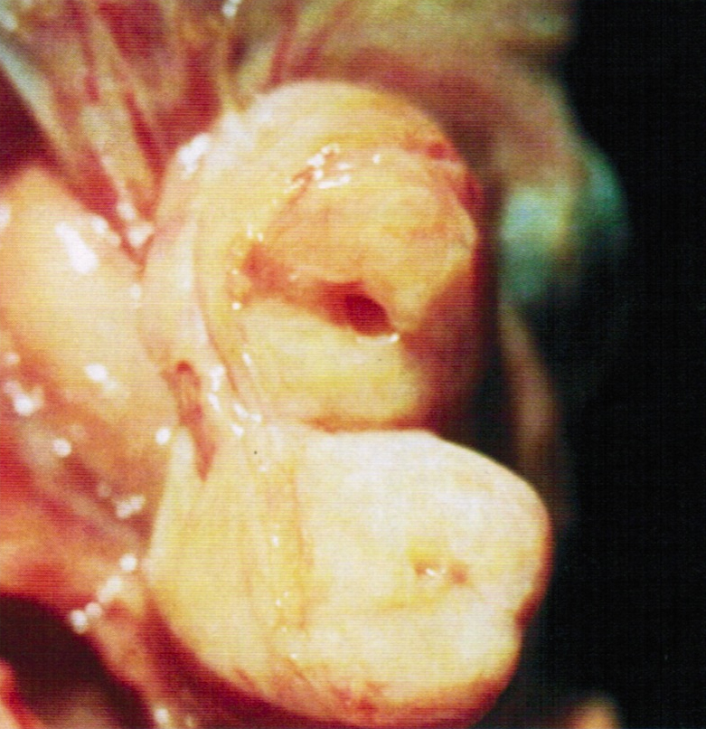
- Cross section of the nodules reveal gray, yellow or brown tissue punctuated by small cysts that surround the original tubal lumen (StatPearls: Salpingitis Isthmica Nodosa [Accessed 22 June 2023])
- Nodosity of the isthmus may give the uterus a horned appearance
Gross images
AFIP images
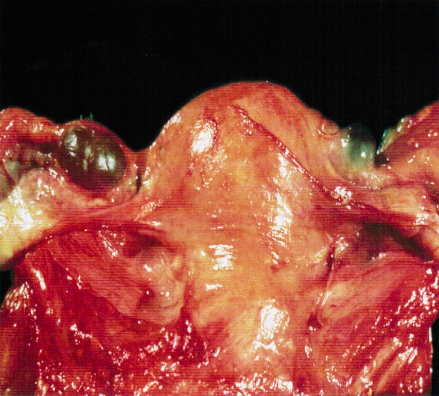
Dark, nodular swelling
Marked thickening
Images hosted on other servers:

Nodules at isthmus
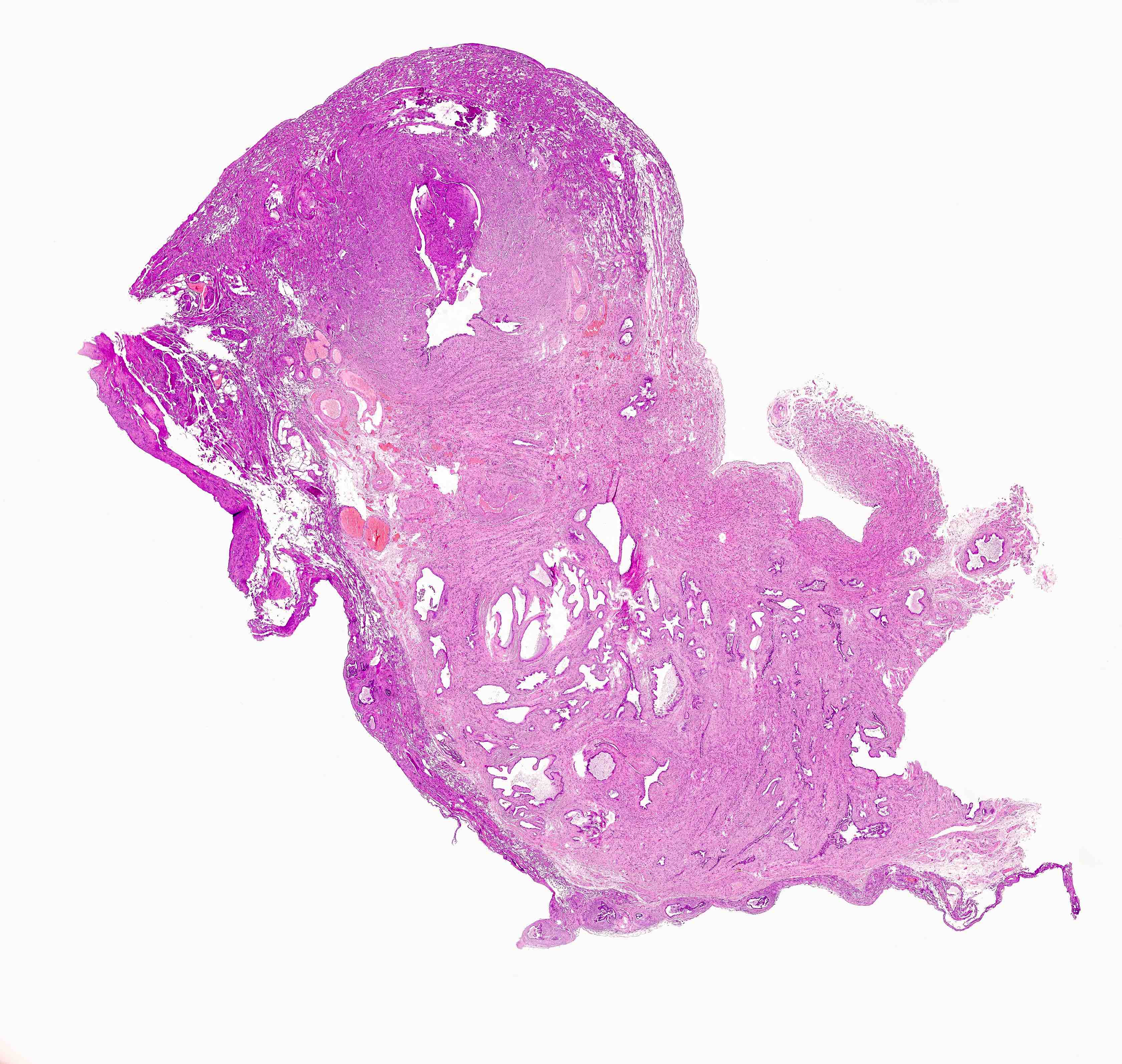
Microscopic (histologic) description
- Regularly spaced glands lined by normal appearing tubal epithelium surrounded by smooth muscle or fibrous tissue
- Tubal lumina are true diverticula that communicate with the central tubal lumen but do not connect with the serosa
- Glands can become cystically dilated (Am J Clin Pathol 1951;21:212)
- No significant atypia, scarring or associated inflammatory / stromal response
- Absence of endometrial stroma differentiates this entity from tubal endometriosis
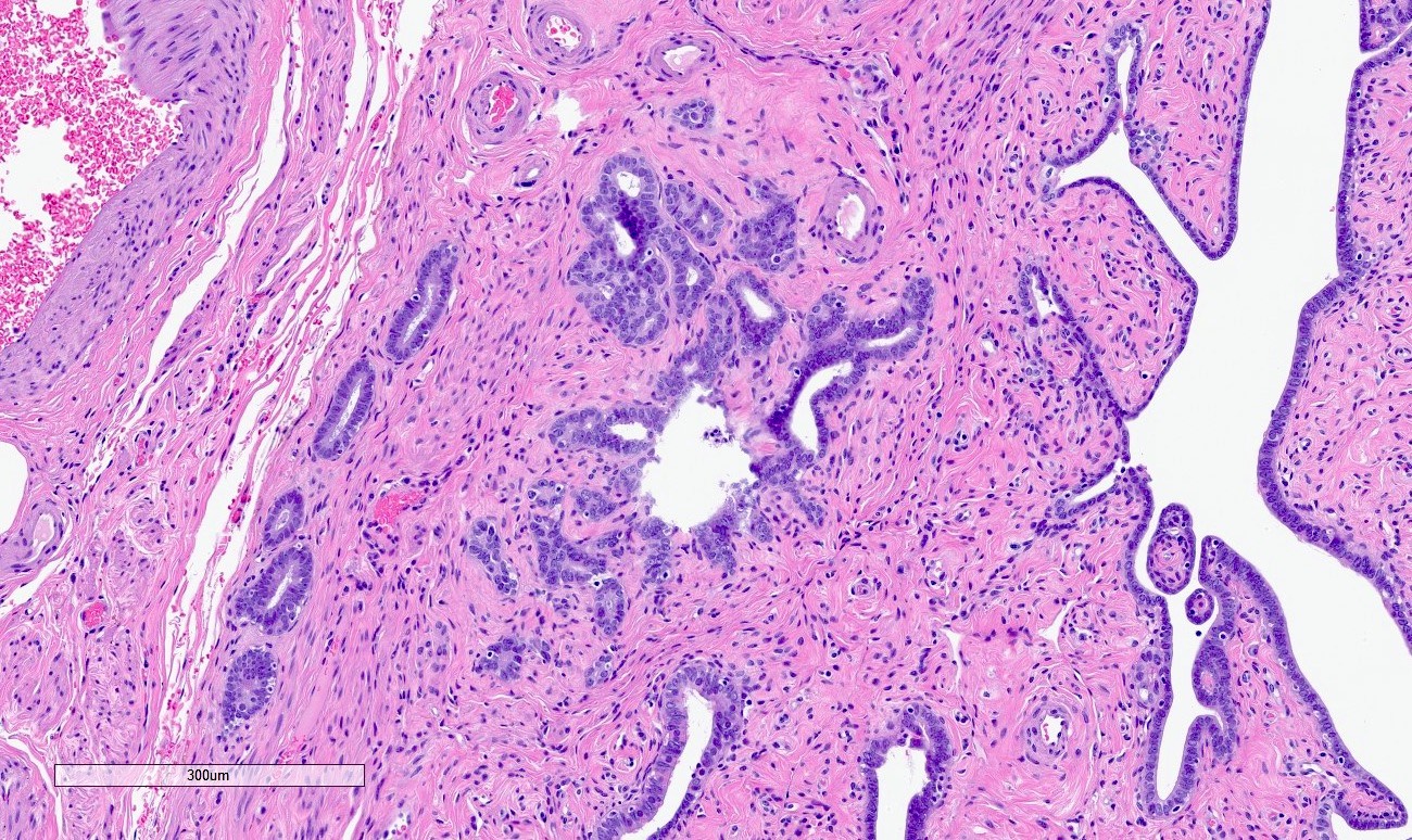
Microscopic (histologic) images
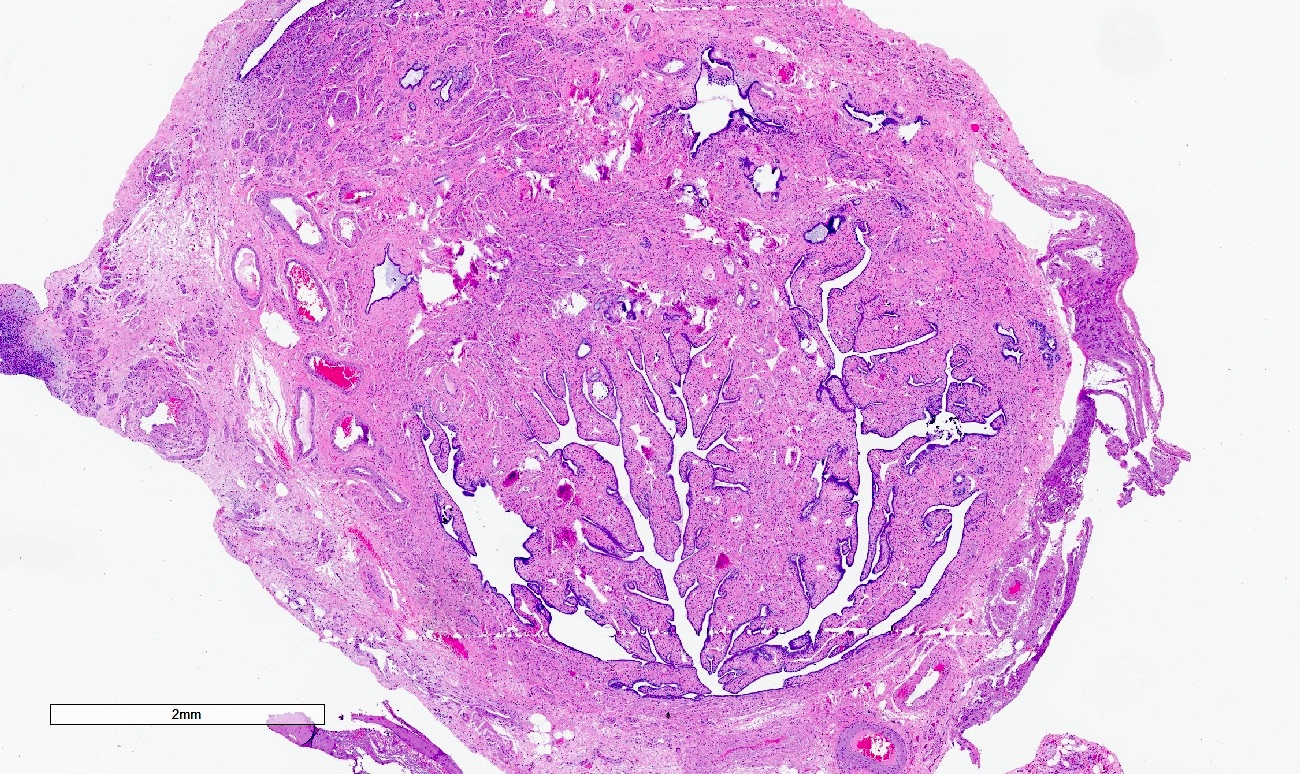
Contributed by Jessica L. Bentz, M.D., AFIP and @Andrew_Fltv on Twitter
Cystically dilated glands in smooth muscle
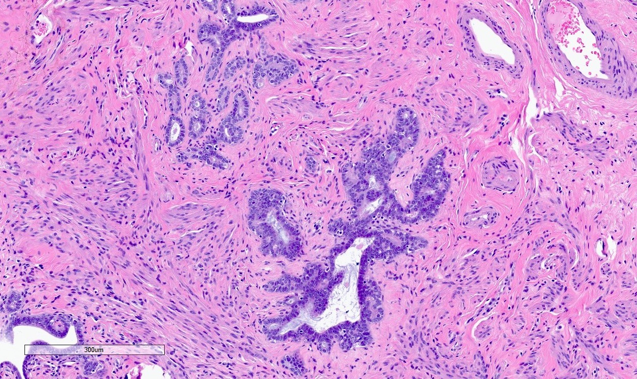
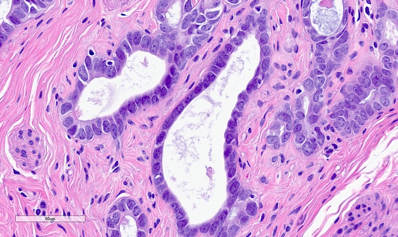
Bland nuclear features
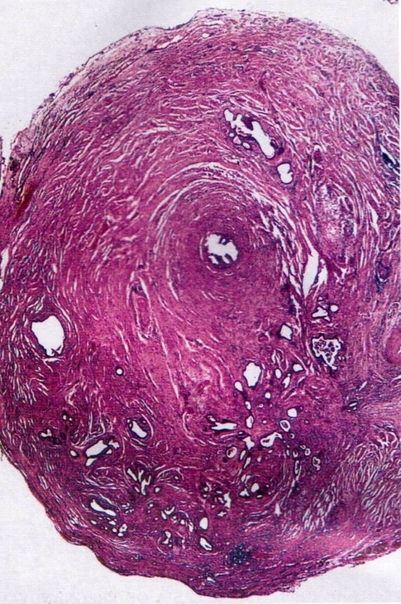
Gland-like structures
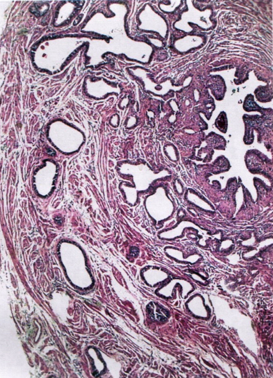
Numerous gland-like structures
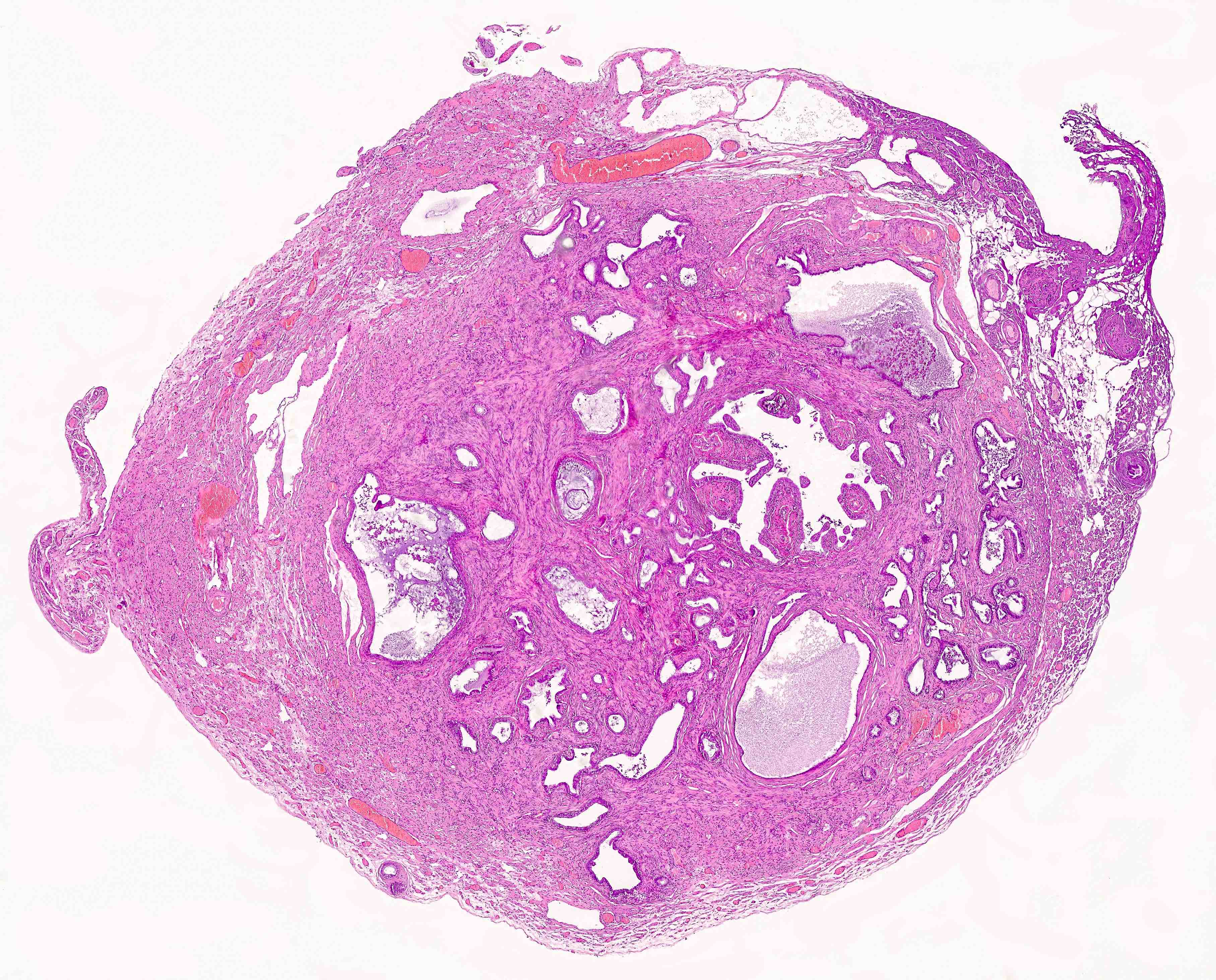
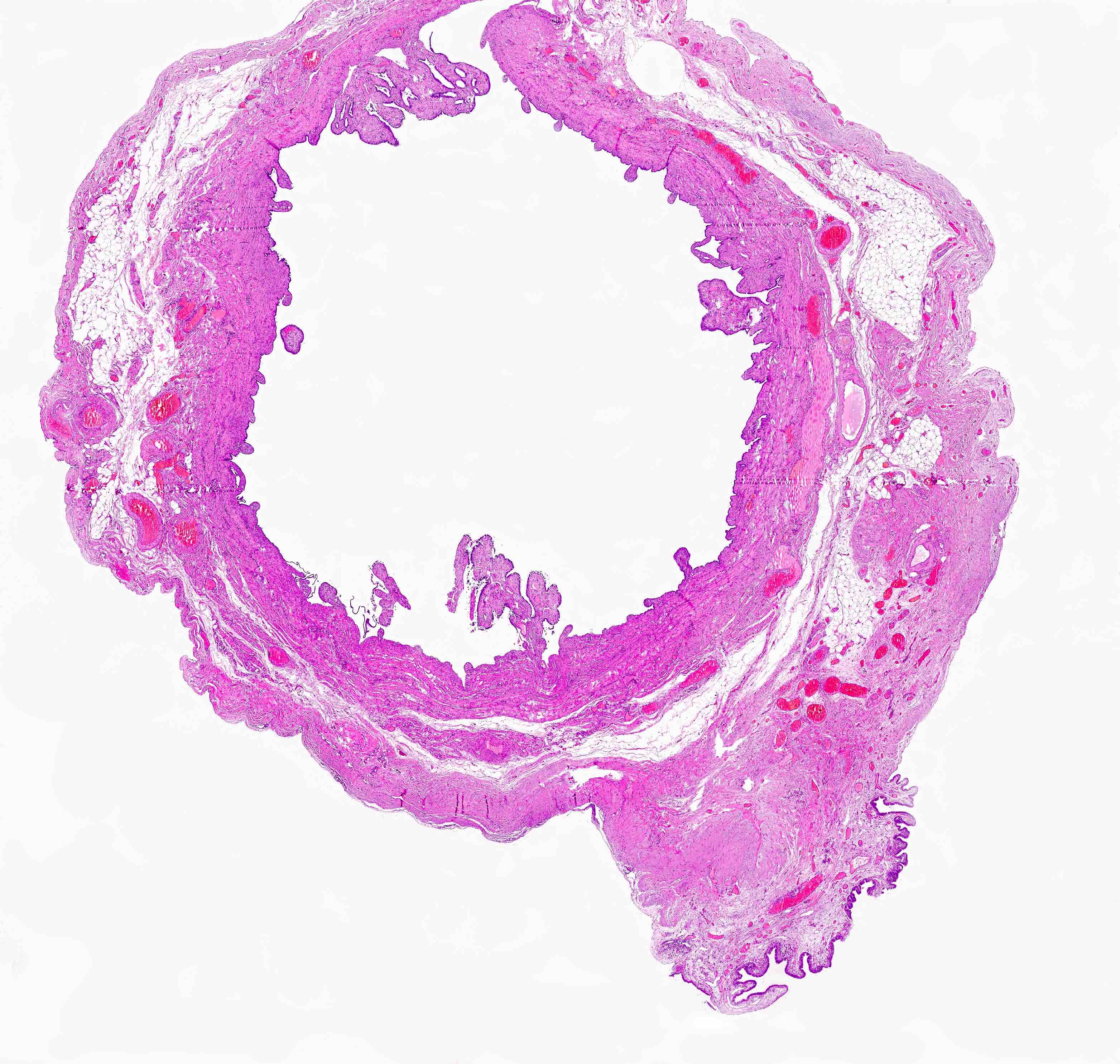
Salpingitis isthmica nodosa
Sample pathology report
- Right fallopian tube, salpingectomy:
- Salpingitis isthmica nodosa
Differential diagnosis
- Endometriosis:
- Glands are surrounded by endometrial stroma with or without hemosiderin laden macrophages
- Adenocarcinoma:
- Desmoplastic or inflammatory response and prominent cytologic atypia
- Adenomatoid tumor:
- Slit-like glands with thin, tapered septa lined by bland flattened to cuboidal cells
Additional references
Board review style question #1
What is the most common clinical sequela associated with salpingitis isthmica nodosa?
- Ectopic pregnancy
- Endometrioid adenocarcinoma
- Endometriosis
- Fitz-Hugh-Curtis syndrome
- Serous tubal intraepithelial carcinoma (STIC)
Board review style answer #1
A. Ectopic pregnancy. The most commonly documented and reported clinical sequela of salpingitis isthmica nodosa (SIN) is ectopic pregnancy. Answers E and B are incorrect because SIN is not a premalignant or malignant lesion. Answer C is incorrect because endometriosis is not a known clinical consequence of SIN. Answer D is incorrect because Fitz-Hugh-Curtis syndrome results from pelvic inflammatory disease, which may be seen in patients with SIN.
Comment Here
Reference: Salpingitis isthmica nodosa
Comment Here
Reference: Salpingitis isthmica nodosa
Board review style question #2
A 48 year old woman with a history of infertility undergoes a hysterectomy with bilateral salpingectomy for abnormal uterine bleeding. What is the most likely diagnosis in her right fallopian tube?
- Endometrioid adenocarcinoma
- Endometriosis
- Endosalpingiosis
- Salpingitis isthmica nodosa (SIN)
- Serous cystadenofibroma
Board review style answer #2
D. Salpingitis isthmica nodosa (SIN). SIN is the correct answer with multiple cystically dilated glands in the muscular wall of the tube and is common in patients with a history of infertility. Answer A is incorrect because endometrioid adenocarcinoma will have nuclear atypia and glandular complexity. Answer B is incorrect because endometriosis may appear as dilated glands in the muscular wall but will contain surrounding endometrial stroma, differentiating this entity from SIN (which does not contain associated endometrial stroma). Answer C is incorrect because endosalpingiosis is usually found in the peritoneum. Answer E is incorrect because serous cystadenofibroma will be grossly described as a cystic mass lesion, more commonly found on the ovary.
Comment Here
Reference: Salpingitis isthmica nodosa
Comment Here
Reference: Salpingitis isthmica nodosa
