
Table of Contents
Definition / general | Essential features | Case reports | Microscopic (histologic) description | Microscopic (histologic) images | Positive stains | Negative stainsCite this page: Lynch D. Crystal storing histiocytosis. PathologyOutlines.com website. https://www.pathologyoutlines.com/topic/hematologycsh.html. Accessed April 25th, 2024.
Definition / general
- Rare; crystalline material accumulates in cytoplasm of histiocytes; usually kappa light chain origin (Histopathology 2016;68:482)
- Adults are affected with a wide age range; men and women affected nearly equally
- Most commonly affects the head and neck, lung, kidney, bone marrow and lymph nodes, although nearly any site may be involved (Head Neck Pathol 2012;6:111)
- Strongly associated with underlying plasma cell neoplasm or lymphoma with plasma cell differentiation
- A minority of cases are associated with nonneoplastic causes such as infections and autoimmune disease
Essential features
- Rare disease with crystalline material in cytoplasm of histiocytes
- May affect any organ system
- Strongly associated with underlying lymphoid or plasma cell neoplasm
Case reports
- 38 year old woman with seizures (Clin Neuropathol 2014;33:23)
- 50 year old woman with lung mass and rheumatoid arthritis (Arch Pathol Lab Med 2005;129:1159)
- 51 year old woman with upper lip and cheek tumor (Head Neck Pathol 2012;6:111)
- 73 year old man with ascites, weight loss and fatigue (Blood 2002;100:1817)
Microscopic (histologic) description
- Cytologically benign histiocytes with abundant cytoplasm filled with many refractile eosinophilic crystals which may be needle-like or rhomboid in shape
- Neoplastic lymphocytes and plasma cells may be present
- Histiocytes often compose the majority of cellular elements, potentially obscuring an underlying neoplasm
Microscopic (histologic) images
Contributed by David Lynch, M.D.
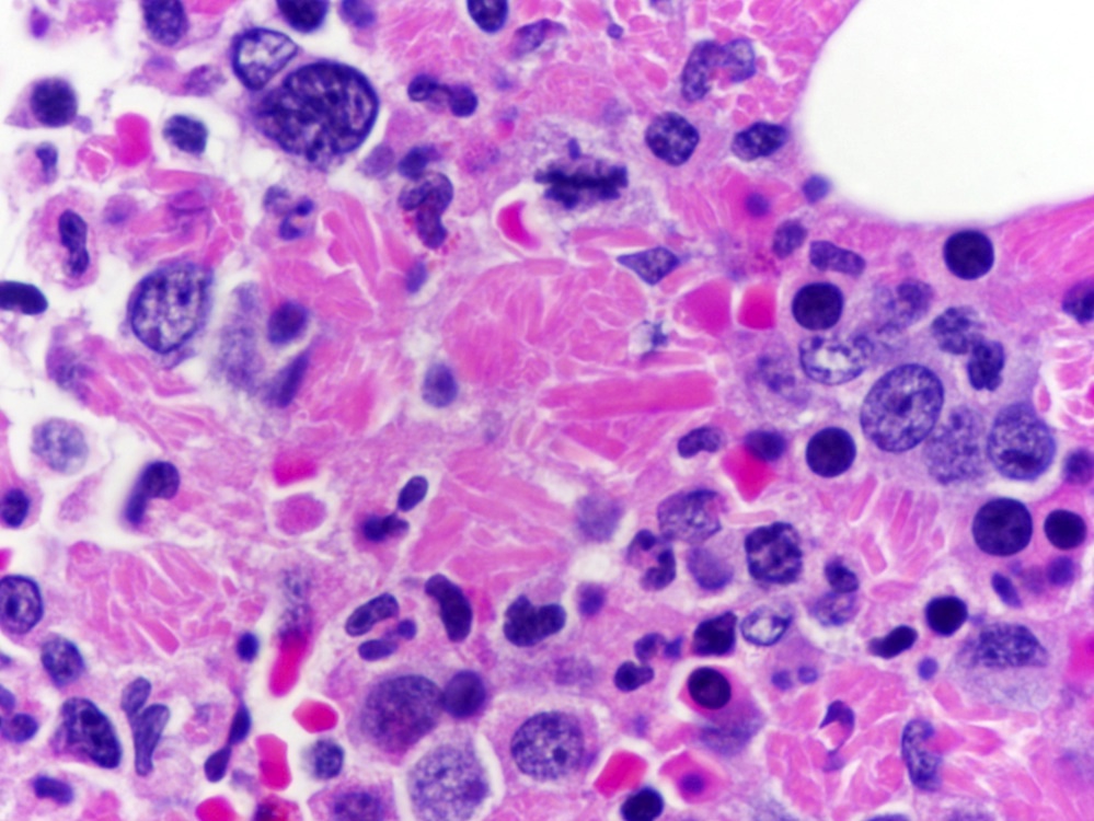
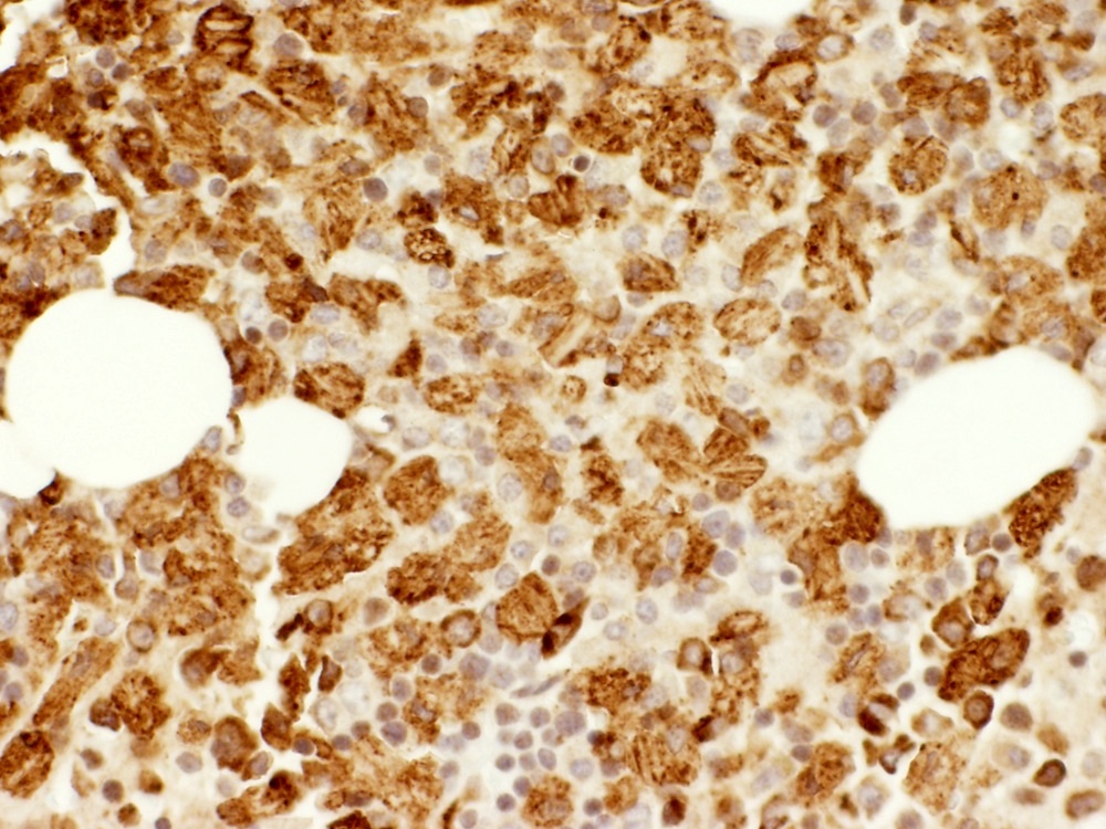
Bone marrow core and CD68
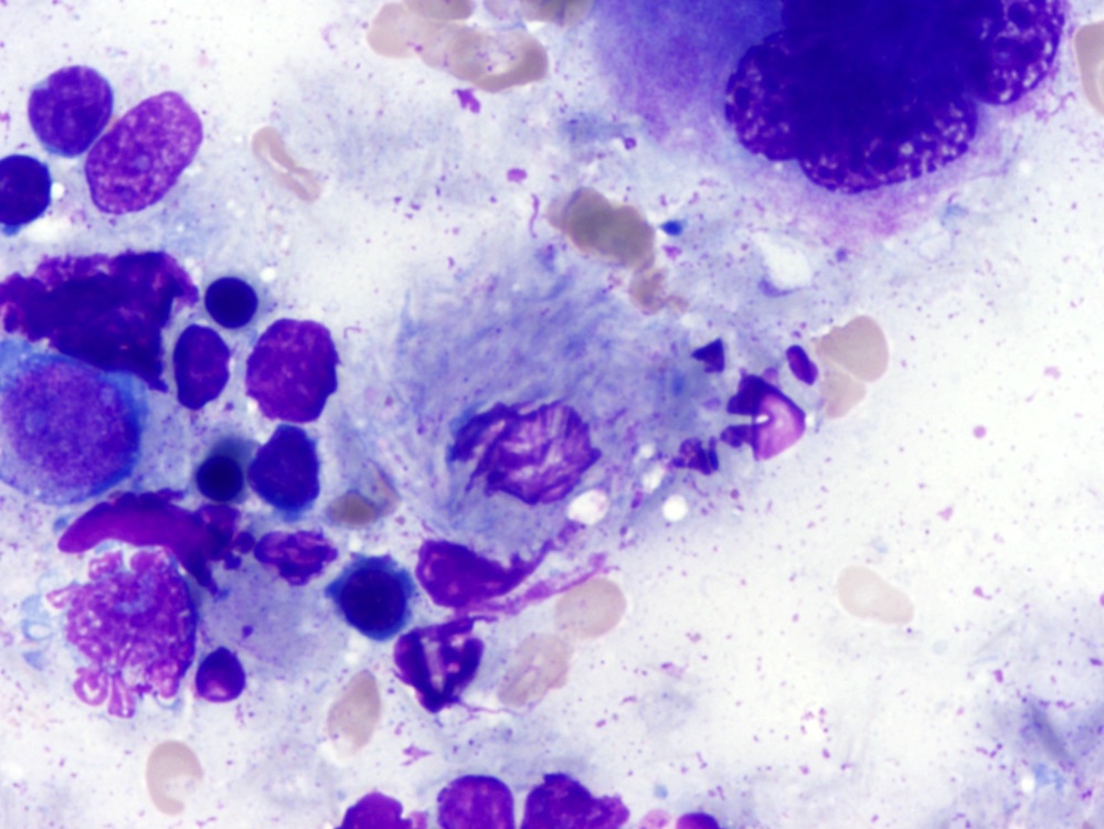
Bone marrow aspirate
Positive stains
Negative stains
