

Kidney nontumor / medical renal
Glomerular disease
Other hereditary renal disease
Collagen type III glomerulopathy
Author: Kriselle Lao, M.D.

Last author update: 18 November 2018
Last staff update: 28 August 2023
Copyright: 2003-2024, PathologyOutlines.com, Inc.
PubMed Search: Collagen type III glomerulopathy
Table of Contents
Definition / general | Essential features | Epidemiology | Sites | Etiology | Clinical features | Laboratory | Case reports | Treatment | Microscopic (histologic) description | Microscopic (histologic) images | Positive stains | Negative stains | Immunofluorescence description | Immunofluorescence images | Electron microscopy description | Electron microscopy images | Differential diagnosis | Board review style question #1 | Board review style answer #1Cite this page: Lao K. Collagen type III glomerulopathy. PathologyOutlines.com website. https://www.pathologyoutlines.com/topic/kidneycollagentypeIIIglomerulopathy.html. Accessed April 20th, 2024.
Definition / general
- Also called collagenofibrotic glomerulopathy
- Idiopathic deposition of type III collagen in glomeruli, normally absent in kidneys
Essential features
- Deposition of abnormally curved and serpentine type III collagen fibers in mesangial and subendothelial regions
Epidemiology
- Rare (< 100 cases in the literature)
- No sex predilection
- All ages (youngest: 3 months)
- Most cases reported from Japan and China
- Most pediatric cases from Europe
- References: Pediatr Nephrol 1993;7:354, Clin Kidney J 2012;5:7, Histopathology 2015;67:568, Exp Ther Med 2015;10:1445, Clin Kidney J 2015;8:543, Diagn Pathol 2009;4:33, Am J Kidney Dis 1999;33:123
Sites
- Renal limited
- Occasional systemic deposition (Am J Kidney Dis 1999;33:123)
Etiology
- Sporadic in adults
- Autosomal recessive in children
- Unknown mutation, type III collagen gene (COL3A1) normal
- References: Clin Kidney J 2015;8:543, BMC Vet Res 2013;9:218, Adv Chronic Kidney Dis 2012;19:101
Clinical features
- Proteinuria is a cardinal sign, sometimes nephrotic range (30 - 60%)
- Microscopic hematuria, chronic anemia (50 - 75%) and hypertension are common
- Variable progression
- Occasional stable disease but generally protracted course with increasing proteinuria and progressive renal failure
- In children, may be associated with hemolytic uremic syndrome (HUS); precipitous course more likely in children, especially if superimposed HUS
- References: Histopathology 2015;67:568, Clin Kidney J 2012;5:7, Am J Kidney Dis 2007;49:499, Indian J Nephrol 2011;21:52, Pediatr Nephrol 1993;7:354
Laboratory
- 10 - 100x increase in levels of procollagen type III peptide (PIIINP), not specific
- Serum hyaluronan may be increased to > 1000x, more specific (Intern Med 2014;53:1801)
Case reports
- 6 year old boy with inherited factor H deficiency and collagen type III glomerulopathy (Pediatr Nephrol 1995;9:11)
- 26 year old man with simultaneous Hodgkin lymphoma (Saudi J Kidney Dis Transpl 2011;22:126)
- 49 year old woman with simultaneous hepatic perisinusoidal deposition (Nephron 1993;63:183)
- 38 cases of collagenofibrotic glomerulopathy (Clin Kidney J 2012;5:7)
- Patient with collagenofibrotic glomerulopathy (Am J Kidney Dis 1999;33:123)
Treatment
- No curative treatment available; one case with reportedly no recurrence after renal transplant (Adv Chronic Kidney Dis 2012;19:101)
Microscopic (histologic) description
- Diffuse increase in mesangial matrix and generalized widening of glomerular capillary walls with pale eosinophilic material; may not be obvious in pediatric cases
- Negative or weakly positive with periodic acid Schiff, negative with methenamine silver, blue with Masson trichrome
Microscopic (histologic) images
Contributed by Joseph Grande, M.D., Ph.D.
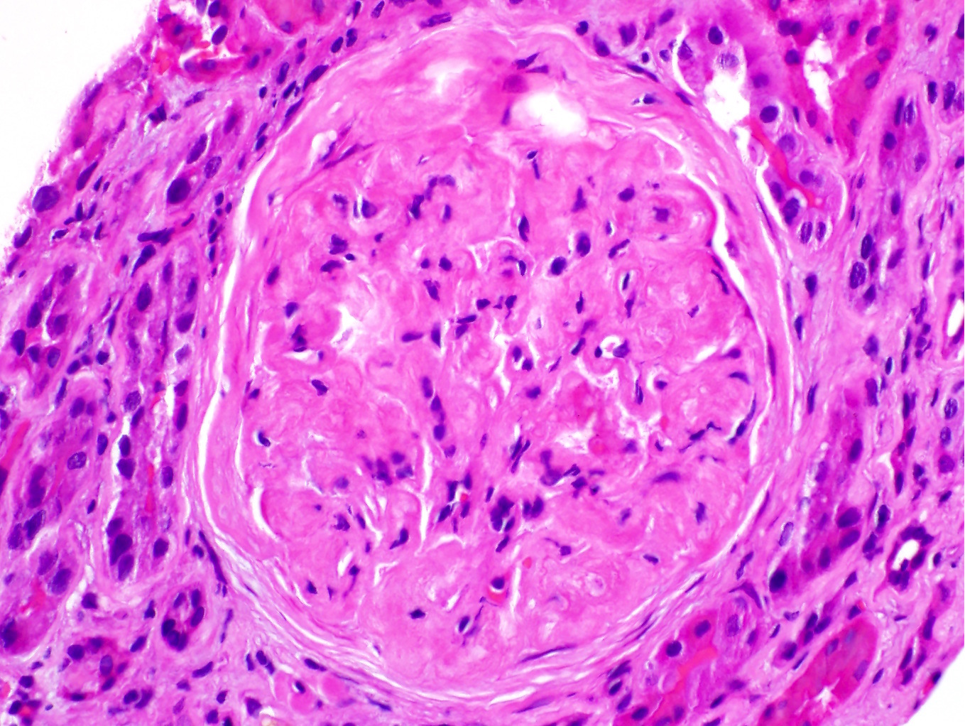
Lobular mesangial expansion
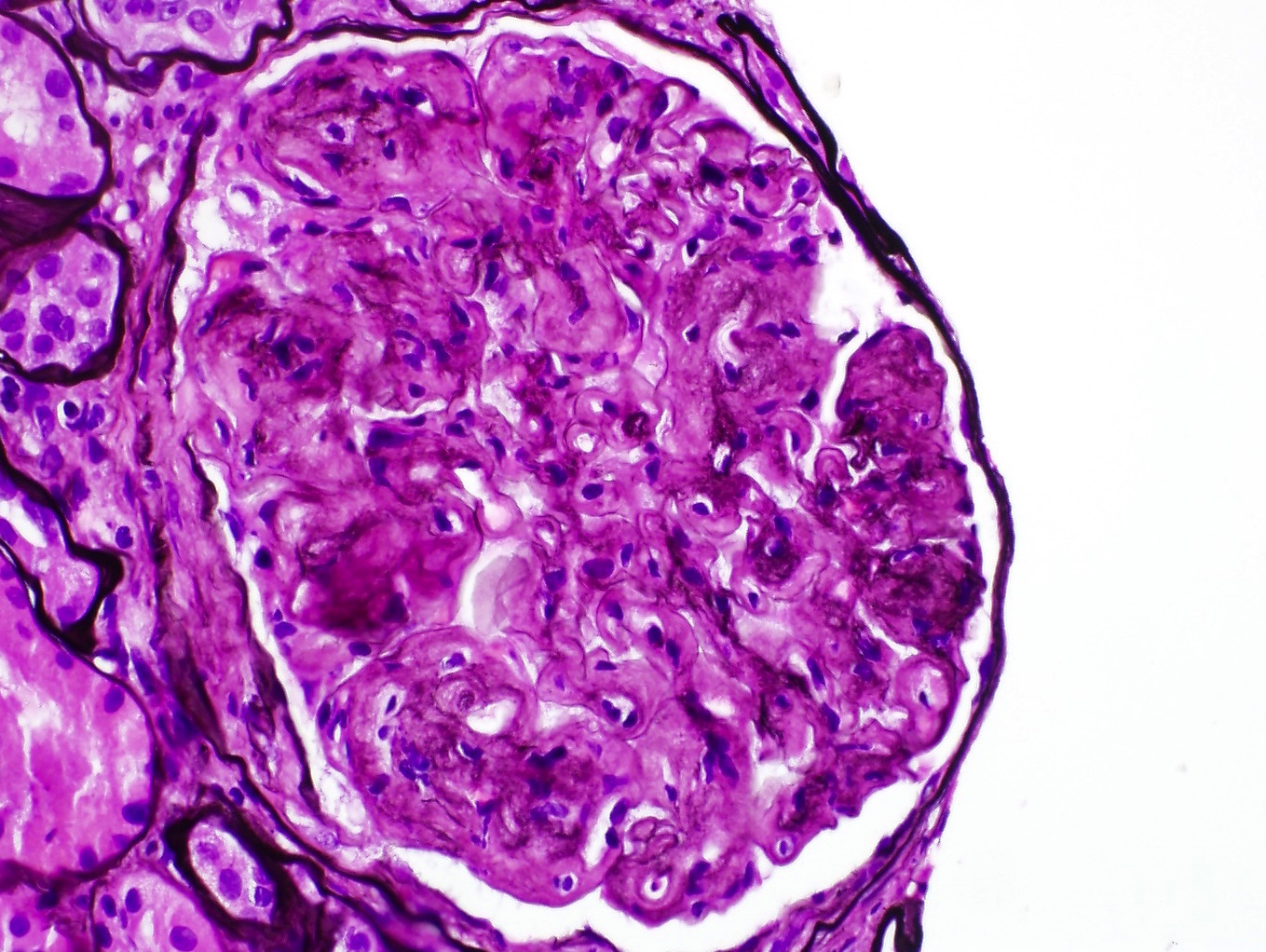
Silver negative material deposits

PAS weak / negative deposits

Trichrome stains deposits blue
Images hosted on other servers:

Enlarged glomerulus with hyaline cap deposits

Lobular appearance

With Hodgkin's lymphoma

Silver stain

Congo red stain
Positive stains
- Strong collagen type III staining in capillary loops and mesangium (normally absent in kidneys)
Negative stains
Immunofluorescence description
- Negative; nonspecific IgM and C3 entrapment
Immunofluorescence images
Images hosted on other servers:

Deposits were negative for all IgG, IgM and complements
Electron microscopy description
- Large accumulation of collagen fibrils in subendothelial glomerular basement membrane and mesangial matrix (Ultrastruct Pathol 2010;34:68)
- Banded with 60 nm periodicity
- Serpentine, curved, frayed
- More prominent curvilinear structure and better detail on phosphotungstic acid or tannic acid lead staining
Electron microscopy images
Contributed by Joseph Grande, M.D., Ph.D.

Extensive mesangial deposits
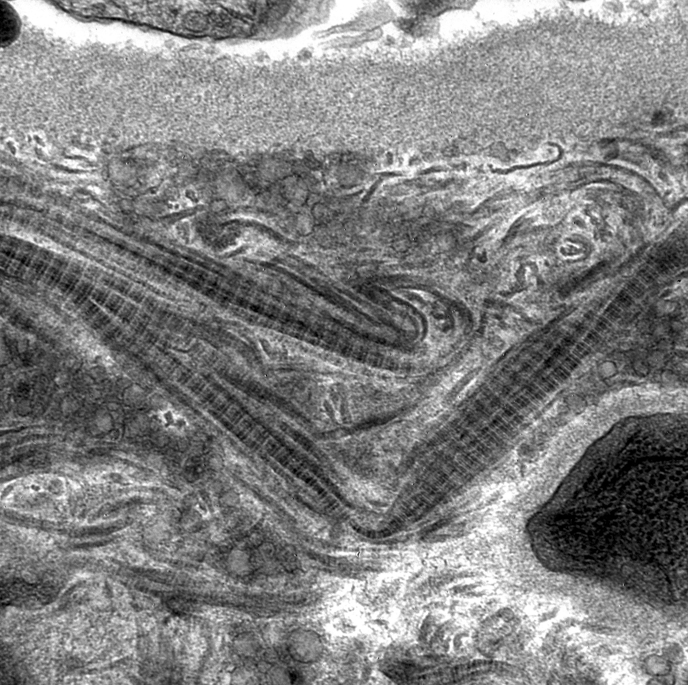
Higher power of subendothelial deposit
Images hosted on other servers:

Abundant subendothelial deposits of large fibers
Differential diagnosis
- Nail-patella syndrome: has other clinical features (e.g. bone / nail abnormalities), sparser fibers, typically located in lamina densa of glomerular basement membrane versus more subendothelial or mesangial location in collagenofibrotic glomerulopathy
Board review style question #1
- Onset of collagen type III glomerulopathy in childhood is associated with what manifestation?
- Aplastic anemia
- Hemolytic uremic syndrome
- Warm autoimmune hemolytic anemia
- Idiopathic thrombocytopenic purpura
Board review style answer #1
