

Kidney nontumor / medical renal
Other
Miscellaneous
Urolithiasis (stones)
Author: Nikhil Sangle, M.D.

Last author update: 3 December 2012
Last staff update: 20 September 2023
Copyright: 2002-2024, PathologyOutlines.com, Inc.
PubMed Search: Urolithiasis stones human
Table of Contents
Definition / general | Clinical features | Types of stones | Diagrams / tables | Case reports | Gross imagesCite this page: Sangle N. Urolithiasis (stones). PathologyOutlines.com website. https://www.pathologyoutlines.com/topic/kidneyurolithiasis.html. Accessed April 25th, 2024.
Definition / general
- Stones within collecting system of kidney are present in 5 - 10% of Americans; most commonly in men ages 20 - 49
- Due to supersaturation of stone constituents, decreased urine volume or deficiency of crystal inhibitors in urine
Clinical features
- 80% unilateral, usually in calyces, pelvis or bladder
- Usually only 2 - 3 mm, but with severe, abrupt flank pain and hematuria
- All stones contain an organic matrix of mucoprotein
Types of stones
- Calcium oxalate / phosphate (75%): due to hypercalciuria (idiopathic, 50%), hypercalcemia (infants may have high Vitamin D levels, Iran J Kidney Dis 2012;6:186), hyperoxaluria (in vegetarians with oxalate rich diet), hyperuricosuria (hyperparathyroidism, bone disease, sarcoidosis) and rarely primary hyperoxaluria (Arch Pathol Lab Med 2002;126:1250); oxalate crystals are highlighted by polarized light; are accompanied by foreign body giant cells and macrophages
- Struvite (triple stones, magnesium ammonium phosphate, 15%): due to urea splitting bacteria (Proteus, Staphylococcus); produce staghorn calculi
- Uric acid (6%): due to hyperuricemia, chemotherapy for leukemia, uricosuric drugs or excess dietary proteins; 50% lack elevated uric acid in blood / urine; may be due to acidic pH; radiolucent; may become staghorn calculi if in renal pelvis; elongated and rectangular crystals in collecting tubules or doubly refractile crystals in interstitium with giant cell reaction
- Cysteine (1%): due to genetic defects in cystine transport (Orphanet J Rare Dis 2012;7:19); autosomal recessive, affects 1 per 20,000; yellow-brown and radioopaque stones form at low urinary pH; crystals are flat hexagons in urine
- Ammonium acid urate: rare, but more common in Asia (Kaohsiung J Med Sci 2012;28:259)
- Stone granuloma: complication of ureteral stone fragmentation and instrumentation
- Xanthinuria (rare): autosomal recessive, due to deficient xanthine oxidase, causing excessive xanthine levels and stones in 1/3 with this disorder
Diagrams / tables
Images hosted on other servers:

Passage of a calculus (stone)
Case reports
- 48 year old man with nephrectomy for renal mass due to urolithiasis caused by 2,8-dihydroxyadenine crystals (Hum Pathol 1992;23:1081)
Gross images
Contributed by Jian-Hua Qiao, M.D.
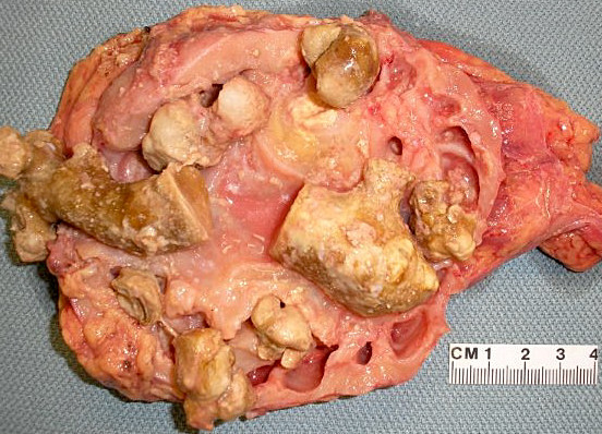
Multiple kidney stones
