
Larynx, hypopharynx & trachea
Benign tumors / nonneoplastic
Laryngeal cysts
Author: Kanwalpreet Kaur, M.D., D.M.
Editorial Board Member: Ruta Gupta, M.D.
Deputy Editor-in-Chief: Kelly Magliocca, D.D.S., M.P.H.

Last author update: 8 April 2024
Last staff update: 8 April 2024
Copyright: 2002-2024, PathologyOutlines.com, Inc.
PubMed Search: Laryngeal cysts
Table of Contents
Definition / general | Essential features | Terminology | ICD coding | Epidemiology | Sites | Pathophysiology | Etiology | Diagrams / tables | Clinical features | Diagnosis | Radiology description | Radiology images | Prognostic factors | Case reports | Treatment | Clinical images | Gross description | Gross images | Microscopic (histologic) description | Microscopic (histologic) images | Sample pathology report | Differential diagnosis | Additional references | Board review style question #1 | Board review style answer #1 | Board review style question #2 | Board review style answer #2Cite this page: Kaur K. Laryngeal cysts. PathologyOutlines.com website. https://www.pathologyoutlines.com/topic/larynxcyst.html. Accessed April 20th, 2024.
Definition / general
- Laryngeal cysts are fluid filled, mucosa lined lesions that are unconnected to the laryngeal ventricle
Essential features
- Rare benign lesions in larynx; can affect both children and adults
- Mostly asymptomatic but can cause respiratory distress
- Computed tomography (CT): initial investigation of choice
- Histology is mandatory for definite diagnosis
- Oncocytic cysts are associated with recurrence particularly when multifocal, so long term follow up is recommended
- Numerous proposed classifications
Terminology
- Cystic laryngeal mass, saccule, laryngocele (not recommended)
ICD coding
- ICD-10: D14.1 - benign neoplasm of larynx
Epidemiology
- No gender predilection; oncocytic cysts more common in women (Eur Arch Otorhinolaryngol 2021;278:3381)
- In adults, more common after sixth decade
- Accounts for 5 - 10% of benign laryngeal lesions in adults
- 1.8 in 100,000 newborns
Sites
- Can occur in any part of the larynx but are more frequent in epiglottis and vallecula
Pathophysiology
- See Clinical features for classifications
- Ductal cyst
- Mucus retention cyst caused by obstruction of mucus gland ducts within mucus membrane; represents 75% of all laryngeal cysts
- Saccular cyst
- Saccule is a membranous sac in the larynx, located between the false vocal cords and the inner surface of the thyroid cartilage, which contains mucous secreting glands
- Obstruction of the mucous gland ducts of the laryngeal saccule with subsequent accumulation of secretions results in a submucosal cyst
- Represents 25% of all laryngeal cysts; further classified as anterior or lateral
- Lateral saccular cysts are predominantly congenital, formed during embryogenesis due to aberrant entrapment of developing saccule by third and fifth brachial arches; these remain quiescent until a trigger such as an upper respiratory tract infection causes expansion of the cyst
- References: Int J Phonosurg Laryngol 2016;6:53, Otolaryngol Head Neck Surg 2017;157:928
Etiology
- Oncocytic cysts more common in old age and in chronic smokers
Diagrams / tables
Images hosted on other servers:

Coronal cross section of larynx showing saccule
Clinical features
- Nonspecific
- Hoarseness, foreign body sensation, dyspnea (Otolaryngol Head Neck Surg 2017;157:928)
- If located near the orifice of the ventricle or subglottic region, symptoms can range from stridor to significant respiratory obstruction
- Those in the vallecula can cause feeding problems
- Numerous proposed classifications
- DeSanto classification (Laryngoscope 1970;80:145)
- Ductal
- Saccular
- No importance to histology
- No separate class of congenital cysts
- Arens classification (Eur Arch Otorhinolaryngol 1997;254:430)
- Congenital
- Retention
- Inclusion cysts
- Forte classification of congenital cysts (Laryngoscope 2004;114:1123)
- Type I: confined to larynx, lined by endoderm
- Type II: extends beyond larynx
- Type IIa: composed of endoderm only
- Type IIb: composed of endoderm and mesoderm elements in cyst wall
- Newman classification (Am J Clin Pathol 1984;81:715)
- Epithelial cyst (includes ductal and saccular)
- Tonsillar cyst
- Oncocytic cyst
- DeSanto classification (Laryngoscope 1970;80:145)
Diagnosis
- Laryngoscopy
Radiology description
- Used to determine the location, size, extent and anatomical relationships
- Computed tomography (CT)
- Initial investigation of choice
- Well defined, fluid attenuation, nonenhancing rounded lesion
- Magnetic resonance imaging (MRI)
- Gold standard (Pediatr Pulmonol 2019;54:478)
- Well defined, fluid signal intensity, nonenhancing rounded lesion
- Endolaryngeal ultrasound (EUS) and optical coherence tomography (OCT)
- These are newer techniques that may improve preoperative evaluation of cysts
Radiology images
Images hosted on other servers:

MRI
Prognostic factors
- Oncocytic cysts are associated with multiplicity and recurrence (Otolaryngol Head Neck Surg 2017;157:928)
Case reports
- 5 year old girl with dysphonia, moderate inspiratory dyspnea (J Med Life 2016;9:199)
- 34 year old man with a laryngeal cyst (Medicine (Baltimore) 2018;97:e11280)
- 64 year old man with laryngeal mass (Clin Pract 2016;6:822)
- 86 year old woman with oncocytic cyst (Forensic Sci Med Pathol 2022;18:554)
Treatment
- Complete excision by endoscopic excision, microlaryngeal surgery, laser excision or marsupialization
- Recurrence is associated with remnant of the cyst wall; therefore, total excision is recommended
Clinical images
Images hosted on other servers:


Spherical bulges covered by smooth epithelium
Gross description
- Specimens are rarely received intact, so the following described features are from endoscopic findings
- Ductal cyst
- Usually measures < 1 cm in diameter
- Tends to be superficial (lies within mucus membrane)
- Saccular cyst
- Submucosal, ranges in size from 1 to > 7 cm in greatest dimension
- Reference: Medicine (Baltimore) 2018;97:e11280
Gross images
Images hosted on other servers:

Cyst in glottic region
Microscopic (histologic) description
- Histology is critical for definitive diagnosis
- Epithelial cyst
- Lined by respiratory or squamous epithelium or a flattened epithelium
- Epithelial lining may be papillary in architecture
- May contain keratin or mucin
- May have a lymphoid component in cyst wall
- Includes both ductal and saccular cysts; differentiation of ductal from saccular cysts is not possible histologically, so differentiation rests on clinical parameters
- Tonsillar cyst
- Resembles palatine tonsillar crypt
- Lined by stratified squamous epithelium
- Lymphoid follicles with germinal centers seen in cyst wall
- Oncocytic cyst
- Unilocular or multilocular cyst lined by uniform oncocytic epithelial cells
- May have prominent intraluminal papillary architecture or hyperplastic solid nests
- Unlike Warthin tumor, doesn't have bilayered oncocytes and lymphoid stroma
- Oncocytes originate from metaplasia of seromucinous gland ducts in response to aging and chronic smoking resulting in disturbance of mitochondrial enzyme organization
- Originates in ventricles or false vocal cords, presents as supraglottic mass
- Also called oncocytic papillary cystadenoma, oncocytic adenomatous hyperplasia, oncocytoma
- No atypia or invasion; mitosis: rare (Eur Arch Otorhinolaryngol 2021;278:3381)
Microscopic (histologic) images
Contributed by Arpita Jindal, M.D.
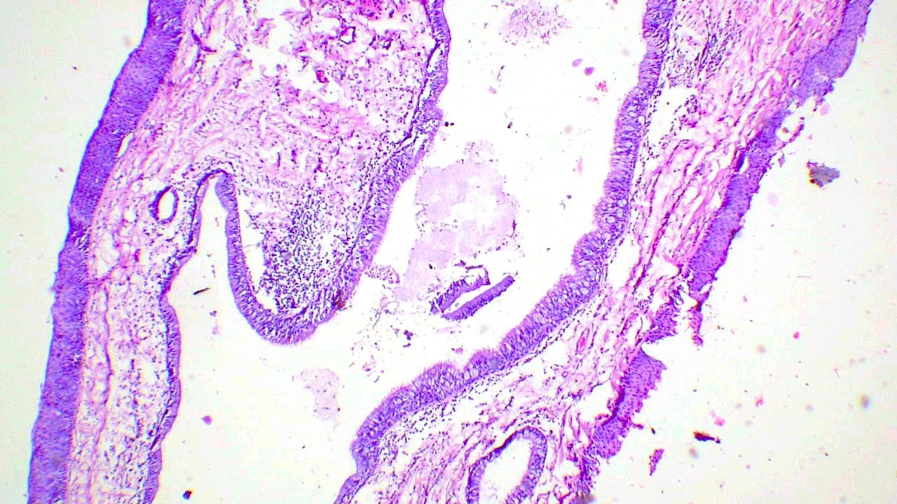
Pseudostratified columnar epithelium lined cyst

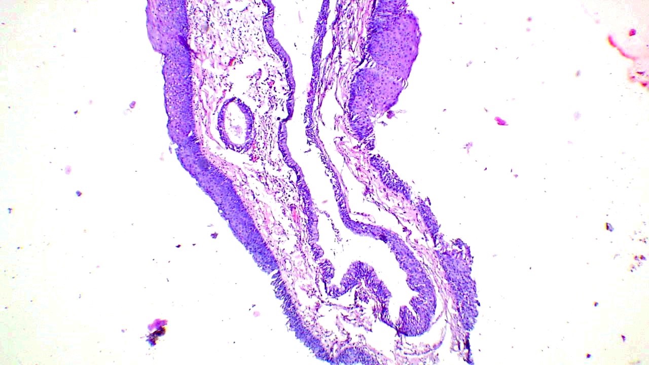
Cyst lining showing squamous metaplasia

Intraluminal infolding with focal lymphoid collection
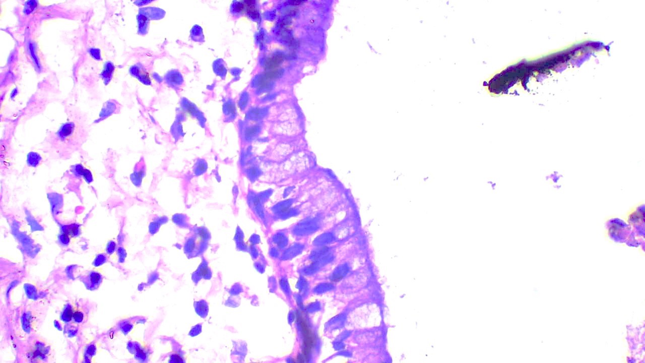
Columnar mucinous lining, basally located nuclei
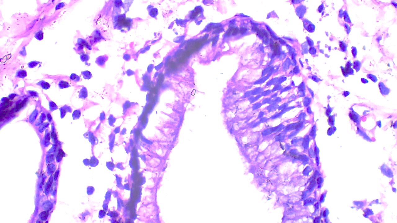
Pseudostratified columnar epithelium with cilia
Sample pathology report
- Larynx, epiglottic cyst, excision:
- Consistent with laryngeal epithelial cyst, lined by stratified squamous epithelium
Differential diagnosis
- Laryngocele:
- Dilatation of saccule
- Different from saccular cyst because these are distended with air and communicate with the laryngeal lumen
- Thyroglossal cyst:
- Difficult to distinguish from saccular cyst if deficient in thyroid tissue
- Differentiated on basis of exact anatomical location of cyst, as thyroglossal duct cysts are related with the hyoid bone and located in the midline of the neck
Additional references
Board review style question #1
A 61 year old woman presented with hoarseness of voice and was found to have a nodular lesion in the larynx with the microscopy shown above. What is the diagnosis?
- Laryngeal epithelial cyst
- Laryngocele
- Squamous papilloma
- Vocal cord polyp
Board review style answer #1
A. Laryngeal epithelial cyst. The section shows a distended cystic lesion lined by columnar epithelial cells with focal squamous metaplasia. The lesion is entirely confined within the larynx with no connection to lumen or dysplasia of overlying squamous epithelium. Answer B is incorrect because laryngocele is an air filled cavity communicating with the laryngeal lumen. Answer C is incorrect because squamous cell papilloma is an exophytic lesion lined by hyperplastic stratified squamous epithelium. Answer D is incorrect because vocal cord nodule / polyp is a surface lesion with no intralaryngeal extension.
Comment Here
Reference: Laryngeal cysts
Comment Here
Reference: Laryngeal cysts
Board review style question #2
Which type of laryngeal cyst needs follow up for a longer period of time after surgical excision?
- Ductal cyst
- Oncocytic cyst
- Saccular cyst
- Tonsillar cyst
Board review style answer #2
B. Oncocytic cyst. Oncocytic cysts need longer follow up because of their association with recurrence and multifocality. Anwers A, C and D are incorrect as surgical excision alone is sufficient and these cysts are not known to be associated with recurrence.
Comment Here
Reference: Laryngeal cysts
Comment Here
Reference: Laryngeal cysts
