
Table of Contents
Definition / general | Diagnosis | Case reports | Microscopic (histologic) description | Microscopic (histologic) images | Positive stains | Negative stains | Electron microscopy images | Molecular / cytogenetics description | Differential diagnosisCite this page: Mihova, D. M5a. PathologyOutlines.com website. https://www.pathologyoutlines.com/topic/leukemiaacutemonocyticleukemiam5a.html. Accessed April 25th, 2024.
Definition / general
- Acute monoblastic leukemia (M5a)
- 5 - 8% of AML
- Children and young adults
Diagnosis
- 80%+ of monocyte lineage cells are monoblasts
Case reports
- 66 year old man with erythropoietin dependent transformation of refractory anemia with ringed sideroblasts into acute monoblastic leukemia (Blood 2001;98:3492)
- 73 year old woman with coexisting mantle cell lymphoma (Leuk Lymphoma 2005;46:1813)
- 82 year old man with acute monoblastic leukemia following granular lymphocyte proliferative disorder (Rinsho Ketsueki 2011;52:1870)
Microscopic (histologic) description
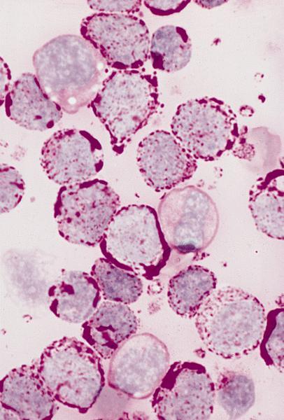
- Hypercellular marrow with large number of monoblasts
- Monoblasts are large with moderately abundant intensely basophilic cytoplasm, variably basophilic and delicate azurophilic granules but no / rare Auer rods
- May have pseudopods or vacuoles
- Have round nuclei and lacy chromatin with one or more prominent nucleoli but no folds
- Promonocytes have abundant less basophilic cytoplasm with obvious azurophilic granules and nuclei have delicate folds
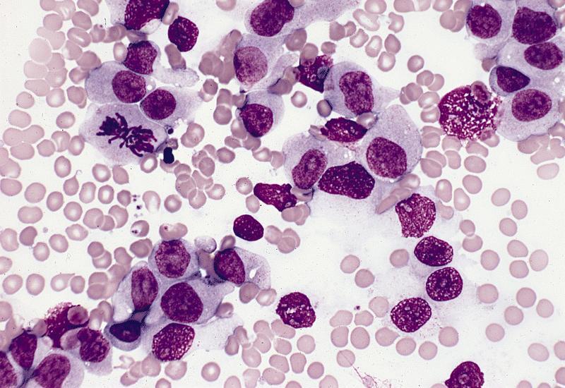
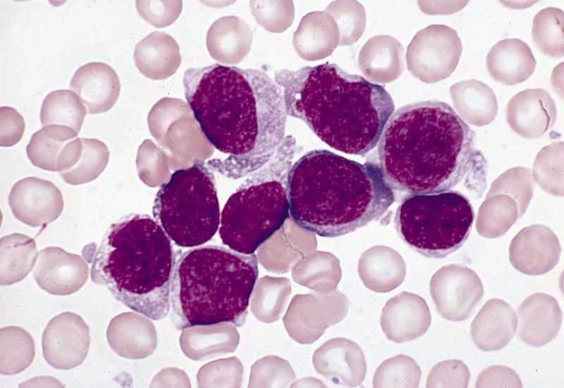
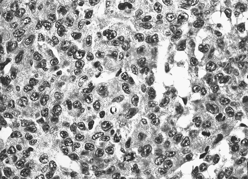
Microscopic (histologic) images
AFIP images

Abundant cytoplasm with azurophilic granules
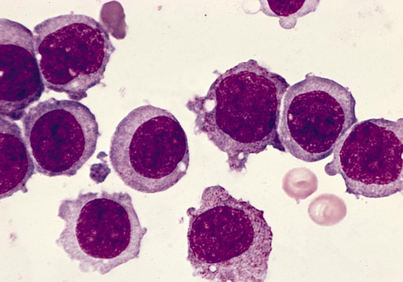
Monoblasts are large with abundant cytoplasm
Some monoblasts also show pseudopods
Monoblasts have variable cytoplasm
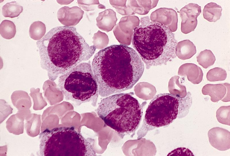
Monoblasts and promonocytes
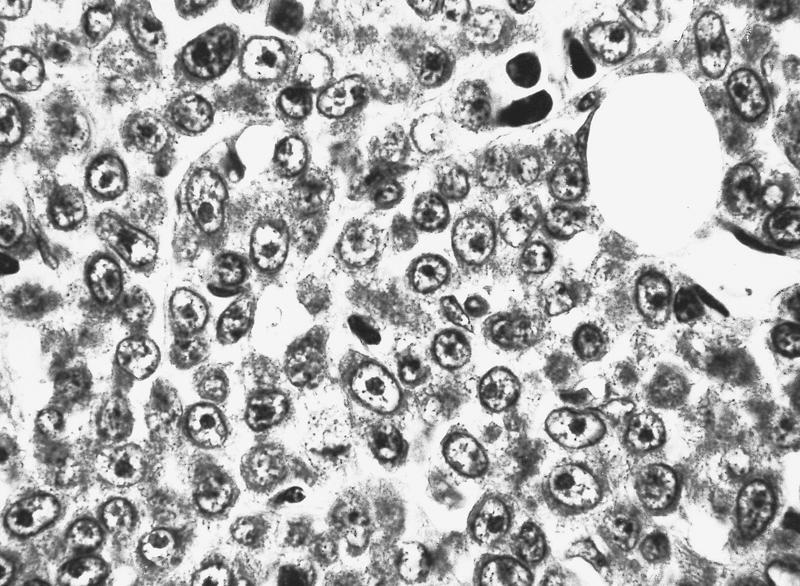
Marrow completely replaced by monoblasts
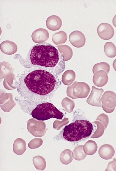
Monoblasts are large with abundant pale cytoplasm
Large monoblasts with abundant cytoplasm
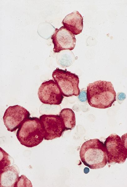
Nonspecific esterase positive
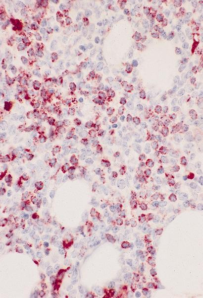
CD68 #1 (KP-1) positive
PAS positive
Negative stains
Electron microscopy images
AFIP images
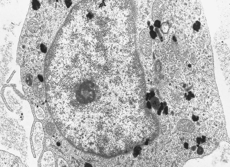
Scattered electron dense deposits
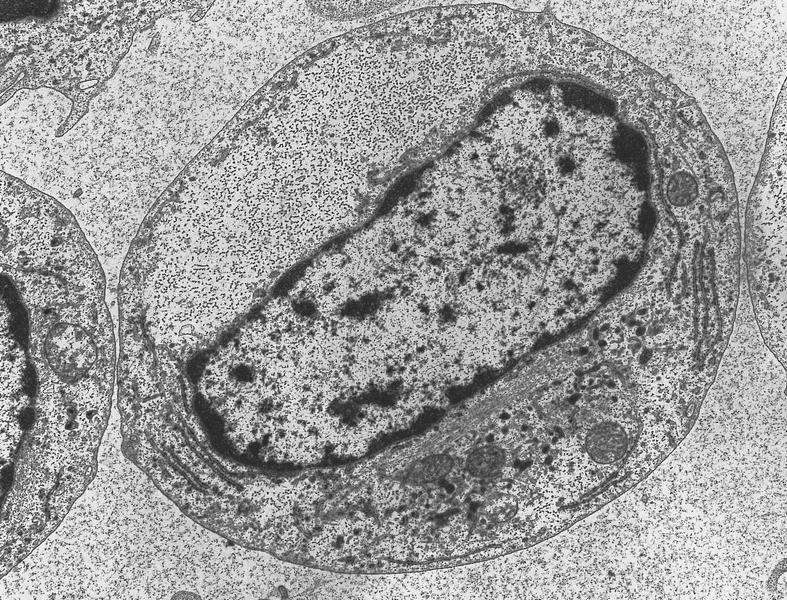
Cytoplasm contains focal area of glycogen deposition
Molecular / cytogenetics description
- 75% have cytogenetics abnormalities, including 11q23 in 30% (these cases should be classified as a recurrent genetic abnormality)
- FLT3 mutations in 7%
