

Lung
Mesenchymal tumors
Primary pulmonary myxoid sarcoma with EWSR1::CREB1 fusion
Editorial Board Member: Jefree J. Schulte, M.D.
Editor-in-Chief: Debra L. Zynger, M.D.


Last author update: 8 August 2022
Last staff update: 8 August 2022
Copyright: 2019-2024, PathologyOutlines.com, Inc.
PubMed Search: Primary pulmonary myxoid sarcoma

Table of Contents
Definition / general | Essential features | Terminology | ICD coding | Epidemiology | Sites | Pathophysiology | Etiology | Diagnosis | Radiology description | Radiology images | Prognostic factors | Case reports | Treatment | Gross description | Microscopic (histologic) description | Microscopic (histologic) images | Positive stains | Negative stains | Electron microscopy description | Molecular / cytogenetics description | Sample pathology report | Differential diagnosis | Board review style question #1 | Board review style answer #1 | Board review style question #2 | Board review style answer #2Cite this page: Gui H, Zhang PJL. Primary pulmonary myxoid sarcoma with EWSR1::CREB1 fusion. PathologyOutlines.com website. https://www.pathologyoutlines.com/topic/lungtumorprimpulmmyxsarcoma.html. Accessed April 25th, 2024.
Definition / general
- Low grade tumor
- Typically in (or close to) large airways
- Lobulated tumor with spindle and stellate cells arranged in lace-like strands and cords within prominent myxoid stroma
Essential features
- Endobronchial nodular mass with reticular / lace-like growth pattern and abundant myxoid stroma
- Expresses EMA and vimentin, negative for most other markers
- FISH positive for EWSR1 rearrangement (85%)
Terminology
- Primary pulmonary myxoid sarcoma
ICD coding
Epidemiology
- Mean age = 46.3 years; F:M = 1.4:1 (24 cases)
- 40% with cough, hemoptysis, chest pain; 20% present as incidental lung mass (Am J Surg Pathol 2011;35:1722)
Sites
- Bilateral lungs, 76% within or close to a large bronchus (Int J Surg Pathol 2017;25:518)
Pathophysiology
- Probably originates from primitive mesenchymal cells with myofibroblastic or fibroblastic differentiation (Virchows Arch 2014;465:453)
- Controversial relationship to myxoid angiomatoid fibrous histiocytoma with similar EWSR1 fusion genes (Histopathology 2014;65:144)
- Primary pulmonary myxoid sarcoma (PPMS) and myxoid variant of angiomatoid fibrous histiocytoma may represent a continuum with overlapping histologic, immunohistochemical and genetic features (Histopathology 2014;65:144, Am J Surg Pathol 2020;44:1535)
Etiology
- Smoking history in 80% (Am J Surg Pathol 2011;35:1722)
Diagnosis
- Relies on histology, immunohistochemistry and FISH in biopsy or surgical resection
Radiology description
- Mass related to bronchus, predominantly endobronchial
Radiology images
Contributed by Hongxing (Simon) Gui, M.D., Ph.D.
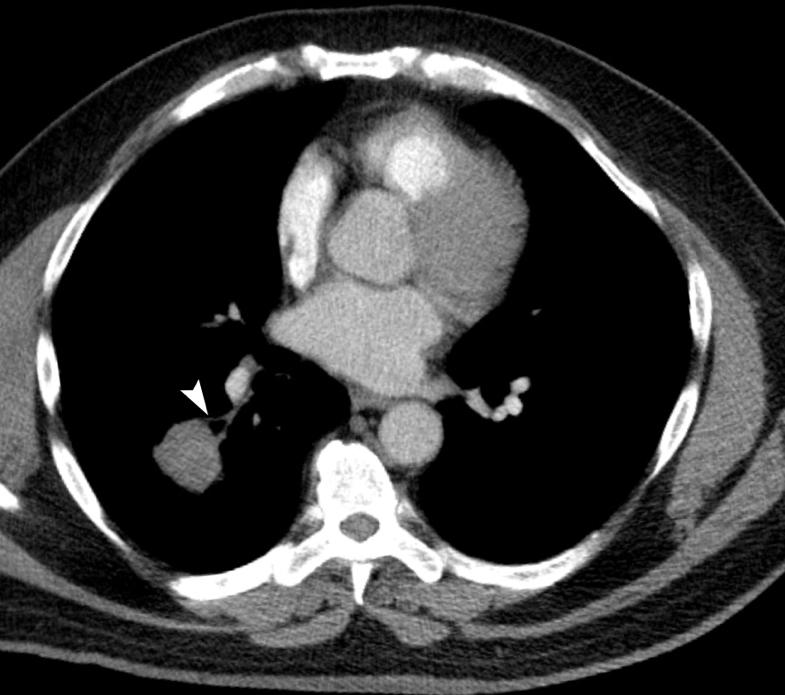
Endobronchial mass
Prognostic factors
- Patients with EWSR1 rearrangement have favorable prognosis; wild type EWSR1 portends poor clinical outcome (Pol J Pathol 2017;68:261)
Case reports
- 28 year old men and 66 year old woman with features overlapping with angiomatoid fibrous histiocytoma (Histopathology 2014;65:144)
- 29 year old woman with mass in a major fissure of the left lung without parenchymal invasion (Thorac Cancer 2017;8:535)
- 32 year old woman with tumor adjacent to the right main bronchus (Pathology 2017;49:792)
- 44 and 49 year old men with both components of PPMS and angiomatoid fibrous histiocytoma (Am J Surg Pathol 2020;44:1535)
- 48 year old man with a huge mass extending into the right main bronchus (Pol J Pathol 2017;68:261)
- 80 year old woman with an endobronchial mass in the left main bronchus (Int J Surg Pathol 2017;25:518)
Treatment
- Surgical resection (Am J Surg Pathol 2011;35:1722)
Gross description
- Generally < 4 cm, well circumscribed or nodular
- Glistening or gelatinous cut surface, white-gray to yellow (Am J Surg Pathol 2011;35:1722)
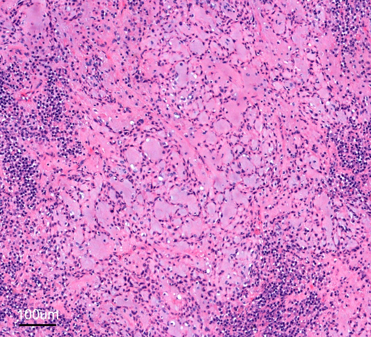
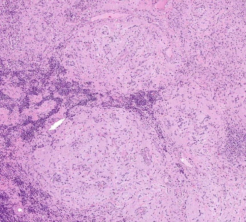
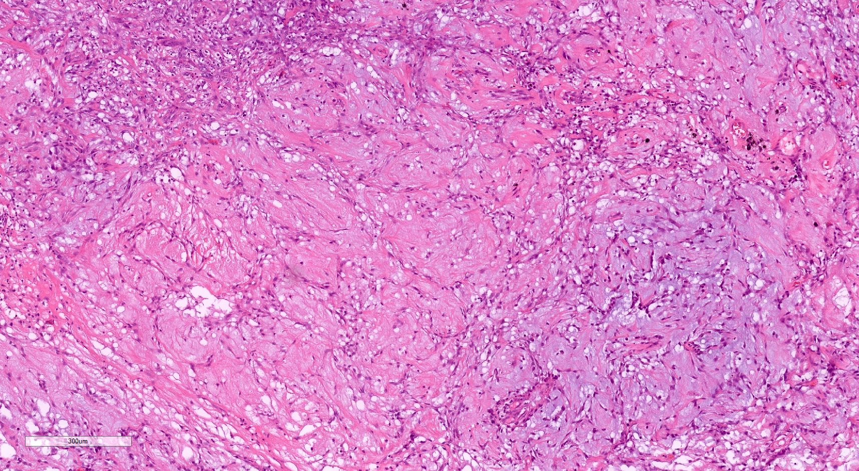
Microscopic (histologic) description
- Lobulated architecture, reticular or lace-like pattern with anastomosing cords and strands, abundant myxoid stroma, often lightly basophilic (Am J Surg Pathol 2011;35:1722)
- Oval, spindle to polygonal cells with minimal to moderate atypia, infrequent mitotic figures with the majority < 5/10 high power fields
- Admixed lymphoplasmacytic infiltrate
- Some cells are chondrocyte-like or mimicking physaliferous cells in chordoma (Diagn Pathol 2020;15:15)
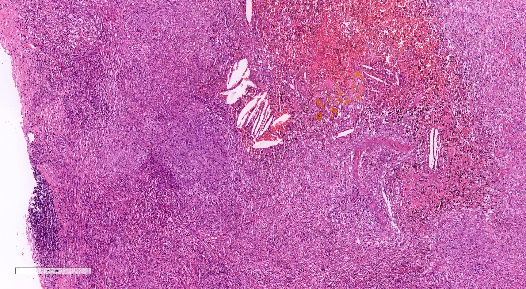
Microscopic (histologic) images
Contributed by Hongxing (Simon) Gui, M.D., Ph.D.
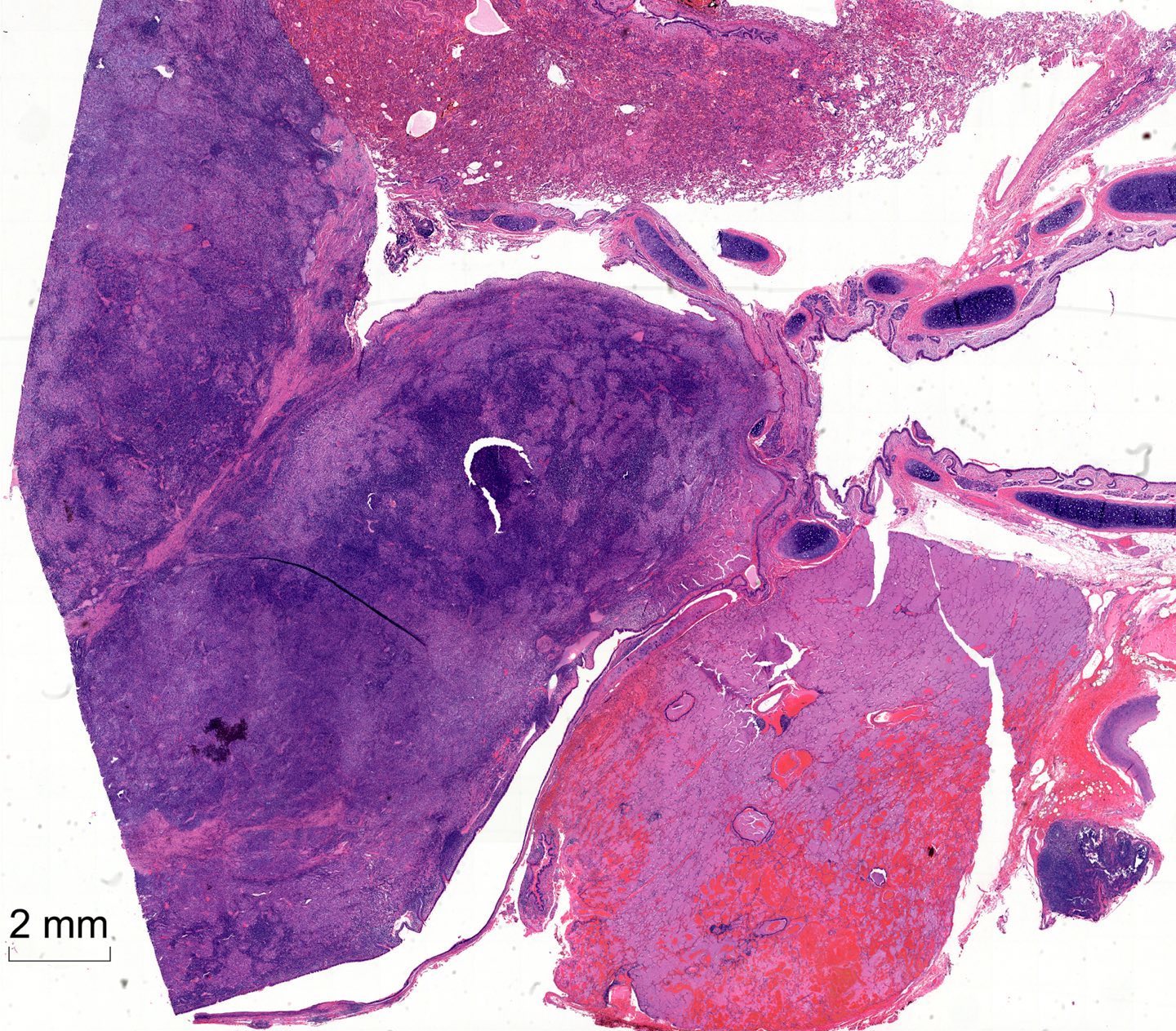
Nodular mass
Reticular pattern
Multinodular growth
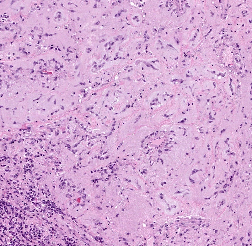
Spindle cells

Hybrid patterns
Angiomatoid fibrous histiocytoma area
Spindle cells in lacy pattern
Positive stains
- Vimentin, EMA, CD99 (weak focal) (Pol J Pathol 2017;68:261)
- Stroma is positive for Alcian blue stain, which is sensitive to hyaluronidase treatment (Am J Surg Pathol 2011;35:1722)
Negative stains
- S100, cytokeratin, p63, CD34, CD31, SMA, MSA, desmin (rarely positive), TTF1, calretinin
Electron microscopy description
- Focal dense plaques present in the plasma membrane, external lamina and intermediate junctions are rarely observed (Virchows Arch 2014;465:453)
Molecular / cytogenetics description
- ~85% of tumors showed t(2;22)(q33;q12) with EWSR1::CREB1 fusion gene detected by FISH or PCR (Int J Surg Pathol 2017;25:518)
- First report of a case with EWSR1::ATF1 fusion gene (Am J Surg Pathol 2020;44:1535)
- Second case (Int J Surg Pathol 2022 Apr 24 [Epub ahead of print])
Sample pathology report
- Lung, right lower lobe, lobectomy:
- Primary pulmonary myxoid sarcoma (see comment)
- Comment: H&E sections demonstrate a well circumscribed endobronchial tumor composed of lobules of spindled and stellate cells arranged in a reticular pattern within a myxoid background. A chronic inflammatory infiltrate is seen throughout the tumor. Immunohistochemically, the tumor cells are positive for EMA and negative for AE1 / AE3, CAM 5.2, pancytokeratin, p40, desmin, SMA, CD34, TTF1, KIT, vimentin, CD1a and S100. A FISH test for EWSR1 was positive for rearrangement in the tumor cells. The morphology in combination with the FISH test result confirm the above diagnosis.
Differential diagnosis
- Extraskeletal myxoid chondrosarcoma:
- History of soft tissue primary, rarely arising intrathoracic as a primary
- S100+
- FISH positive for EWSR1::NR4A3 fusion or others
- Myxoid angiomatoid fibrous histiocytoma:
- Peripheral lymphoid cuff
- Whorled or storiform growth patterns common
- 50% desmin+
- FISH positive for EWSR1::ATF1 (more common), EWSR1::CREB1 and FUS::ATF1
- Inflammatory myofibroblastic tumor:
- Myoepithelioma:
- Clear and plasmacytoid cells
- Positive for myoepithelial markers (cytokeratins, p63, S100, calponin and SMA)
Board review style question #1
Which of the following is a good prognostic factor for this primary pulmonary lung lesion?
- Brisk lymphoplasmacytic infiltrate
- FISH positive for EWSR1 translocation
- Lack of tumor necrosis
- Middle age onset
- Size > 5 cm
Board review style answer #1
B. FISH positive for EWSR1 translocation. Primary pulmonary myxoid sarcoma tumors (shown in photo) with EWSR1 rearrangement have a relatively good prognosis.
Comment Here
Reference: Primary pulmonary myxoid sarcoma with EWSR1::CREB1 fusion
Comment Here
Reference: Primary pulmonary myxoid sarcoma with EWSR1::CREB1 fusion
Board review style question #2
Which of the following entities is the closest mimicker of primary pulmonary myxoid sarcoma?
- Epithelioid hemangioendothelioma
- Extraskeletal myxoid chondrosarcoma
- Inflammatory myofibroblastic tumor
- Myoepithelioma
- Myxoid angiomatoid fibrous histiocytoma
Board review style answer #2
E. Myxoid angiomatoid fibrous histiocytoma. Myxoid angiomatoid fibrous histiocytoma and primary pulmonary myxoid sarcoma have many overlapping features.
Comment Here
Reference: Primary pulmonary myxoid sarcoma with EWSR1::CREB1 fusion
Comment Here
Reference: Primary pulmonary myxoid sarcoma with EWSR1::CREB1 fusion
