
Lymph nodes & spleen, nonlymphoma
Lymph nodes-vascular lesions
Lymphangiomyomatosis
Author: Sheren Younes, M.D., Ph.D.

Last author update: 1 February 2016
Last staff update: 10 November 2023
Copyright: 2003-2024, PathologyOutlines.com, Inc.
PubMed Search: Lymphangiomyomatosis
Table of Contents
Definition / general | Terminology | Epidemiology | Etiology | Diagrams / tables | Case reports | Microscopic (histologic) description | Microscopic (histologic) images | Cytology description | Positive stains | Differential diagnosis | Additional referencesCite this page: Younes S. Lymphangiomyomatosis. PathologyOutlines.com website. https://www.pathologyoutlines.com/topic/lymphnodeslymphangiomyomatosis.html. Accessed April 26th, 2024.
Definition / general
- Multisystem disorder affecting mainly middle age females, causing pulmonary and extrapulmonary disease
- Its pathologic features result from the proliferation of neoplastic cells (LAM cells), which have characteristics of both smooth muscle cells and melanocytes
- It was originally classified as benign but now is considered a "low grade, destructive metastasizing neoplasm" (Am J Respir Crit Care Med 2012;186:1210)
Terminology
- Also called lymphangioleiomyomatosis
Epidemiology
- Estimated median transplant free survival time for LAM patients in the U.S. is 29 years from onset of symptoms and 23 years from diagnosis
- Estimated 10 year survival transplant free is 86% (Lung 2013;191:35)
- About 80% of women with TSC develop cystic disease in lungs (Chest 2013;144:578), compared to 13% of male patients (Clin Radiol 2011;66:625)
- Relationship between pulmonary and extrapulmonary LAM needs further investigation
Etiology
- Associated with tuberous sclerosis complex (TSC), an autosomal dominant disorder caused by mutations in the TSC1 or TSC2 genes, characterized by intellectual disability, autism, seizures, facial angiofibroma, cortical tubers, cardiac rhabdomyoma, asrocytoma
Diagrams / tables
Images hosted on other servers:

Diagnostic criteria
Case reports
- 47, 59 and 71 year old women with extrapulmonary lymphangioleiomyomatosis in pelvic and paraaortic lymph nodes (Int J Gynecol Pathol 2011;30:470)
- 55 year old woman with extrapulmonary lymphangioleiomyomatosis (Case of the Month #522)
- 59 year old woman with "malignant" uterine perivascular epithelioid cell tumor, pelvic lymph node lymphangioleiomyomatosis and gynecological pecomatosis (Int J Gynecol Pathol 2008;27:86)
- Extrapulmonary lymphangioleiomyomatosis in pelvic lymph nodes associated with uterine cancer (Zhonghua Bing Li Xue Za Zhi 2015;44:527)
- Incidental pelvic and paraaortic lymph node lymphangioleiomyomatosis (Am J Surg Pathol 2015;39:1015)
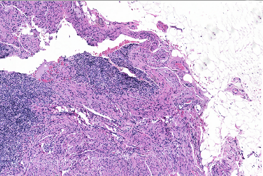
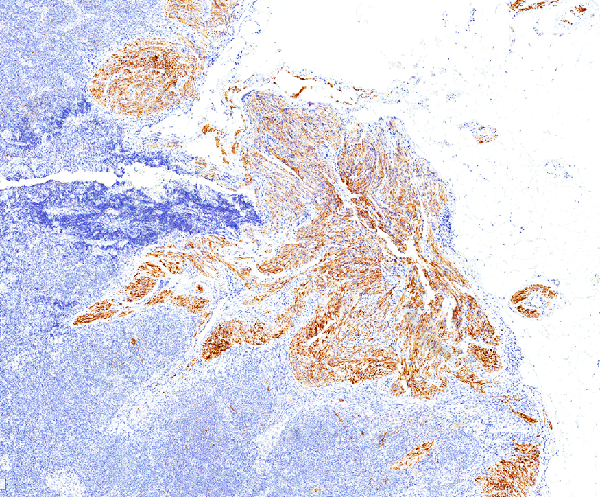
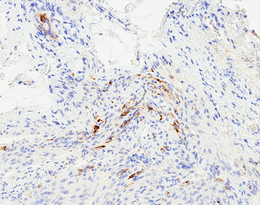
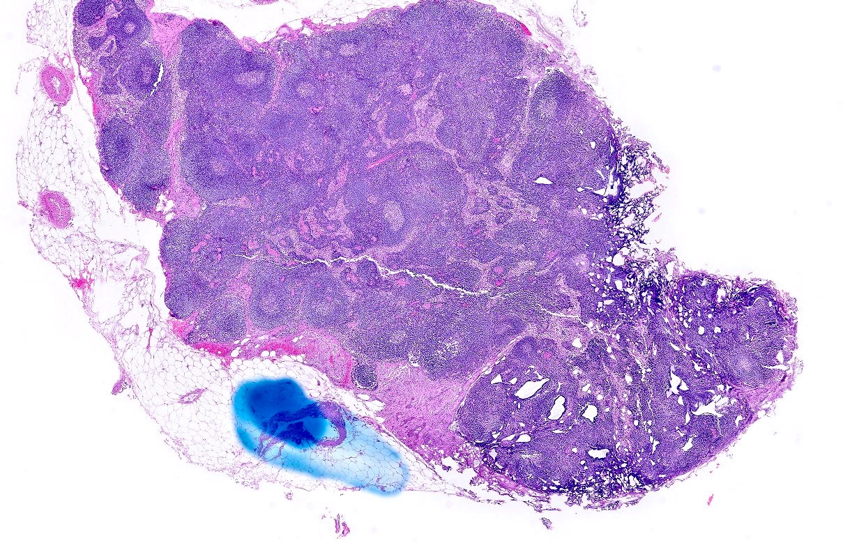
Microscopic (histologic) description
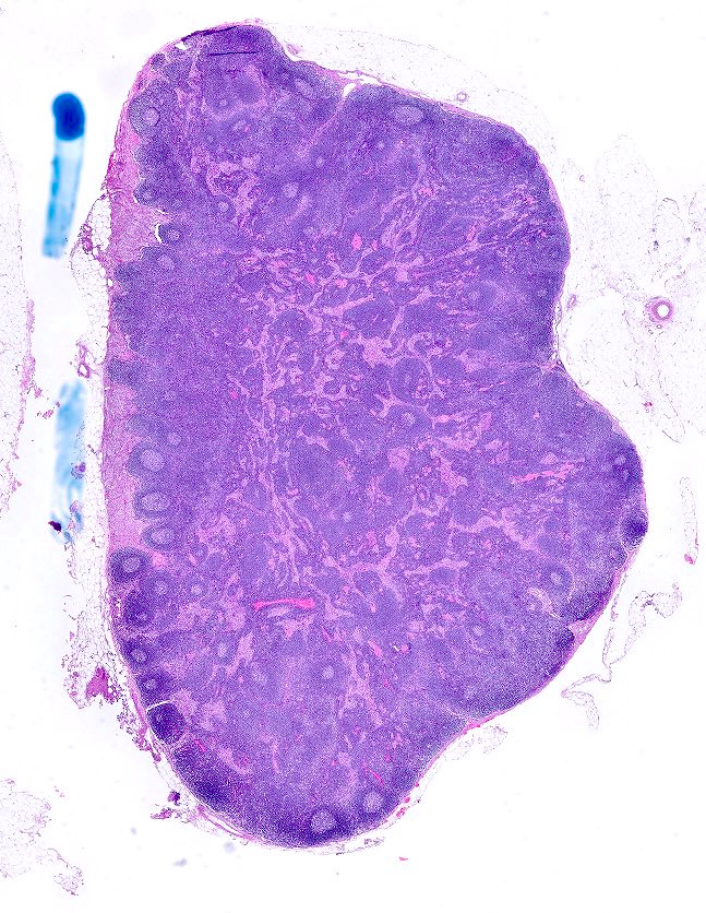
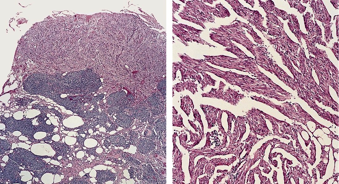
- Single or multiple lymph nodes, median size is 3.5 cm
- Mostly affecting nodal parenchyma but can be seen in hilum, subcapsular sinuses or extranodal extension
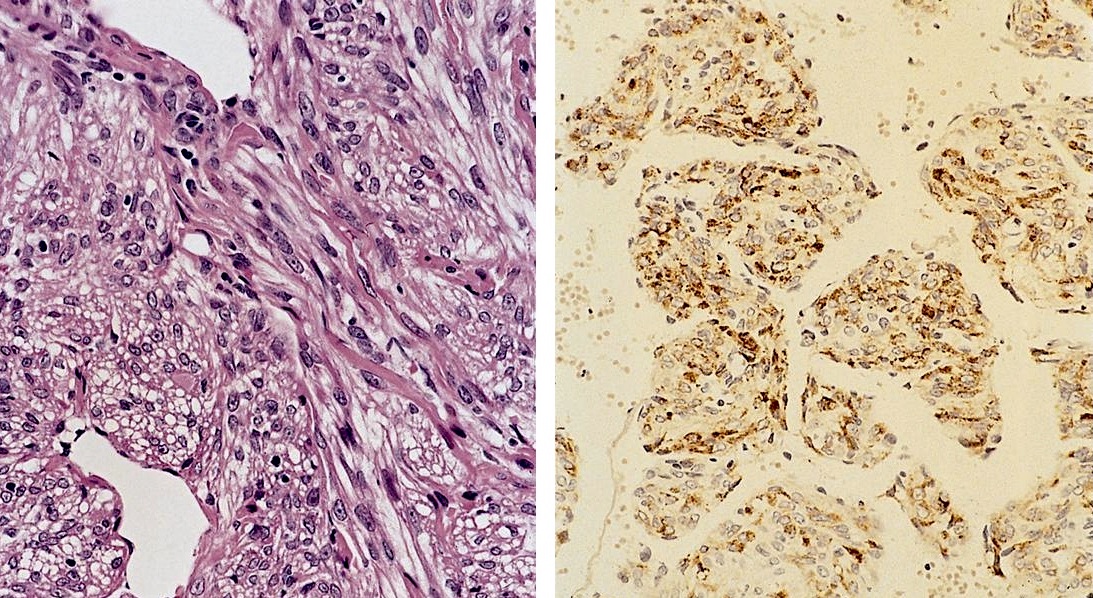
- LAM cells are either spindle or epitheliod
- Epithelioid cells are arranged in nests or swirls, separated by clefts which resemble lymphatic spaces
- Spindled cells show fascicular growth with some nesting and less prominent lymphatic channels
- No atypia, no necrosis, no mitosis
Microscopic (histologic) images
Contributed by Anna Sarah Erem, M.D. and Gulisa Turashvili, M.D., Ph.D. (Case #522), @katcollmd on Twitter and AFIP


Extrapulmonary lymphangioleiomyomatosis
Lymphangiomyomatosis


Lymphangiomyomatosis
Lymph node
Involving pelvic lymph node
Cytology description
- Relatively large monomorphic spindle cells (Acta Cytol 1997;41:877)
Positive stains
Differential diagnosis
- Metastatic well differentiated leiomyosarcoma: no prominent vascular channels, more atypia and mitotic figures, HMB45-
Additional references
