
Lymph nodes & spleen, nonlymphoma
Ectopic tissue / inclusions
Other ectopic tissue / inclusions
Last author update: 1 June 2006
Last staff update: 28 March 2024
Copyright: 2003-2024, PathologyOutlines.com, Inc.
PubMed Search: Lymph node inclusions

Table of Contents
Extramedullary hematopoiesis | Megakaryocytes | Müllerian inclusions | Nevus cells | Salivary gland | Smooth muscle | Squamous epithelium | ThymusCite this page: DePond WD, Balakrishna J, Sharabi A. Other ectopic tissue / inclusions. PathologyOutlines.com website. https://www.pathologyoutlines.com/topic/lymphnodesotherectopic.html. Accessed April 18th, 2024.
Extramedullary hematopoiesis
Definition / general
Case reports
Microscopic (histologic) images
Contributed by Nikhil Sangle, M.D., Sherif Shabaan, M.D. and Julie M. Jorns, M.D. (Case #537)


Differential diagnosis
- Rarely associated with myeloproliferative disorders but also in healthy patients (Diagn Cytopathol 1993;9:522, Arch Otolaryngol 1982;108:523)
Case reports
- 26 year old woman presented with modified Bloom Richardson grade 2 invasive ductal carcinoma and ipsilateral axillary lymph node metastasis at diagnosis; her cancer was hormone receptor positive, HER2 equivocal (2+) on IHC (Case of the month #537)
Microscopic (histologic) images
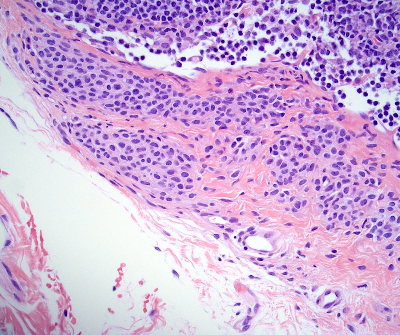
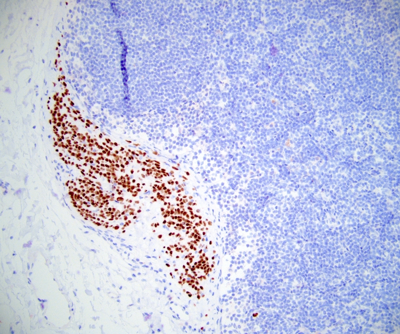
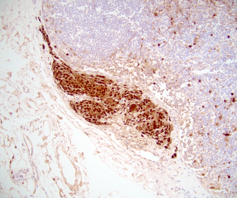
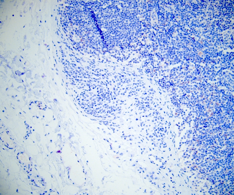
Contributed by Nikhil Sangle, M.D., Sherif Shabaan, M.D. and Julie M. Jorns, M.D. (Case #537)
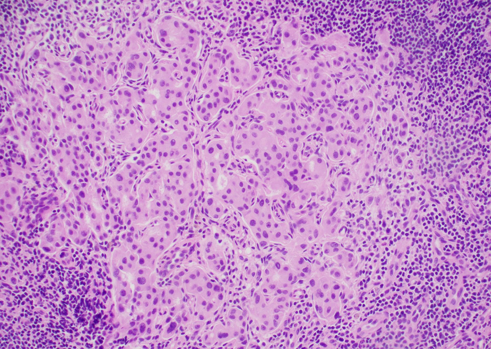
Liver with extramedullary hematopoiesis
H&E SLN capsule and parenchyma
H&E Another region of the SLN
Metastatic carcinoma
Cytokeratin AE1 / AE3
Differential diagnosis
Megakaryocytes
Definition / general
Case reports
Microscopic (histologic) images
Images hosted on other servers:

Positive stains
Negative stains
Differential diagnosis
- Often associated with extramedullary hematopoiesis when present in lymph nodes
Case reports
- 35 year old woman with megakaryocytes mimicking metastatic breast carcinoma (Arch Pathol Lab Med 2002;126:618)
Microscopic (histologic) images
Images hosted on other servers:
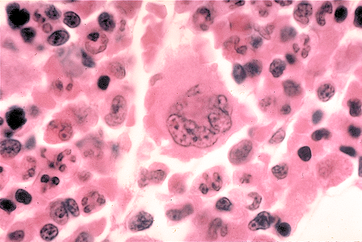
Megakaryocytes in bone marrow
Positive stains
Negative stains
Differential diagnosis
- Multinucleated histiocytes:
- Larger, more cytoplasm, vesicular nuclei that are not multilobated
- CD68+
Müllerian inclusions
Definition / general
Terminology
Sites
Pathophysiology
Clinical features
Case reports
Treatment
Gross description
Microscopic (histologic) description
Microscopic (histologic) images
AFIP images
Positive stains
Differential diagnosis
Additional references
- Benign glandular inclusions lined by salpingeal or ovarian epithelium with no endometrial stroma or hemosiderophages
Terminology
- Also called endosalpingiosis
Sites
- Predominantly pelvic and paraaortic lymph nodes
- Present in 5 - 41% of intra-abdominal nodes of women, 20% of women with gynecologic malignancies in paraaortic / pelvic nodes (Am J Obstet Gynecol 1978;130:813, Gynecol Oncol 2000;78:242)
Pathophysiology
- Theories include:
- Passage of epithelial cells through the tubal ostium for implantation in the peritoneum following peeling, followed by transportation by the lymphatic route to reach the pelvic lymph node
- Metaplastic origin from the peritoneum
- Foci of endometriosis
- Vestiges of paramesonephricus ducts
- Metastases from ovarian serous borderline tumors (Am J Surg Pathol 2000;24:710)
Clinical features
- Incidental finding / enlarged lymph nodes
Case reports
- 64 year old woman with endosalpingiosis with psammoma bodies within an intramammary lymph node presented as microcalcifications on mammography (Arch Pathol Lab Med 1995;119:841)
- Coexisting with breast metastatic carcinoma in an axillary lymph node (Virchows Arch 2005;446:467)
Treatment
- Benign process, with no clinical significance
Gross description
- Enlarged lymph nodes
Microscopic (histologic) description
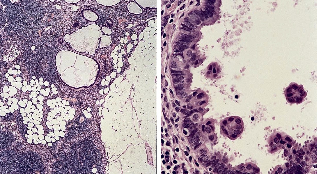
- Glandular / cystic inclusions lined by cuboidal / columnar cells with a Müllerian or coelomic appearance commonly found in the capsule
- Psammoma bodies are common
- May undergo squamous metaplasia
- No / rare mitotic figures, no / mild atypia, no desmoplastic stroma, no endometrial stroma
Microscopic (histologic) images
AFIP images
In pelvic lymph node
Positive stains
Differential diagnosis
- Metastasis from borderline / malignant ovarian tumors or uterine carcinomas
Additional references
- J Clin Oncol 2006;24:2013, Ioachim: Ioachim's Lymph Node Pathology, 4th Edition, 2008, Rosai: Rosai and Ackerman's Surgical Pathology, 10th Edition, 2011, Robboy: Robboy's Pathology of the Female Reproductive Tract, 2nd Edition, 2008, Dunphy: Frozen Section Library - Lymph Nodes, 2012, Weiss: Lymph Nodes, 1st Edition, 2008, University of Pittsburgh: Endometriosis and Bland Glandular Inclusions [Accessed 12 April 2023]
Nevus cells
Definition / general
Microscopic (histologic) description
Microscopic (histologic) images
Contributed by Debra L. Zynger, M.D.
Positive stains
Negative stains
Differential diagnosis
- Incidence in axillary nodes is 7% per patient and 0.5% per node in one study (Am J Clin Pathol 1994;102:102)
- Presence in sentinel nodes in melanoma patients is associated with
- Cutaneous nevi (Am J Clin Pathol 2004;121:58)
- Congenital cutaneous nevi (Am J Dermatopathol 2002;24:1)
- May represent benign metastases from intradermal nevus in area of lymphatic drainage (Am J Clin Pathol 1985;84:220)
Microscopic (histologic) description
- Single cells, linear arrangements or aggregates of small, round / oval nevus cells with moderate pale / clear cytoplasm, round nuclei with fine chromatin, no prominent nucleoli or pleomorphism, usually within fibrous capsule and trabeculae but also within nodal parenchyma or surrounding a small vessel (Am J Surg Pathol 2003;27:673, Am J Surg Pathol 1996;20:834)
Microscopic (histologic) images
Contributed by Debra L. Zynger, M.D.
Nests within capsule
Bland cells
SOX10
S100
AE1/3
Positive stains
Negative stains
- HMB45 (occasionally is very focal), Ki-67 (< 1%, Am J Surg Pathol 2002;26:1351)
Differential diagnosis
- Blue nevus:
- Also spindled / dendritic cells with heavy pigmentation
- Metastatic carcinoma or melanoma:
- Usually not confined to capsule, atypia, mitotic figures, different immunostaining
- Spitz nevus:
- Larger cells with abundant eosinophilic cytoplasm
- Vesicular nuclei with prominent nucleoli
Salivary gland
Definition / general
Terminology
Epidemiology
Sites
Pathophysiology
Clinical features
Prognostic factors
Case reports
Treatment
Clinical images
Images hosted on other servers:

Gross description
Microscopic (histologic) description
Microscopic (histologic) images
AFIP images

Images hosted on other servers:


Differential diagnosis
Additional references
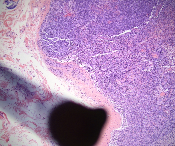
- Benign salivary gland structures in lymph nodes
- Includes ducts and acini
Terminology
- Salivary gland inclusions
- Ectopic salivary tissue
- Heterotropic salivary gland tissue
Epidemiology
- Common incidental finding
Sites
- Most common in upper cervical lymph nodes
- Rarely - thoracic, mediastinal lymph nodes
Pathophysiology
- Considered a normal event related to the embryology of the region
Clinical features
- None - incidental finding
- Enlarged lymph nodes
Prognostic factors
- Benign with no significance
- May undergo diseases of salivary glands including neoplastic processes - Warthin tumor is most common tumor
Case reports
- 55 year old woman with sialometaplasia arising in ectopic salivary gland ductal inclusions (J Clin Pathol 2004;57:1335)
- 60 year old man with salivary gland tissue in cervical lymph nodes (Laryngorhinootologie 2007;86:44)
- 68 year old woman with benign salivary gland tissue inclusion in a pulmonary hilar lymph node (Int J Surg Pathol 2011;19:382)
- Proliferative sialometaplasia arising in an intraparotid lymph node (Am J Clin Pathol 1986;86:116)
Treatment
- Usually none but may be indicated based on clinical situation
Clinical images
Images hosted on other servers:
Circumscribed mass lesions due to
sialometaplasia arising in ectopic
salivary gland ductal inclusions
Gross description
- Normal / enlarged lymph nodes
Microscopic (histologic) description
- Both ducts and acini are usually present
- Benign ducts are lined by double layer of epithelium and myoepithelium
- Benign acini have zymogen granules
Microscopic (histologic) images
AFIP images
Salivary gland inclusion in lymph node
Images hosted on other servers:
Inclusions undergoing sialometaplasia show extensive squamous metaplasia
Inclusions with sialometaplasia: SMA
Differential diagnosis
Additional references
Smooth muscle
Definition / general
Sites
Etiology
Clinical features
Diagnosis
Case reports
Gross description
Microscopic (histologic) description
Microscopic (histologic) images
Images hosted on other servers:

Positive stains
Negative stains
Differential diagnosis
Additional references
- Topic discusses the presence of smooth muscle within the capsule and hilum of normal lymph nodes
- Common, incidental finding in lymph nodes of hilar smooth muscle proliferation, more commonly in males than females (Virchows Arch A Pathol Anat Histopathol 1985;406:261)
Sites
- Superficial, inguinal and axillary lymph nodes are most commonly involved
Etiology
- Suggested etiological factors include back pressure in efferent lymphatics, local inflammation
Clinical features
- Asymptomatic, with no apparent clinical significance, but may have enlarged lymph nodes
Diagnosis
- Biopsy
- Immunohistochemistry for smooth muscle to confirm
Case reports
- 6 year old boy with intranodal leiomyoma (Pediatr Dev Pathol 2014;17:118)
Gross description
- Enlarged lymph nodes with firm, tan cut surface
Microscopic (histologic) description
- Nodal architecture is well preserved
- Muscle fibers run parallel to capsule or hilar blood vessels
- Varied patterns of hilar changes:
- Smooth muscle fibers may be densely interwoven within fibrous tissue and hug the hilar nodal capsule
- Smooth muscle fibers may form a loose network extending in all directions within hilar adipose tissue
- Smooth muscle may surround individual islands of fat cells
Microscopic (histologic) images
Images hosted on other servers:
Angiomyomatous hamartoma of lymph node
Positive stains
Negative stains
Differential diagnosis
- Angiomyomatous hamartoma (Korean J Path 2011;45:S58)
- Benign metastasizing leiomyoma
- Inflammatory myofibroblastic tumor
- Intranodal leiomyoma
- Lymphangioleiomyomatosis
- Pallisading myofibroblastoma
- Vascular transformation of lymph node sinuses
Additional references
Squamous epithelium
Definition / general
Case reports
Microscopic (histologic) description
Differential diagnosis
- Also called lymphoepithelial cyst
- Most common in upper cervical nodes
- Depending on location, may be a branchial pouch derivative or derive from metaplastic calyceal urothelium (Hum Pathol 1990;21:1239)
Case reports
- 33 and 64 year old women with benign heterotopic epithelial inclusions in axillary lymph nodes (Arch Pathol Lab Med 2003;127:e25)
- 48 year old woman with axillary intranodal cysts associated with breast malignancy (Arch Pathol Lab Med 2004;128:361)
- 57 and 75 year old women with cystic lymphoepithelial lesions of peripancreatic nodes (Surg Today 1999;29:467)
- Epithelial inclusion in axillary lymph node associated with a breast carcinoma (Pathol Res Pract 1999;195:263)
Microscopic (histologic) description
- Small cystic structures lined by well differentiated squamous epithelium
Differential diagnosis
- Metastatic well differentiated squamous cell carcinoma:
- Often undergoes cystic change
