Table of Contents
Definition / general | Essential features | Terminology | ICD coding | Epidemiology | Sites | Pathophysiology | Diagrams / tables | Clinical features | Diagnosis | Laboratory | Radiology description | Radiology images | Case reports | Treatment | Clinical images | Gross description | Gross images | Microscopic (histologic) description | Microscopic (histologic) images | Cytology description | Cytology images | Sample pathology report | Differential diagnosis | Additional references | Board review style question #1 | Board review style answer #1 | Board review style question #2 | Board review style answer #2Cite this page: Vaughan VC, Wisell J. Epidermal (epidermoid) type. PathologyOutlines.com website. https://www.pathologyoutlines.com/topic/skintumornonmelanocytickeratinouscystepidermal.html. Accessed April 16th, 2024.
Definition / general
- Benign skin tumor
- Cystic mass containing keratin
Essential features
- Cystic mass with soft white keratin contents
- Histologically cystic mass with squamous epithelium and keratin flakes
Terminology
- Epidermoid cyst, epidermal cyst, epidermal inclusion cyst, infundibular cyst
ICD coding
- ICD-10: L72.0 - epidermal cyst
Epidemiology
- Young and middle aged adults are most often affected (Int J Trichology 2017;9:108)
- M = F
Sites
- Face, neck, trunk, perineal area, cerebellopontine angle
- Less commonly spine, intrapancreatic accessory spleen
Pathophysiology
- Follicular orifice becomes plugged with bacteria and keratin, leading to cystic dilation and entrapment of keratin debris
- Presence of multiple epidermal inclusion cysts has been documented in Gardner syndrome, a variant of familial adenomatous polyposis with benign osteomas and intestinal fibromatoses
- Less frequently, patients may have lipomas, pilomatrixomas (including epidermoid cysts with pilomatrical lining) or leiomyomas
- Multiple and large epidermoid cysts may occur with the use immunosuppressants in the posttransplantation setting, for example, with cyclosporine or tacrolimus (Cutis 1992;50:36, Ann Dermatol 2011;23:S182)
- May complicate penetrating trauma to skin, such as a sewing needle, with resultant implantation of squamous epithelium into the dermis (Turk J Pediatr 2011;53:108)
Clinical features
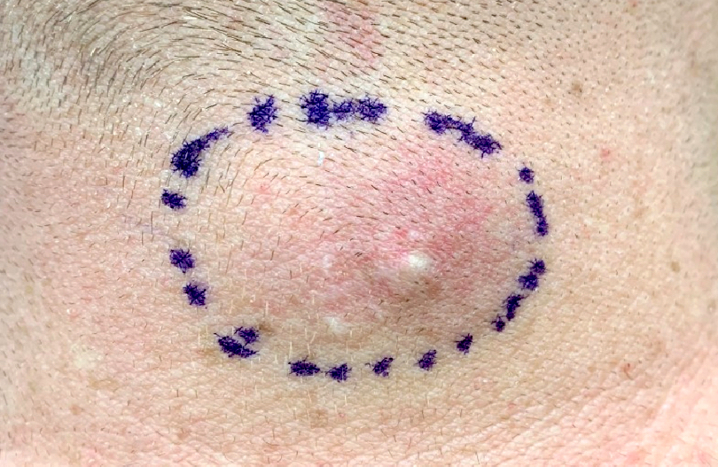
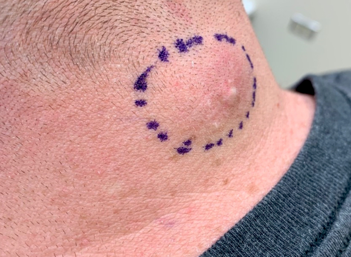
- Present as smooth dome shaped swellings varying in size from a few millimeters to a few centimeters (Ann Dermatol 2017;29:33)
- Usually occur on the face, neck or trunk but can occur anywhere
- Overlying skin may be taut with the pressure of the cyst and have a central punctum
- Generally occurs in postpubertal individuals
- Freely mobile unless ruptured, in which case a foreign body giant cell reaction may make them more adherent to surrounding connective tissue
- May become painful and inflamed with external manipulation
- Cyst contents white, cheesy macerated keratin that may have an odor
Diagnosis
- Often based on clinical examination
- Pathological diagnosis relies on recognizing the cyst wall and contents
- In cases of extensive foreign body giant cell reaction or fibrosis, the classic features may not be visualized as readily and a diligent search for keratin flakes may lead to diagnosis
Laboratory
- No specific laboratory findings
Radiology description
- Ultrasound:
- Subcutaneous rounded structure that is mostly anechoic or hypoechoic with focal present inner echoes (pseudotestis appearance) representing cystic debris
- MRI:
- Fluid-like enhancement
- High intensity of cyst contents on T2 weighted images and peripheral cyst wall enhancement with T1 gadolinium enhancement
- Cyst rupture results in irregular enhancement (AJR Am J Roentgenol 2006;186:961)
Radiology images
Case reports
- 7 year old boy with epidermal inclusion cyst infected by molluscum contagiosum virus (J Dtsch Dermatol Ges 2018;16:1143)
- 40 year old woman with recurrent spinal epidermoid cysts with atypical hyperplasia (Medicine 2017;96:e8950)
- 51 year old man with squamous cell carcinoma arising in a 30 year old perineal epidermal inclusion cyst (World J Surg Oncol 2018;16:155)
- 63 year old man with epidermal inclusion cyst containing a splinter with nonpigmented fungi (J Cutan Pathol 2018;45:954)
- 73 year old man with squamous cell carcinoma arising in an epidermoid cyst with human papillomavirus changes (Clin Exp Dermatol 2018;43:201)
Treatment
- Complete excision of the cyst is curative
Clinical images
Gross description
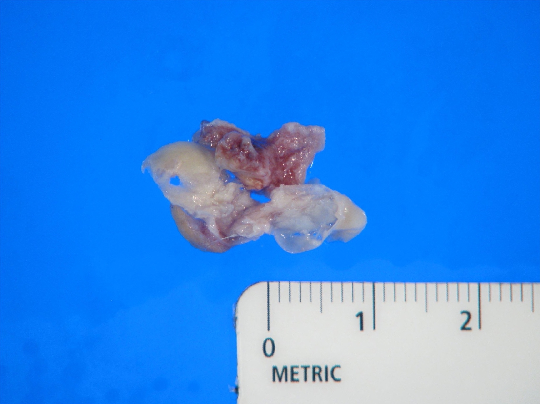
- Pearly glistening cyst with creamy contents
Gross images
Microscopic (histologic) description
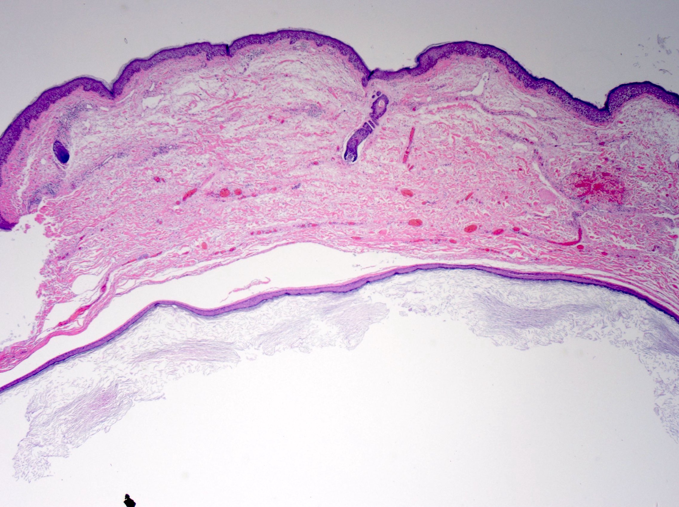
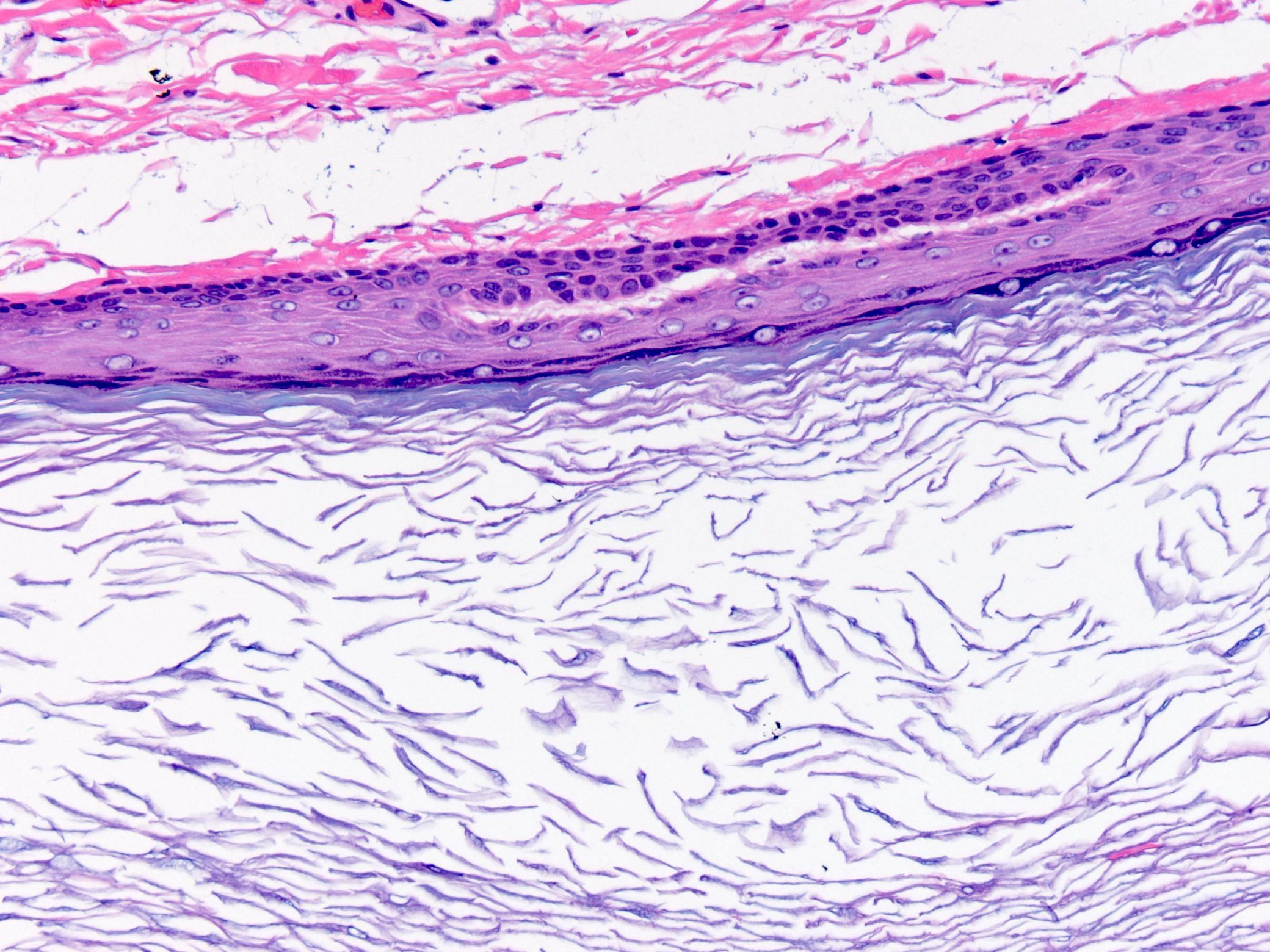
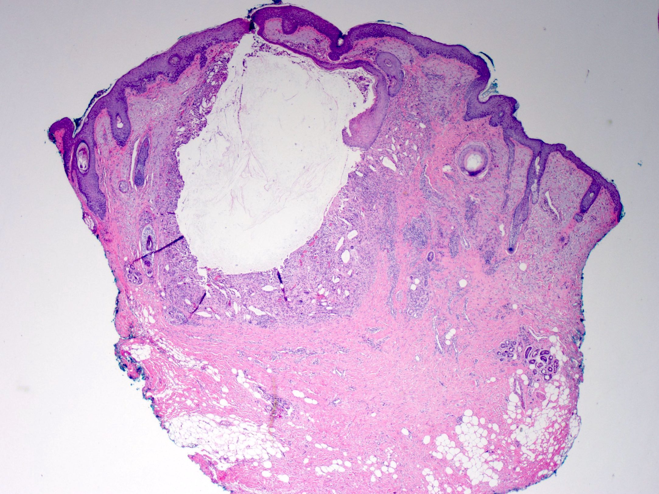
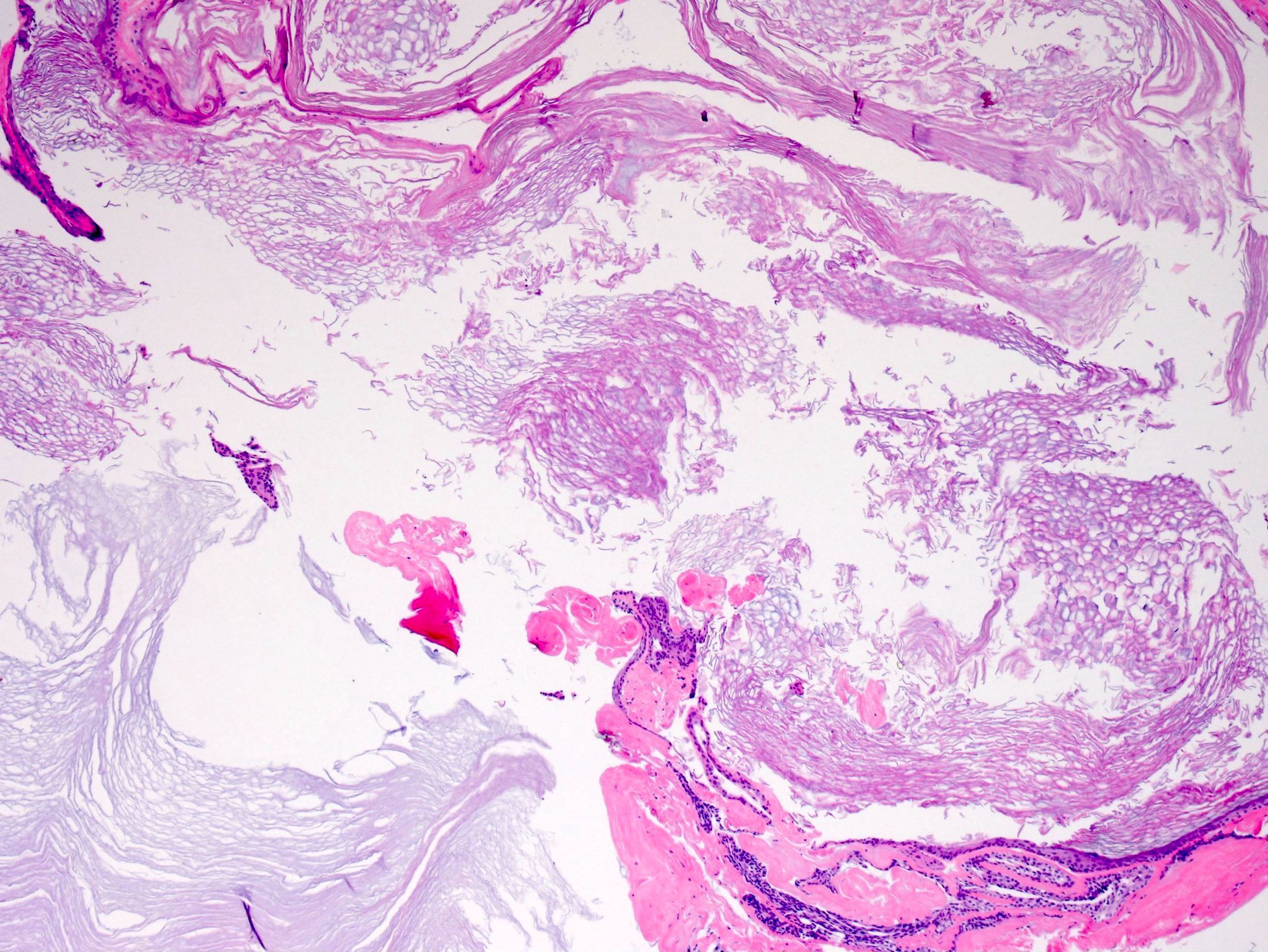
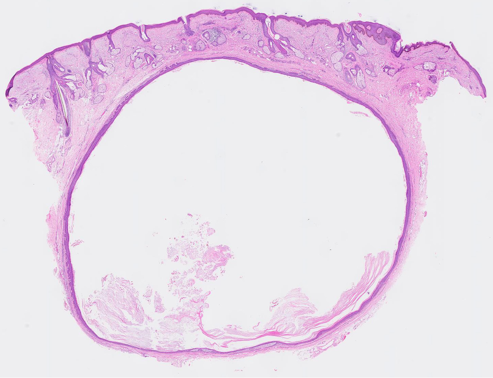
- Subepidermal cyst that may or may not open into the overlying epidermis
- Cyst lining composed of stratified squamous epithelium with a granular layer
- Cyst wall does not contain eccrine glands, sebaceous glands or hair follicles
- Cyst contents composed of abundant keratin flakes
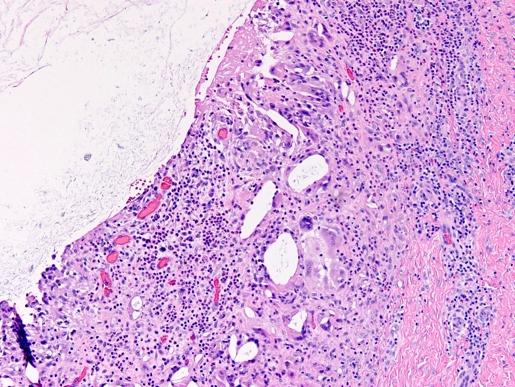
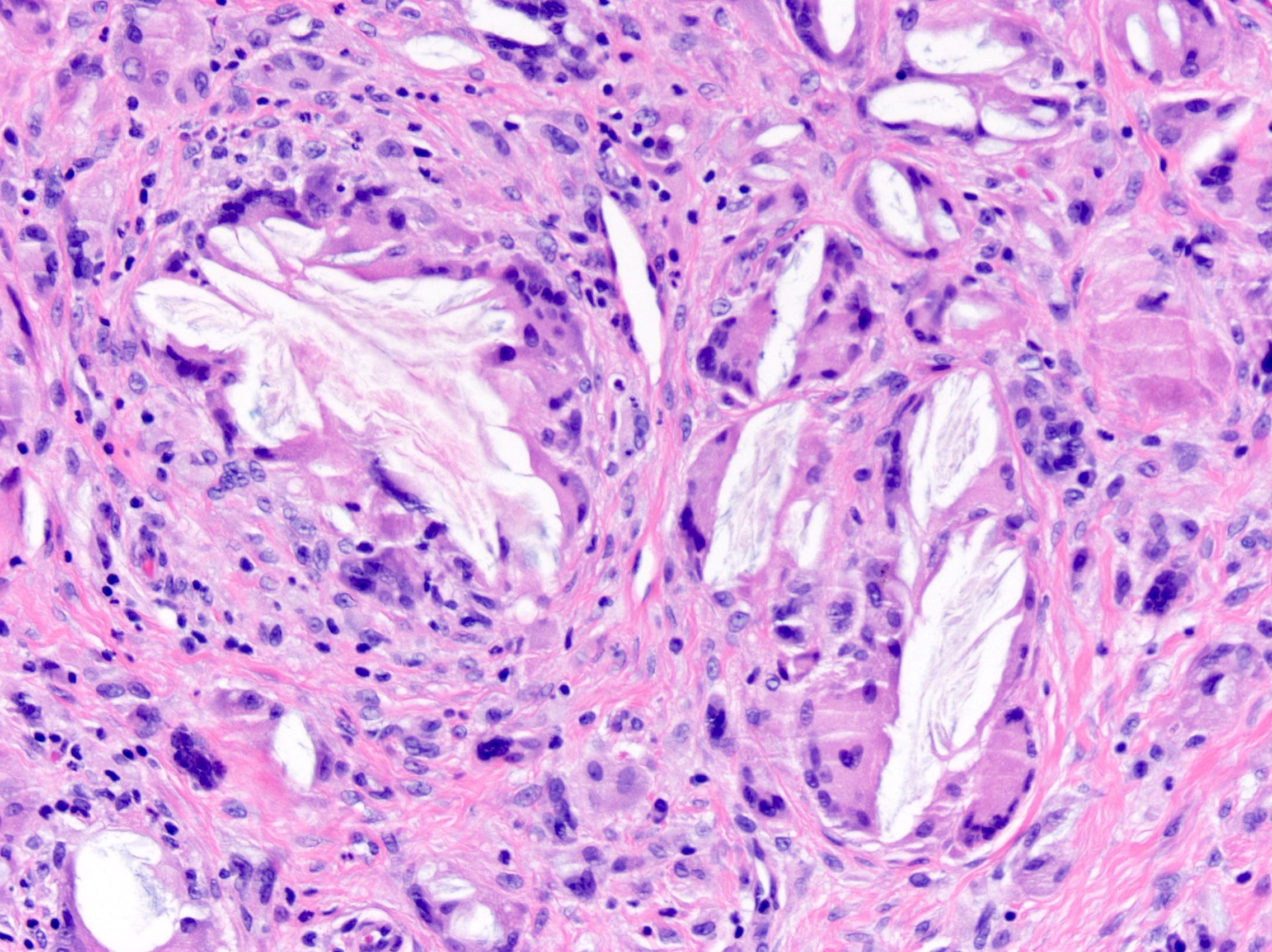
- Foreign body giant cell reaction is frequently present in ruptured cysts
- Melanoma, basal cell carcinoma and squamous cell carcinoma have been reported in association with epidermal inclusion cysts (J Am Acad Dermatol 2013;68:e6, J Cutan Med Surg 2015;19:105)
Microscopic (histologic) images
Contributed by the University of Colorado Department of Pathology, Jijgee Munkhdelger, M.D., Ph.D. and Andrey Bychkov, M.D., Ph.D.
Cytology description
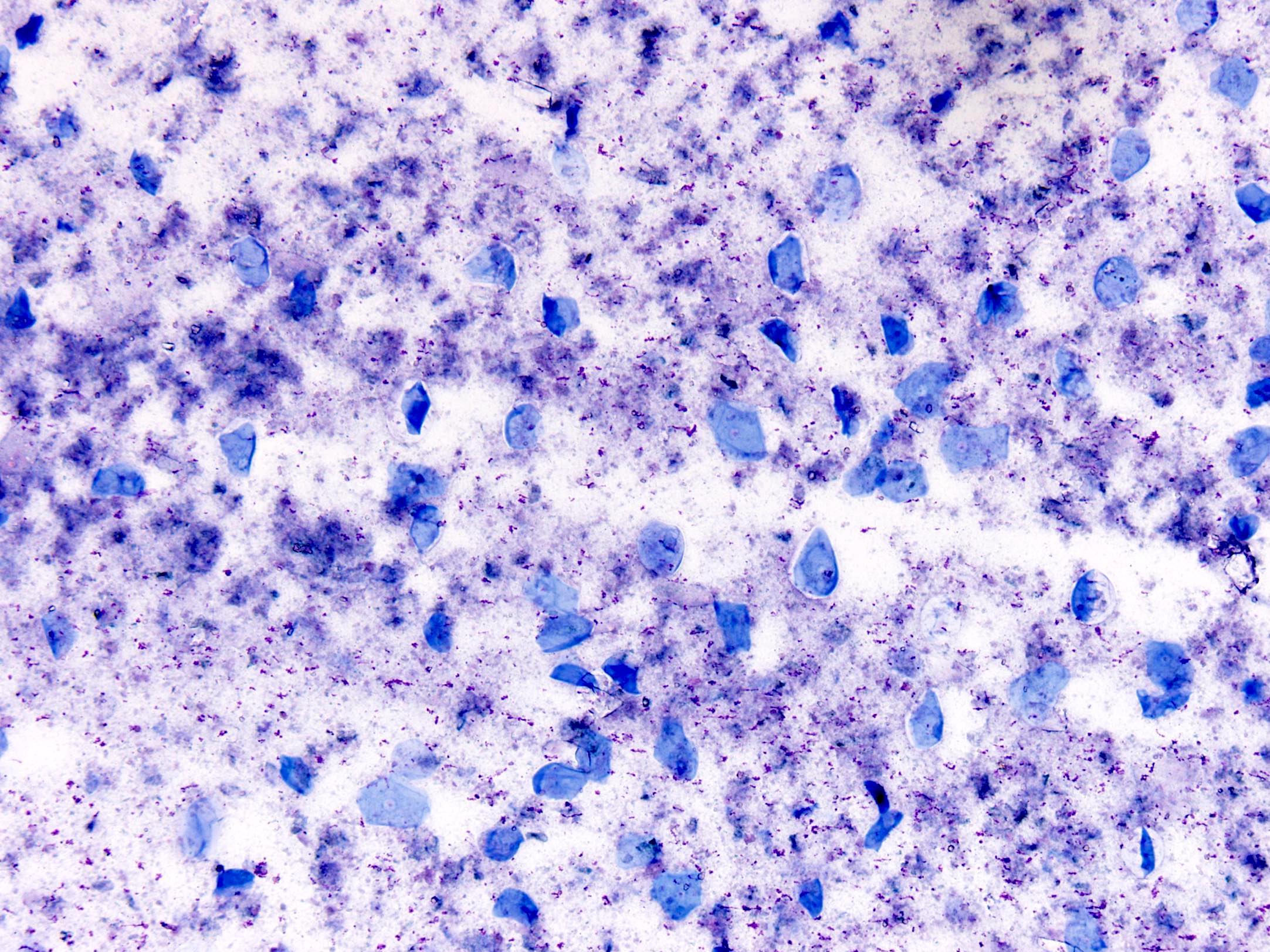
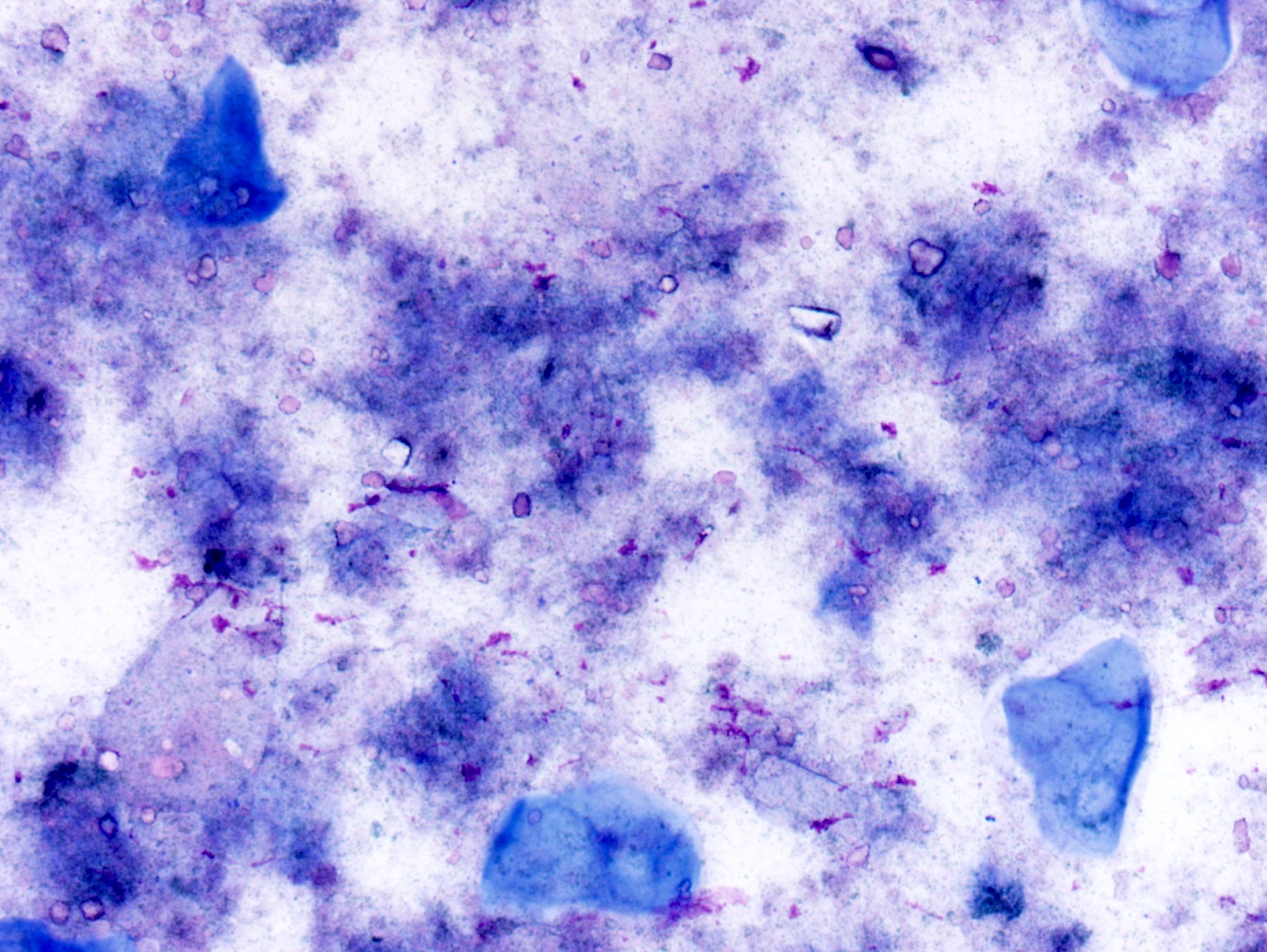
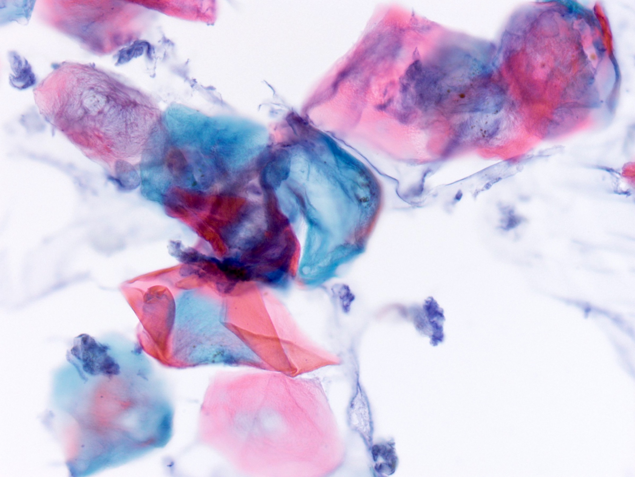
- Anucleate keratinizing squamous cells with some nucleated squamous cells
- Frequent erythrocytes, leukocytes, multinucleated giant cells and cholesterol crystals (J Nat Sci Biol Med 2014;5:460)
Cytology images
Sample pathology report
- Skin, neck, excision:
- Epidermal inclusion cyst
- Microscopic description: Cyst lined by squamous epithelium with granular layer containing lamellated keratin.
Differential diagnosis
- Proliferating epidermoid cyst:
- May show carcinomatous changes, invasion and be locally aggressive
- Trichilemmal (pilar) cyst:
- Lack a granular layer in the cyst lining
- Dense lamellated keratin cyst contents
- More likely to occur on the scalp
- Pilomatricoma:
- Eosinophilic cellular outlines of squamous cells ("ghost cells") in addition to the more basophilic matrical cells
- More likely to occur in a pediatric population
- Hybrid cyst:
- Demonstrates features of both an epidermal and trichilemmal cyst
- Dermoid cyst:
- Similar in appearance
- Has adnexal structures (ie. sebaceous glands) in cyst wall
- Arises at sites of embryonic closure such as the lateral eyebrow
- Pilonidal sinus:
- Sinus tract surrounded by epithelium and mixed inflammation
- Characteristically contains broken hair shafts
- Steatocystoma:
- Compressed sebaceous gland within the cyst wall
- Characteristic wavy eosinophilic crenulated cuticle of the lining
- Odontogenic keratocyst:
- Attenuated squamous epithelium with parakeratosis
- Retraction of epithelium with basal palisade can be a helpful finding
- Often filled with keratin debris like epidermoid cyst
Additional references
Board review style question #1
An elderly man has a soft, round subcutaneous mass on the back of the neck with a central punctum. Which of the following is the most likely diagnosis?
- Angiosarcoma
- Epidermoid cyst
- Lymphadenitis
- Pilar cyst
- Spindle cell lipoma
Board review style answer #1
Board review style question #2
Board review style answer #2


