

Table of Contents
Definition / general | Essential features | Terminology | Pathophysiology | Diagrams / tables | Interpretation | Uses by pathologists | Microscopic (histologic) description | Microscopic (histologic) images | Electron microscopy description | Molecular / cytogenetics description | Molecular / cytogenetics images | Sample pathology report | Board review style question #1 | Board review style answer #1 | Board review style question #2 | Board review style answer #2 | Board review style question #3 | Board review style answer #3Cite this page: Yusuke K. t(X;18). PathologyOutlines.com website. https://www.pathologyoutlines.com/topic/moleculartX18.html. Accessed April 25th, 2024.
Definition / general
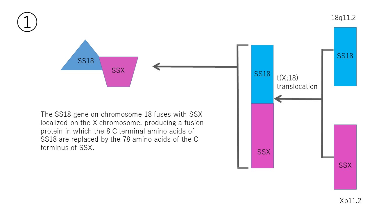
- t(X;18)(p11.2;q11.2) is a characteristic reciprocal translocation found in synovial sarcoma
Essential features
- The chimeric SS18::SSX protein is localized to the nucleus where it interferes with polycomb related machinery, chromatin remodeling complexes and YAP / TAZ Hippo signaling cascade to drive the development of synovial sarcoma (Nat Struct Mol Biol 2020;27:836, Cancer Cell 2018;33:1128, Clin Cancer Res 2019;25:3718)
- RT-PCR and IHC can utilize specific antibodies for the SS18::SSX chimera protein (Appl Immunohistochem Mol Morphol 2018;26:206, Am J Surg Pathol 2020;44:922)
- FISH can be used but may be replaced by IHC for the detection of SS18::SSX in paraffin embedded formalin fixed tissue sections
- It may be practical to use specific antibodies against the SS18::SSX chimeric protein (Am J Surg Pathol 2021;45:582)
- Synovial sarcoma can occur in any part of the body, including sites such as the lungs, heart, kidneys, prostate, gastrointestinal tract, pleura and larynx; detection of the SS18::SSX translocation or the chimeric protein is believed to be specific for synovial sarcoma at all sites, irrespective of the tissue
Terminology
- SS18 gene on chromosome 18 was formerly called the SYT gene
Pathophysiology
- SS18 is a stable subunit of the SWI / SNF nucleosome remodeling complex; SS18 does not bind DNA but acts as a coactivator of transcription by interacting with the SWI / SNF complex for chromatin remodeling (PLoS One 2012;7:e33834)
- SSX genes are a multigene family of which several members are transcribed in normal testes and various human malignant tumors and likely act as transcription modulators (Int J Cancer 2002;101:448)
- Synovial sarcoma SS18::SSX chimera fusion genes usually encode proteins that comprise the first 379 amino acids of SS18 and the first 78 amino acids of the SSXs
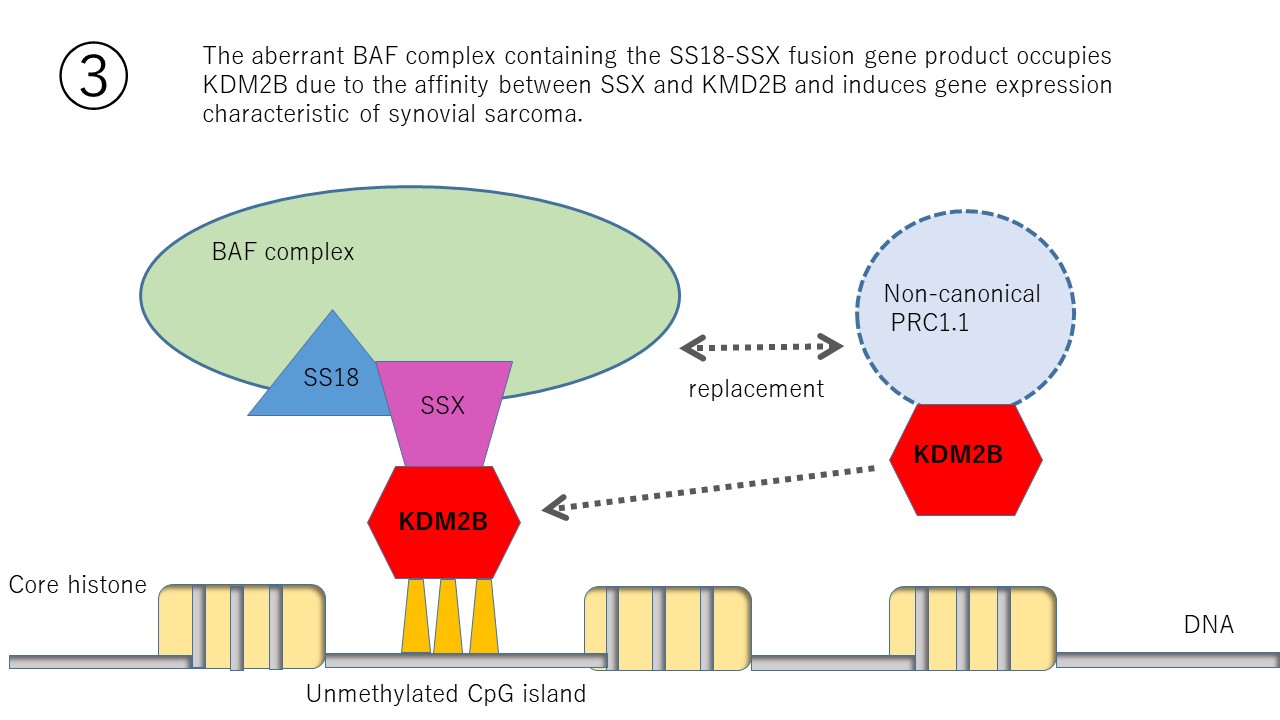
- Banito et al. revealed that the SS18::SSX chimeric protein hijacks KDM2B-PRC1.1, which drives synovial sarcoma as it disrupts the gene silencing mechanism by binding to the DNA region and inducing gene expression specific to synovial sarcoma (Cancer Cell 2018;33:527)
Diagrams / tables
Contributed by Kito Yusuke, M.D., Ph.D.
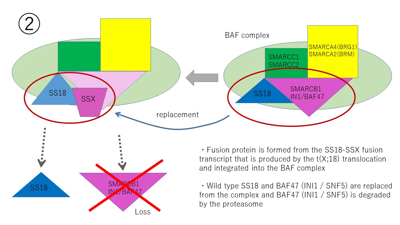
Oncogenic BAF complex
Interpretation
- Signals in FISH are observed in the nucleus
- This fluorescence in situ hybridization (FISH) probe is intended to detect chromosome rearrangements involving the SS18 (SYT) gene located on chromosome 18q11.2
- The Vysis SS18 Dual Color, Break Apart Rearrangement Probe consists of a mixture of 2 FISH DNA probes:
- The first probe, a ~650 kb probe labeled in Spectrum Orange, extends distally from the SS18 gene
- The second probe labeled in Spectrum Green lies 3′ or proximal to the SS18 gene and is 1044 kb in length (Abbott: Vysis LSI SS18 Break Apart FISH Probe Kit [Accessed 22 August 2022])
- A probe was considered to be split when the orange and green signals were separated by a distance greater than the size of one hybridization signal (spectrum orange 650 kb); this cutoff was chosen because the distance between the nonsplit probes is 56 kb
- When 20% or more single nuclei were found to harbor both fusion and split (break apart) signals, the cases were classified as positive; when less than 20% of cells had fusion and split signals, they were scored as negative (Mod Pathol 2007;20:482)
Uses by pathologists
- Diagnosis of synovial sarcoma: demonstrates SS18 rearrangement by FISH
- Any other tumors that show SS18 rearrangement by FISH: microsecretory adenocarcinoma
Microscopic (histologic) description
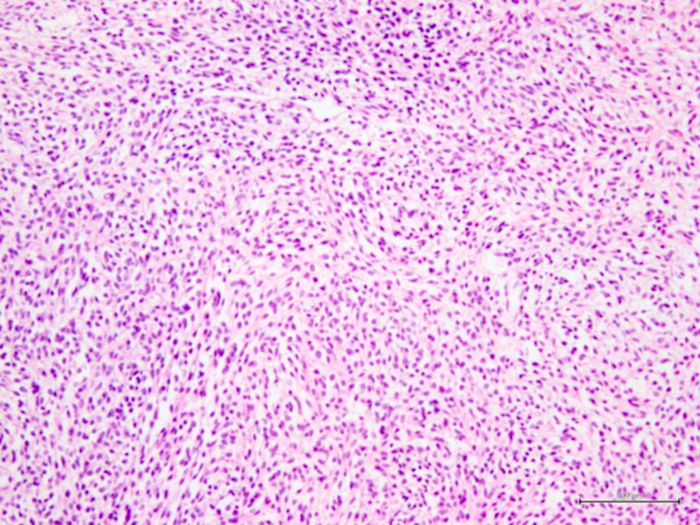
- Synovial sarcoma is characterized by spindle shaped cells with markedly monotonous nuclei (Ann Diagn Pathol 2014;18:369)
- Synovial sarcoma develops not only in soft tissues but also in parenchymal organs and its morphological appearance is similar, regardless of where it occurs (Ann Diagn Pathol 2014;18:369)
- There is no normal counterpart for synovial sarcoma (Ann Diagn Pathol 2014;18:369)
- 3 major types of organization:
- Biphasic type: a classic type with a mixture of distinct epithelial components and spindle cells in varying proportions
- Monophasic fibrous type: only fibrosarcoma-like spindle cells proliferate and no epithelial-like cells are observed
- Poorly differentiated type: very rare type and highly cellular round cells with hyperchromatic nuclei and frequent mitotic activity and necrosis
- Regarding histological and genotypes, the majority of the biphasic types were SS18::SSX1 and most of SS18::SSX2 are the monophasic type (Ann Diagn Pathol 2014;18:369)
Microscopic (histologic) images
Contributed by Kito Yusuke, M.D., Ph.D.
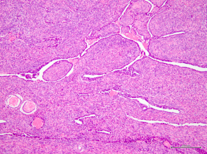
Biphasic type

Monophasic type
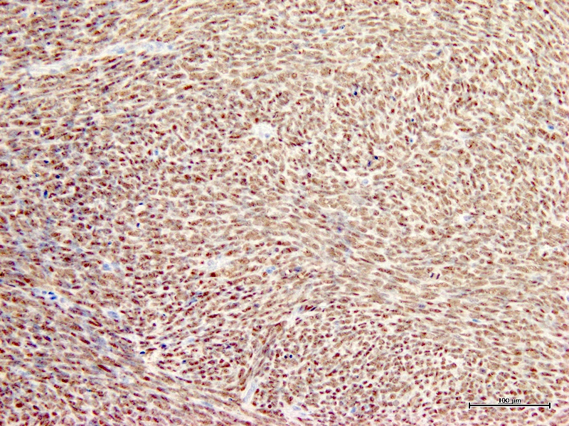
Rabbit monoclonal antibody
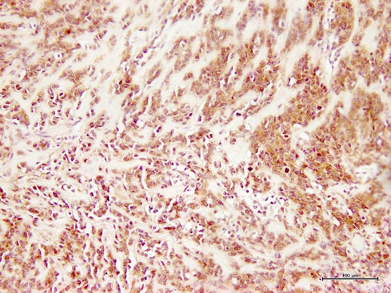
Mouse monoclonal antibody
Electron microscopy description
- Regarding the epithelial characteristics of tumor cells, electron microscopy revealed basement membrane formation and adhesion spots on the epithelial components, while immunostaining showed the expression of cytokeratin and EMA with various molecular weight; this is considered to be a true epithelial differentiation (Ann Diagn Pathol 2014;18:369)
Molecular / cytogenetics description
- t(X;18)(p11.2;q11): SS18::SSX1 fusion in 90%, can detect via RT-PCR
- Also t(X;18)(p11.21;q11): SS18::SSX2 fusion, variants can be detected by optimizing RT-PCR (Mod Pathol 2002;15:679, Hum Pathol 2001;32:105)
- Detection of t(X;18)(p11;q11) by RT-PCR of human synovial sarcoma cell line, HS-SY-II (Lab Invest 1992;67:498)
- In recent years, numerous monoclonal antibodies that specifically recognize SS18::SSX fusion gene products have been reported (Am J Surg Pathol 2020;44:922, Appl Immunohistochem Mol Morphol 2018;26:206)
- IHC could replace molecular genetic or cytogenetic testing in most cases
Molecular / cytogenetics images
Contributed by Kito Yusuke, M.D., Ph.D. and Ruta Gupta, M.D.

Detection of fusion gene
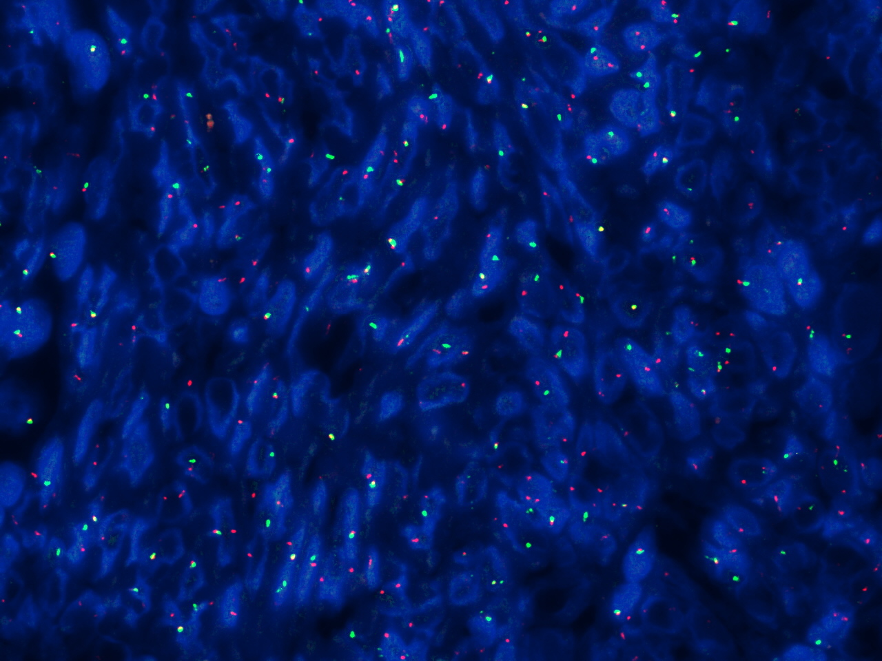
FISH with SS18 break apart probe
Sample pathology report
- Stomach, laparoscopic wedge resection:
- Synovial sarcoma, monophasic, FNCLCC grade 2 (see comment)
- Comment: H&E staining of the histological section showed dense proliferation of rather uniform spindle tumor cells mainly in the submucosa. Tumor cells were mixed with smooth muscle fibers of the muscularis mucosae. Some of them were also present even in the mucosa. There was no necrosis, vascular invasion and apparent mitotic figures. IHC showed that KIT, DOG1, CD34, S100P, desmin and αSMA were also negative. Ki67 labeling index of the tumor cells in the resected specimen was approximately 5%. The tumor cells were shown to be partially positive for AE1 / AE3, partially and weakly positive for CD99 and diffusely positive for vimentin and TLE1 by IHC. Finally, IHC with SS18::SSX fusion specific antibody gave diffuse positive staining to the tumor cells and fusion gene analysis using mRNA extracted from paraffin sections revealed that the tumor had SS18 (SYT)::SSX1 fusion gene. Thus, the tumor was diagnosed as synovial sarcoma, monophasic fibrous type (Diagn Pathol 2021;16:115).
Board review style question #1
Which of the following is a characteristic fusion gene found in this neoplasm?
- CIC::DUX4
- EWSR1::FL1
- NAB2::STAT6
- SS18::SSX
Board review style answer #1
Board review style question #2
Which of the following is the most common synovial sarcoma specific fusion gene?
- SS18::SSX1
- SS18::SSX2
- SS18::SSX3
- SS18::SSX4
Board review style answer #2
A. SS18::SSX1 has a frequency of 65%, SS18::SSX2 has a frequency of 35% and SS18::SSX4 has a frequency of less than 1% (Histopathology 2006;48:13). SS18::SSX3 has never been reported.
Comment Here
Reference: t(X;18)
Comment Here
Reference: t(X;18)
Board review style question #3
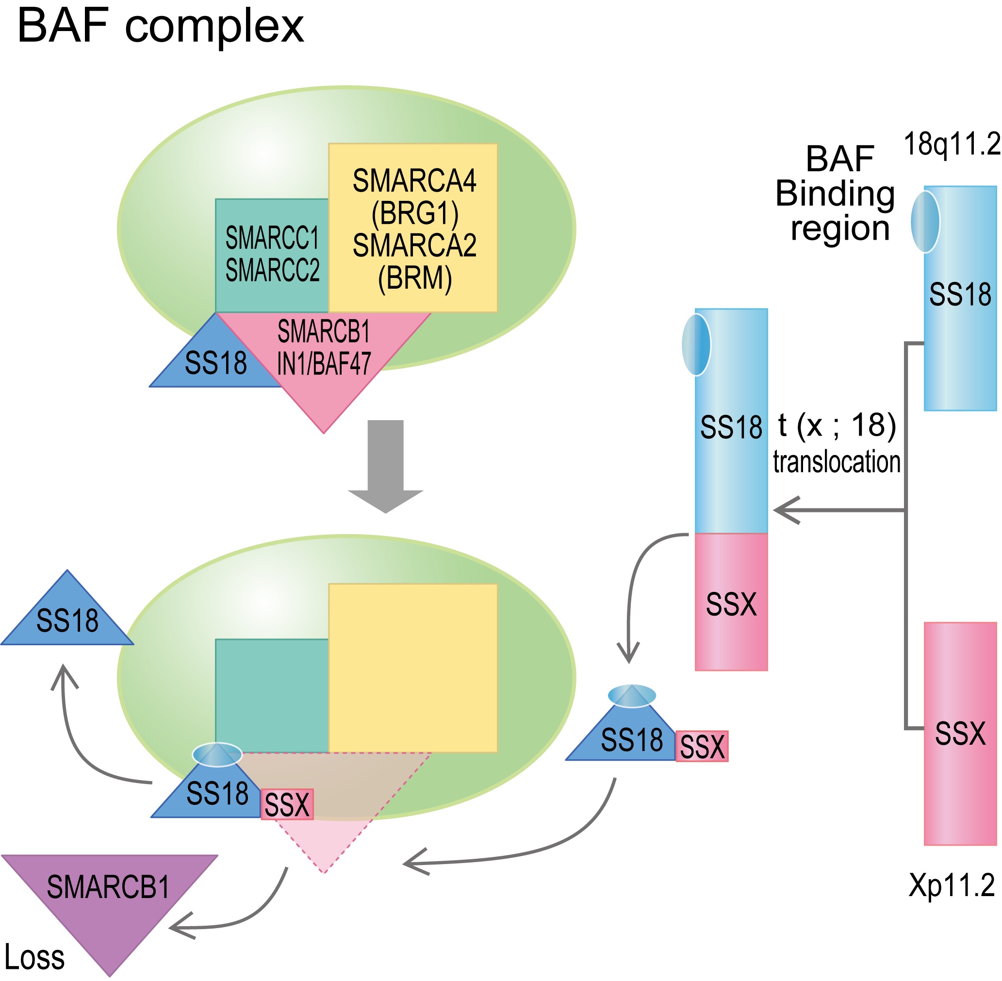
Which protein is one of the normal chromatin remodeling factors that decomposes and disappears when the fusion protein found in synovial sarcoma binds?
- ARID1A / B
- BAF60
- BRG / BRM
- SMARCB1 (INI1)
Board review style answer #3

