

Ovary
Tumor-like lesions
Fibromatosis and massive edema
Author: Shahid Islam, M.D., Ph.D.

Last author update: 1 June 2012
Last staff update: 14 July 2023
Copyright: 2002-2024, PathologyOutlines.com, Inc.
PubMed Search: (Massive edema [title]) ovary
Table of Contents
Definition / general | Gross description | Microscopic (histologic) description | Microscopic (histologic) images | Differential diagnosis | Additional referencesCite this page: Islam S. Fibromatosis and massive edema. PathologyOutlines.com website. https://www.pathologyoutlines.com/topic/ovarymassiveedema.html. Accessed April 25th, 2024.
Definition / general
- Tumor-like enlargement of ovary due to edema fluid
- First described in 1969 (Obstet Gynecol 1969;34:564)
- Patients present with pain, abdominal mass, menstrual irregularities, virilization, precocious puberty, Meigs syndrome (with ascites and pleural effusion, eMedicine: Meigs Syndrome)
- May be due to partial torsion of mesovarium leading to interference of venous / lymphatic drainage
- Usually young women
- Right sided involvement is more common
Gross description
- Marked ovarian enlargement, watery cut surface, no necrosis
Microscopic (histologic) description
- Marked edema of stroma surrounding follicles and clusters of luteinized cells
- Stroma around vessels and in superficial cortical zone is normal
- Variable stromal luteinization
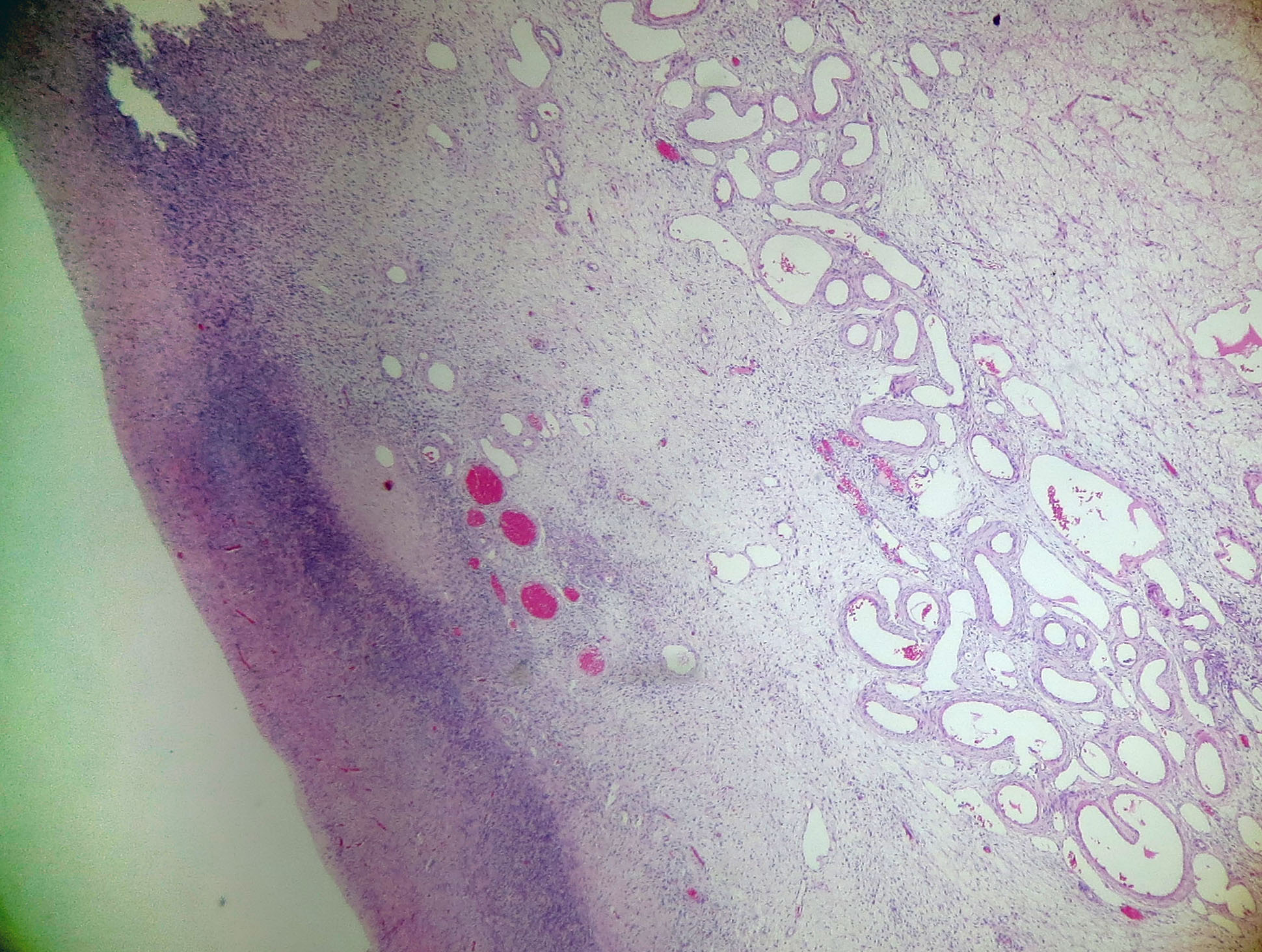
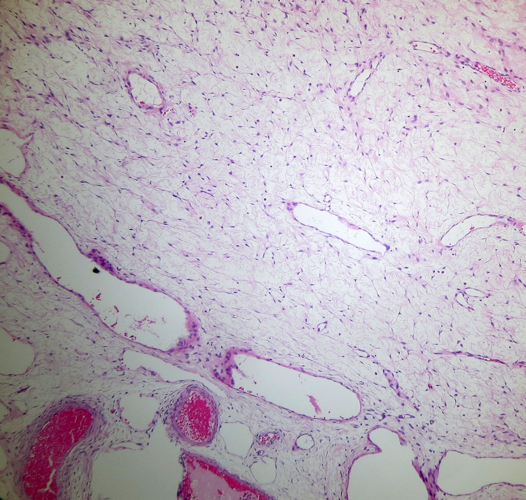
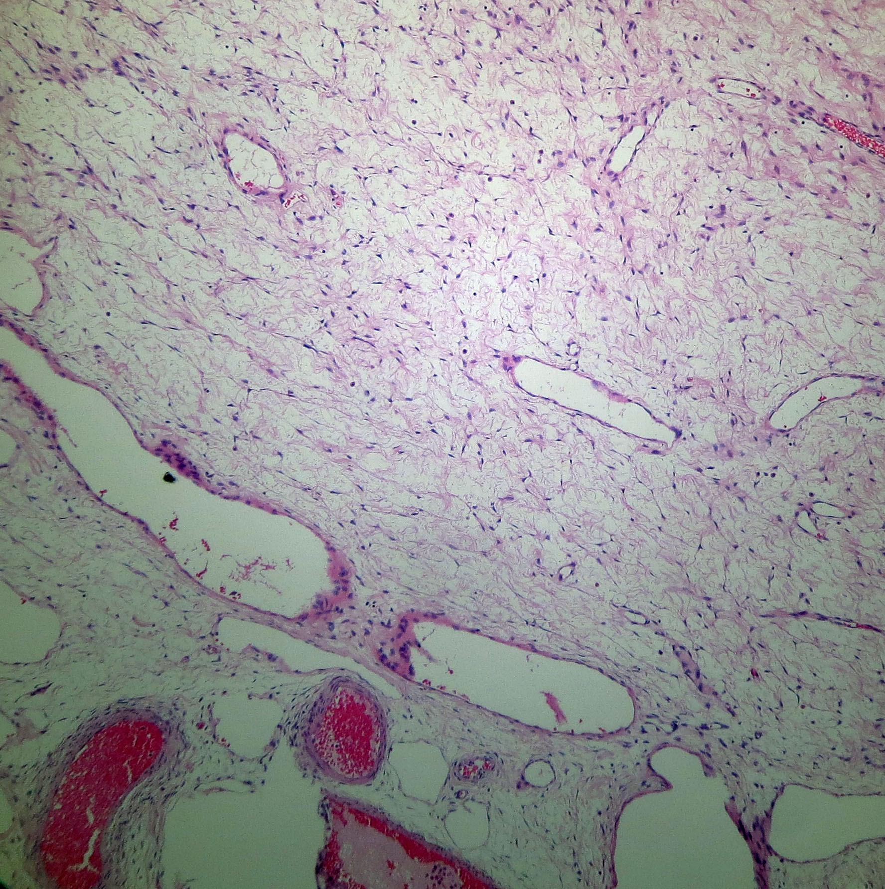
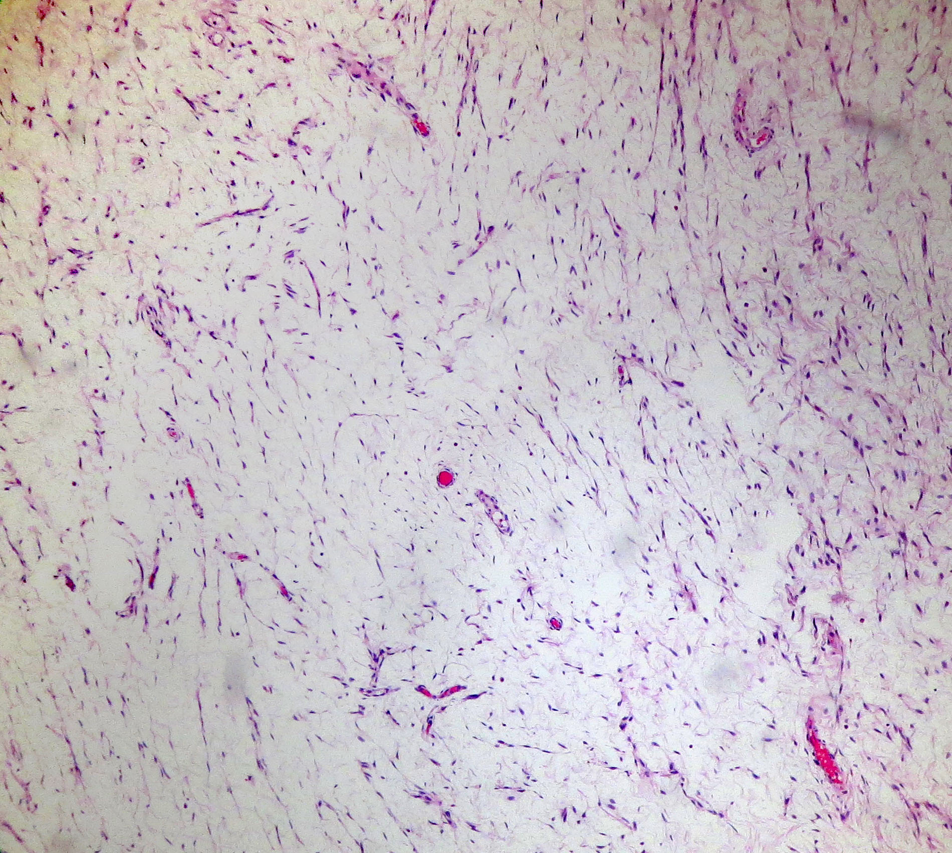
Microscopic (histologic) images
Contributed by Dr. Josehp Christopher Castillo


AFIP images

Clusters of lutein cells lie in the edematous stroma
Images hosted on other servers:


Loose stroma with occasional inflammatory cells
Differential diagnosis
- Fibroma: circumscribed, tumor cells replace ovarian architecture
- Krukenberg tumor: usually bilateral, signet ring cells present
Additional references
