

Ovary
Sex cord stromal tumors
Pure sex cord tumors
Granulosa cell tumor-juvenile
Editorial Board Member: Gulisa Turashvili, M.D., Ph.D.
Deputy Editor-in-Chief: Jennifer A. Bennett, M.D.


Last author update: 6 July 2021
Last staff update: 6 July 2021
Copyright: 2002-2024, PathologyOutlines.com, Inc.
PubMed search: Ovarian granulosa cell tumor juvenile [TI] pathology full text [SB]
Table of Contents
Definition / general | Essential features | ICD coding | Epidemiology | Sites | Pathophysiology | Etiology | Clinical features | Diagnosis | Radiology description | Prognostic factors | Case reports | Treatment | Gross description | Gross images | Frozen section description | Microscopic (histologic) description | Microscopic (histologic) images | Virtual slides | Positive stains | Negative stains | Molecular / cytogenetics description | Sample pathology report | Differential diagnosis | Additional references | Board review style question #1 | Board review style answer #1Cite this page: Huvila J, Gilks CB. Granulosa cell tumor-juvenile. PathologyOutlines.com website. https://www.pathologyoutlines.com/topic/ovarytumorgctjuv.html. Accessed April 25th, 2024.
Definition / general
- Sex cord stromal tumor composed of primitive appearing granulosa cells with follicular and solid growth patterns
Essential features
- Sex cord stromal tumor with primitive granulosa cell differentiation
- Solid and follicular growth
- Almost always occurs in patients younger than 30 years
- Lacks the FOXL2 somatic mutation seen in adult granulosa cell tumor
- Usually stage IA and associated with a favorable prognosis
ICD coding
- ICD-O: 8622/1 - granulosa cell tumor, juvenile
- ICD-10:
- ICD-11: 2F76 & XH2KH2 - granulosa cell tumor, juvenile
Epidemiology
- Mean age 13 years; range 0 - 67 years
- 80% occur before age 20 and 97% before age 30 (Am J Surg Pathol 1984;8:575)
Sites
- Ovary
- Rarely extraovarian (Pediatr Hematol Oncol 2021;38:272)
Pathophysiology
- Unknown
Etiology
- Unknown
Clinical features
- Most prepubertal patients present with sexual precocity due to excessive estrogen production and rarely produce androgens; older patients have nonspecific abdominal swelling and pain (J Endocrinol Invest 2006;29:653, Am J Surg Pathol 1984;8:575)
- Rarely associated with enchondromatosis (Ollier disease), Maffucci syndrome, abnormal karyotype / ambiguous genitalia (Am J Surg Pathol 1984;8:575, Am J Surg Pathol 1985;9:737)
Diagnosis
- Diagnosis is made at the time of removal of an adnexal mass
- Possibility of juvenile granulosa cell tumor may be suspected if there are estrogenic manifestations in a prepubertal patient but histopathological examination is required for diagnosis
Radiology description
- Typically appears on imaging as a large, unilateral, multicystic mass with occasional septae; most contain both solid and cystic components
- Can have a sponge-like appearance on imaging (Radiographics 2014;34:2039)
Prognostic factors
- Of low malignant potential, with a very favorable prognosis for patients with stage IA tumors (which account for a large majority of cases) but a guarded prognosis if there is spread beyond the ovary
Case reports
- Fetus with an androgenic ovarian mass in the third trimester (J Obstet Gynaecol Res 2021;47:2220)
- 2 year old girl with precocious pseudopuberty (J Clin Res Pediatr Endocrinol 2021 Apr 14 [Epub ahead of print])
- 17 year old girl with gender dysphoria (Case Rep Oncol 2020;13:1330)
- 33 year old woman with adnexal mass and Maffucci syndrome (Clin Imaging 2019;56:77)
- 40 year old Japanese woman with late onset juvenile granulosa cell tumor (Acta Med Okayama 2020;74:159)
Treatment
- Large majority of tumors are stage IA and surgical removal, with preservation of fertility if desired, is all that is required for treatment (Acta Obstet Gynecol Scand 2021 May 24 [Epub ahead of print])
- With recurrent or advanced stage tumors, platinum based chemotherapy is typically offered (Acta Obstet Gynecol Scand 2021 May 24 [Epub ahead of print])
Gross description
- Usually unilateral with a smooth surface
- Mean size 12.5 cm (Am J Surg Pathol 1984;8:575)
- Multiloculated, cystic and solid tumor with yellow-white solid areas
- May have hemorrhage and necrosis
Gross images
AFIP Images
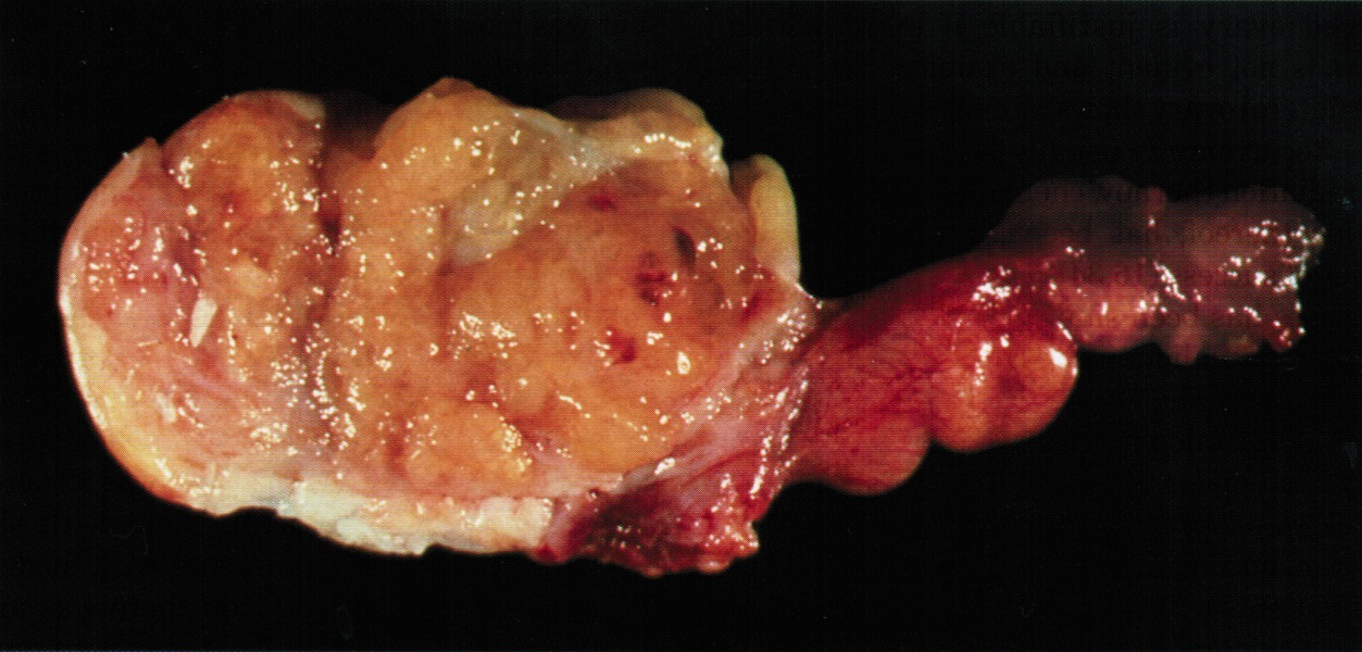
Solid and slightly lobulated
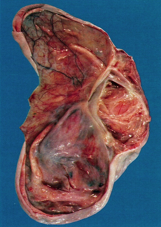
Large locules with smooth linings
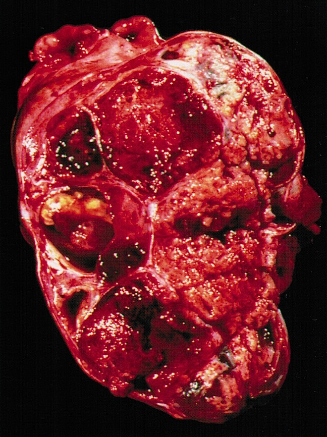
Solid and cystic
Frozen section description
- Smooth surfaced ovarian mass in a young patient
- Presence of follicular differentiation is helpful but definitive diagnosis may have to be deferred to permanent sections
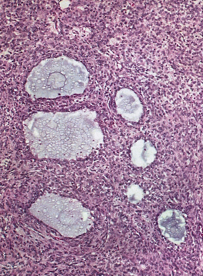
Microscopic (histologic) description
- Diffuse or nodular appearance at low power
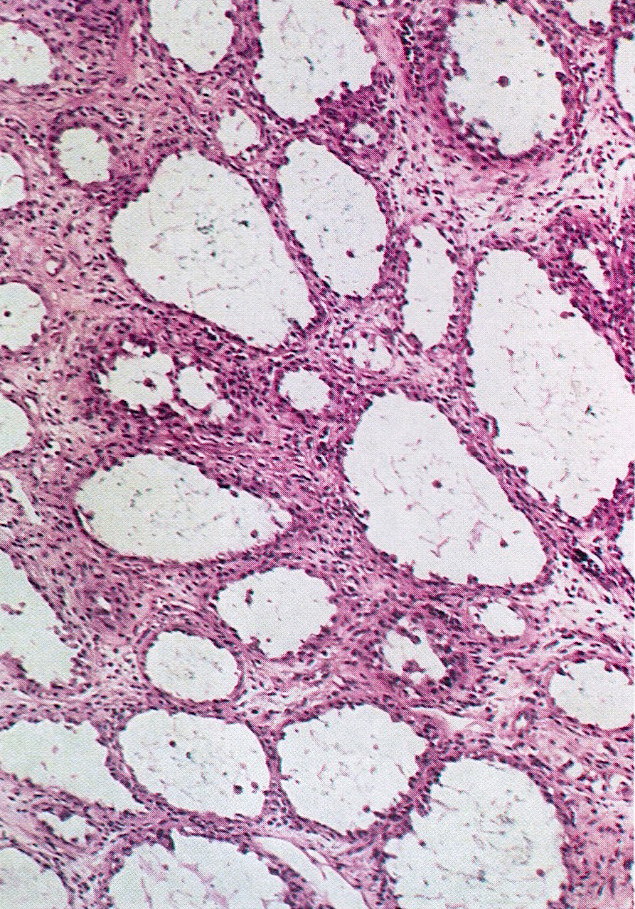
- Macrofollicle and microfollicle formation containing eosinophilic secretions
- Tumor cells are often luteinized
- Round / oval hyperchromatic nuclei with small nucleoli, irregular nuclear contours
- No / rare nuclear grooves; high mitotic rate (mean 11/10 high power fields) (Arch Pathol Lab Med 1989;113:40)
- Bizarre / atypical nuclei are rarely present
- May have pseudopapillary architecture (Am J Surg Pathol 2008;32:581)
Microscopic (histologic) images
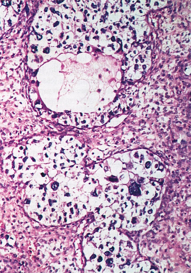
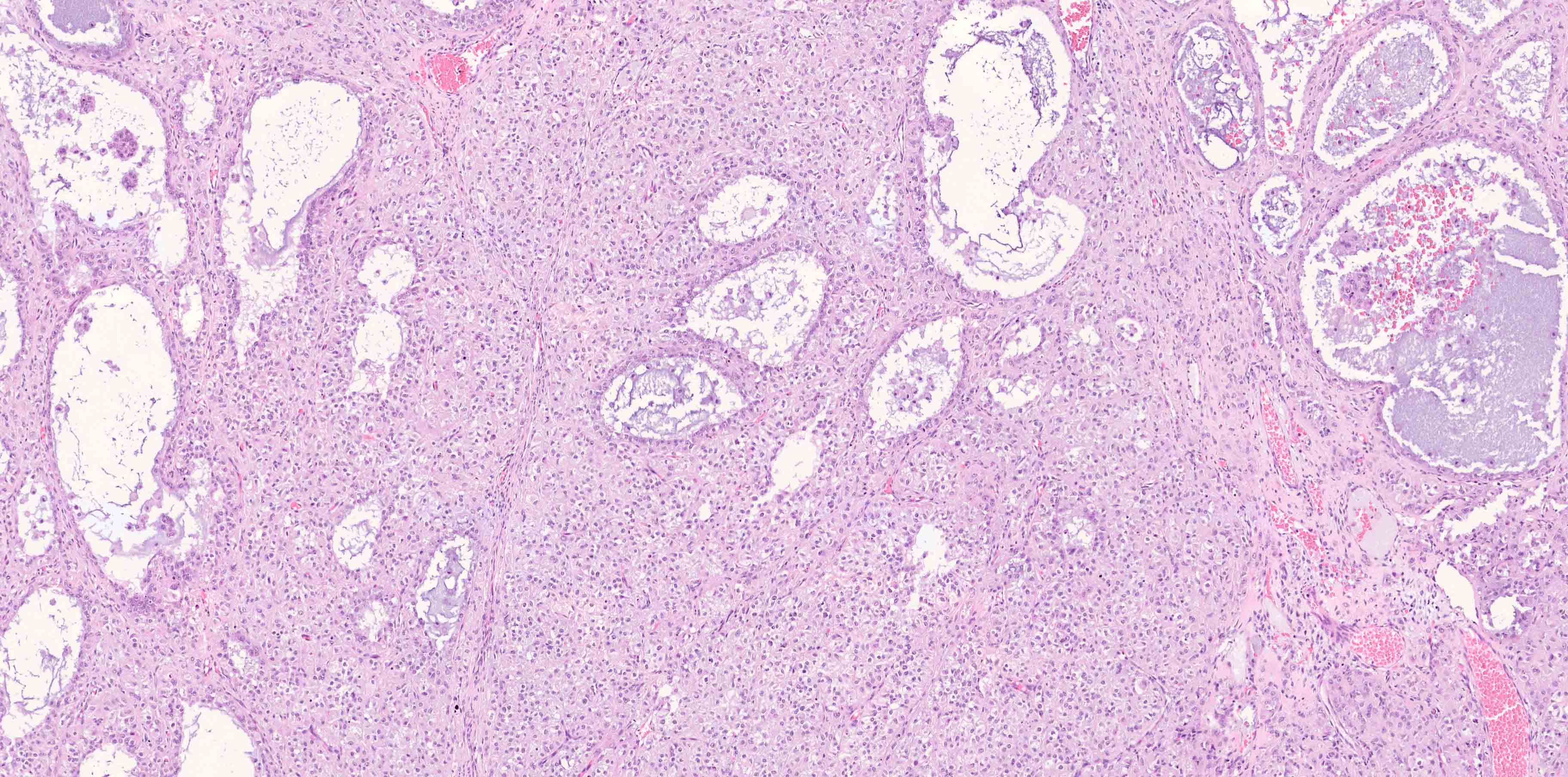
Contributed by Jutta Huvila, M.D., Ph.D. and AFIP
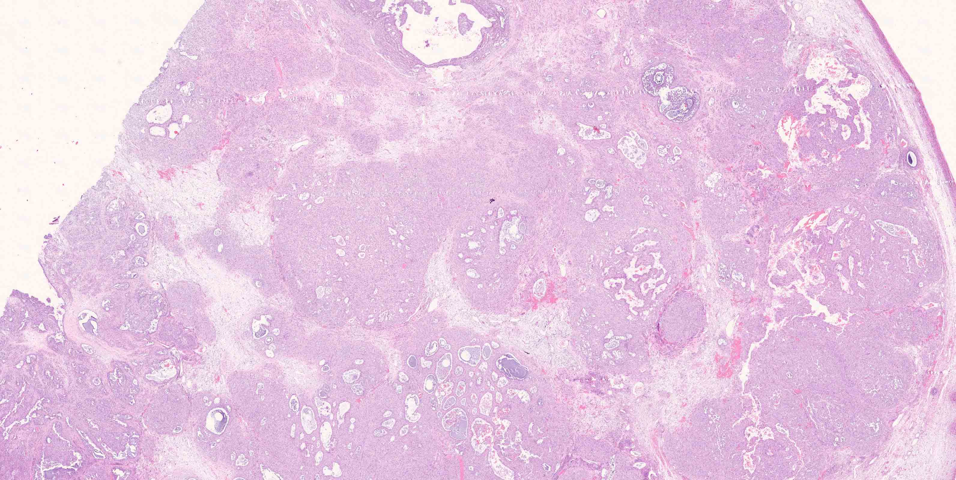
Lobulated growth pattern
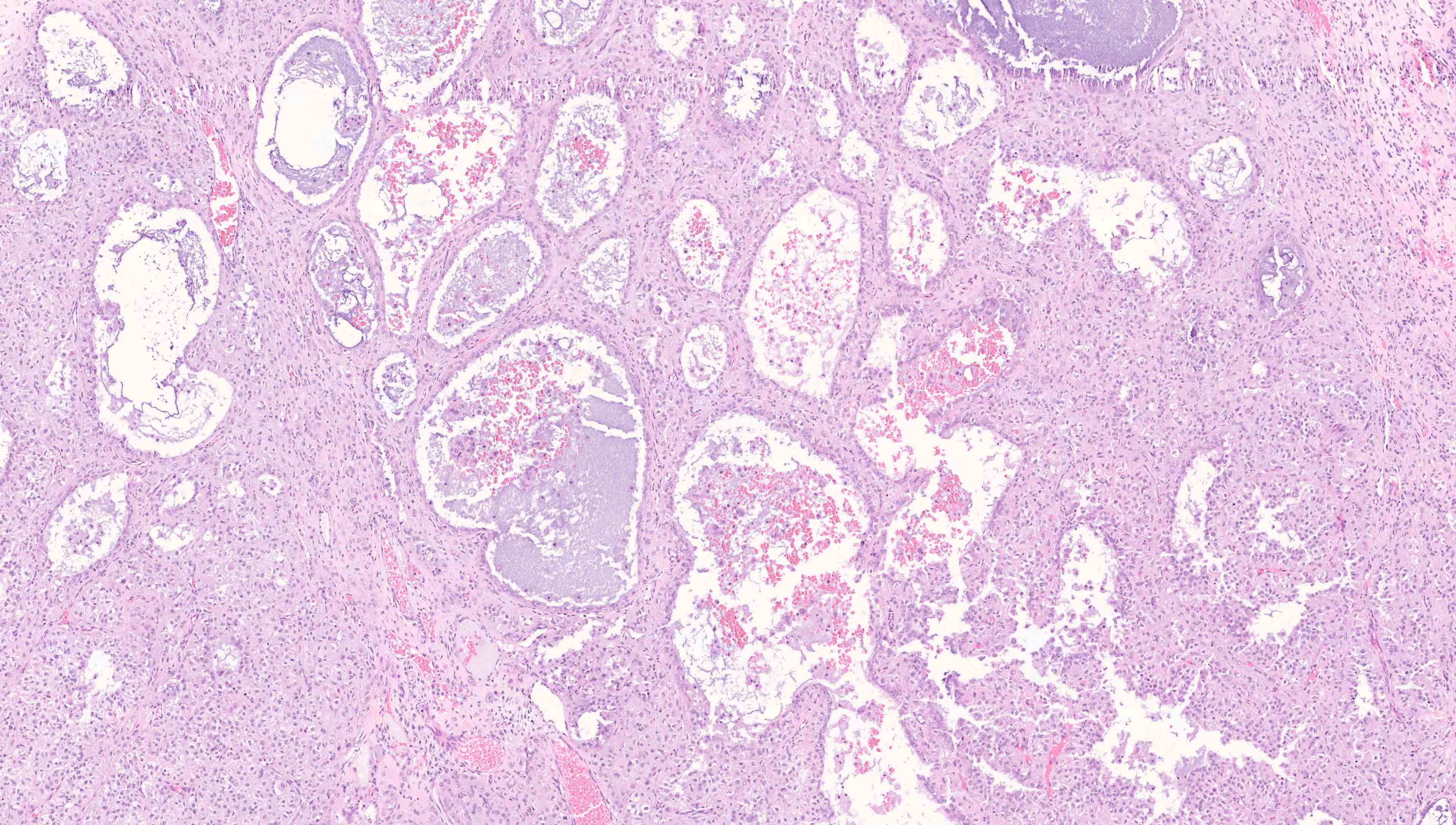
Follicular differentiation
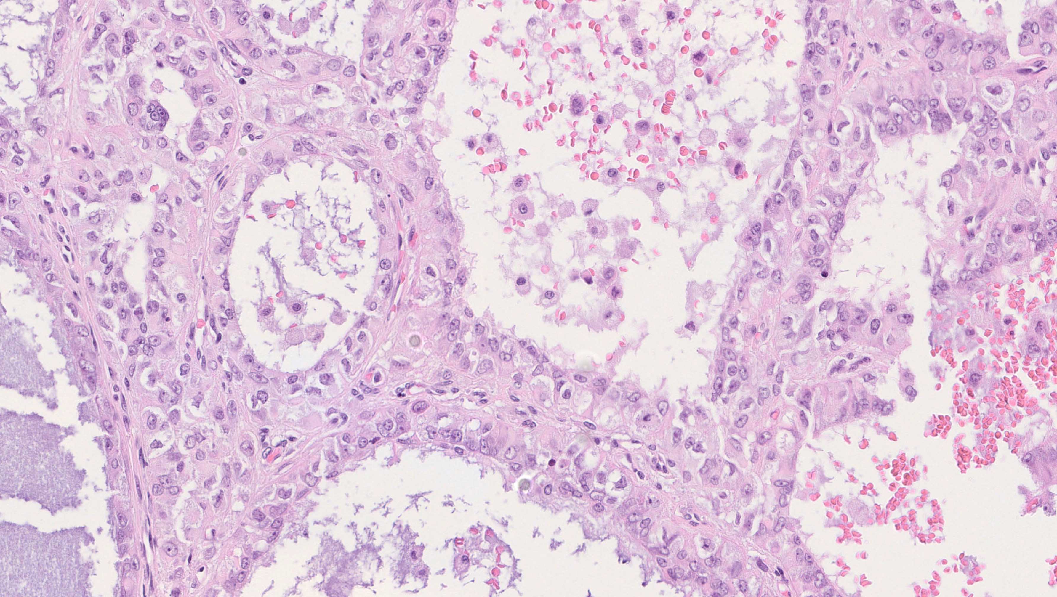
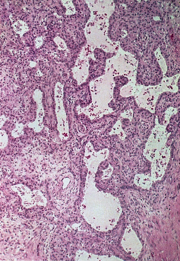
Atypical cells lining follicle
Solid growth
Solid cellular neoplasm with focal follicle formation
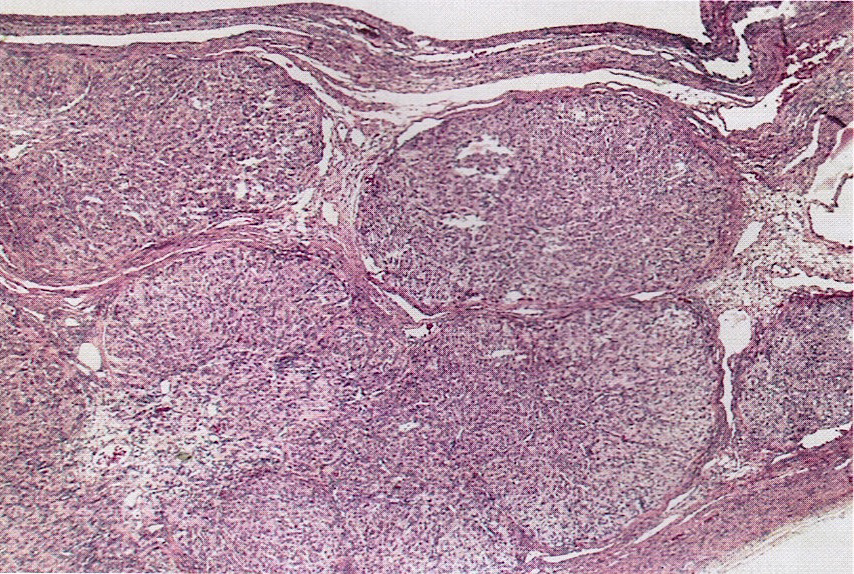
Solid nodules
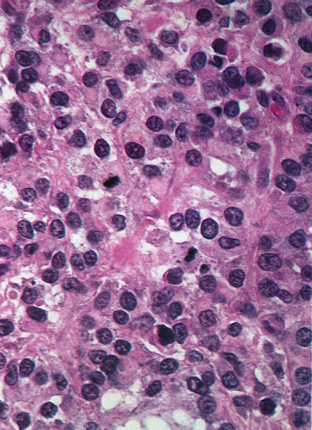
Mitotic figures
Hobnail nuclei
Bizarre nuclei
Virtual slides
Images hosted on other servers:

Lobulated architecture with follicle-like spaces
Positive stains
Negative stains
Molecular / cytogenetics description
- Most have AKT1 mutations (EBioMedicine 2015;2:421)
- GNAS mutations reported in 30% of cases (J Clin Endocrinol Metab 2006;91:1842)
- Consistently negative for FOXL2 (402C > G) mutation, while 5 - 10% have a mutation in DICER1; the DICER1 mutations may be germline, i.e. associated with DICER1 syndrome (Am J Surg Pathol 2021;45:223)
- Somatic mutations in IDH1 or IDH2 are associated with Ollier disease or Maffucci syndrome; juvenile granulosa cell tumor is an uncommon manifestation of both diseases (Am J Med Genet A 2020;182:1093)
Sample pathology report
- Right ovary, oophorectomy:
- Juvenile granulosa cell tumor, negative for ovarian surface involvement or extraovarian spread
Differential diagnosis
- Adult granulosa cell tumor:
- Older age at presentation (but there is some overlap in the age ranges)
- Small uniform cells resembling normal granulosa cells
- FOXL2 mutation in 95% of cases (N Engl J Med 2009;360:2719)
- Small cell carcinoma, hypercalcemic type:
- More primitive appearing cells, with scant cytoplasm or rhabdoid cells
- Loss of BRG1 expression (protein encoded by SMARCA4) (J Pathol 2016;238:389)
- Mutation in SMARCA4 (Nat Genet 2014;46:427)
- Sertoli-Leydig cell tumor:
- Tubules (in well differentiated tumors), cords (intermediate differentiation), sarcomatoid growth (poorly differentiated) or retiform architecture
- Follicle-like spaces occasionally seen admixed with typical sertoliform differentiation (Am J Surg Pathol 2021;45:59)
- Gynandroblastoma:
- Typical Sertoli-Leydig cell tumor component coexisting with separate typical juvenile granulosa cell tumor (Am J Surg Pathol 2021;45:59)
- Yolk sac tumor:
- Clear cell carcinoma:
Additional references
Board review style question #1
A 2 year old girl presents with a unilateral ovarian tumor. Histology shows follicle-like spaces lined by cells with moderate amounts of cytoplasm and showing nuclear atypia and prominent nucleoli. What is the diagnosis?
- Juvenile granulosa cell tumor
- Sertoli-Leydig cell tumor
- Teratoma
- Yolk sac tumor
Board review style answer #1
