

Ovary
Mesenchymal tumors
Leiomyosarcoma
Author: Nalini Gupta, M.D.

Last author update: 1 December 2014
Last staff update: 23 August 2022
Copyright: 2003-2024, PathologyOutlines.com, Inc.
PubMed Search: Leiomyosarcoma ovary
Table of Contents
Definition / general | Sites | Pathophysiology | Clinical features | Prognostic factors | Case reports | Treatment | Gross description | Microscopic (histologic) description | Microscopic (histologic) images | Positive stains | Negative stains | Differential diagnosisCite this page: Gupta N. Leiomyosarcoma. PathologyOutlines.com website. https://www.pathologyoutlines.com/topic/ovarytumorleiomyosarcoma.html. Accessed April 19th, 2024.
Definition / general
- Rare tumor of teratomatous or nonteratomatous origin
- "True" leiomyosarcoma if of nonteratomatous origin
Sites
- Omentum, reteroperitoneum, mesentery
Pathophysiology
- Arises from: malignant degeneration of ovarian leiomyoma; smooth muscle in blood vessel walls in cortical stroma and corpus luteum, muscular attachments of ovarian ligament, wolfian duct remnants, or totipotential ovarian mesenchyme; or from a teratoma
Clinical features
- Mostly postmenopausal patients
- Presents with abdominal pain and mass
- 62% die of disease within mean 24 months (Am J Surg Pathol 2004;28:1436)
Prognostic factors
- Tumor stage, tumor size, grade, mitotic index (Obstet Gynecol Surv 2007;62:480)
Case reports
- 42 year old woman with epithelioid tumor (Gynecol Oncol 2005;97:697)
- 58 year old woman (Arch Pathol Lab Med 1991;115:941)
- 60 year old women (World J Oncol 2001;2:265, Online J Health Allied Scs 2009;8:16)
Treatment
- Multimodality treatment: surgical debulking, postoperative radiotherapy or chemotherapy
- FIGO staging and treatment of ovarian sarcoma is same as for epithelial ovarian carcinoma (Obstet Gynecol Surv 2007;62:480)
Gross description
- Solitary, lobular, soft fleshy solid mass with hemorrhage and cystic degeneration
Microscopic (histologic) description
- Usually 2 of 3: moderate / severe cytologic atypia, 10+ MF/10 HPF, tumor cell necrosis
- Varies from well differentiated to highly pleomorphic sarcoma
Microscopic (histologic) images
AFIP images
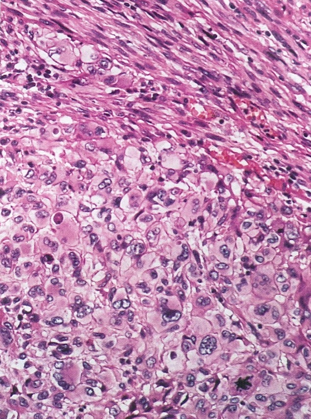
Epithelioid tumor
Images hosted on other servers:

Pleomorphic tumor cells

H-Caldesmon, desmin and smooth muscle actin
Positive stains
Negative stains
Differential diagnosis
- Cellular fibroma: low mitotic activity <10/10HPF
- Krukenberg tumors have stromal reaction, history of GI primary, pan CK+
- Mixed Müllerian tumors: pan CK+ areas
- Sarcomatoid form of sex cord stromal tumors: sex cord elements present
- Undifferentiated carcinomas: pan CK+, H caldesmon-
