

Ovary
Other carcinomas
Carcinosarcoma
Author: Nalini Gupta, M.D.

Last author update: 1 December 2014
Last staff update: 27 October 2023
Copyright: 2003-2024, PathologyOutlines.com, Inc.
PubMed Search: Carcinosarcoma ovary
Table of Contents
Definition / general | Terminology | Epidemiology | Sites | Pathophysiology | Clinical features | Laboratory | Radiology description | Case reports | Treatment | Gross description | Gross images | Microscopic (histologic) description | Microscopic (histologic) images | Positive stains | Negative stains | Differential diagnosisCite this page: Gupta N. Carcinosarcoma. PathologyOutlines.com website. https://www.pathologyoutlines.com/topic/ovarytumormmt.html. Accessed April 23rd, 2024.
Definition / general
- Aggressive tumor with malignant epithelial and sarcomatous components
- Most common in postmenopausal, low parity women
- Very poor prognosis; stage is best predictor and most patients present at advanced stage
Terminology
- Previously called malignant mixed mesodermal tumor, MMMT
Epidemiology
- Mostly postmenopausal females, peaks in sixth decade
Sites
- Uterus, cervix, fallopian tube (rare)
Pathophysiology
- Appear to have epithelial origin (Am J Surg Pathol 1990;14:317, Am J Surg Pathol 1995;19:666)
Clinical features
- Abdominal mass, pain, vaginal bleeding
- Risk factors include advanced age, excess estrogen exposure, nulliparity, prior pelvic irradiation, tamoxifen use (Lancet 2000;356:881)
Laboratory
- No useful biochemical marker (Curr Opin Obstet Gynecol 2006;18:20)
Radiology description
- Pelvic ultrasound and CT: heterogeneous pelvic mass containing solid parts with/without ascites
Case reports
- 52 year old woman (Taiwan J Obstet Gynecol 2010;49:87)
- 70 year old woman (Journal of Medical Cases 2010;1:55)
- Growing into an inguinal hernia sac (Surg Today 2003;33:797)
Treatment
- Surgical cytoreduction with adjuvant chemotherapy (Gynecol Oncol 2000;79:196)
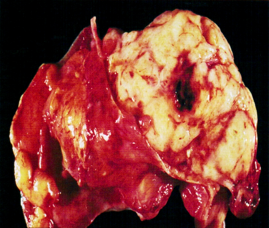
Gross description
- Soft and fleshy mass, often with bleeding and necrosis
Gross images
Contributed by Mona Kandil, M.D., Ph.D. and AFIP

Ovary

Uterus

Omental deposit
Fallopian tube
Microscopic (histologic) description
- Malignant epithelial and sarcomatous elements
- Sarcomatous element can be homologous (nonspecific malignant stroma) or heterologous (malignant elements of a different tissue type, particularly cartilage)
- Often contains cytoplasmic hyaline droplets containing alpha-1-antitrypsin (Hum Pathol 1982;13:930)
- Rarely trophoblastic tissue (Hum Pathol 1988;19:1235)
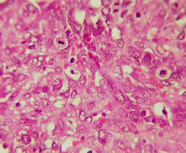
Microscopic (histologic) images
Contributed by Mona Kandil, M.D., Ph.D.
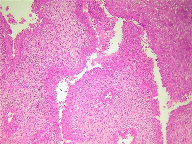
Malignant stroma
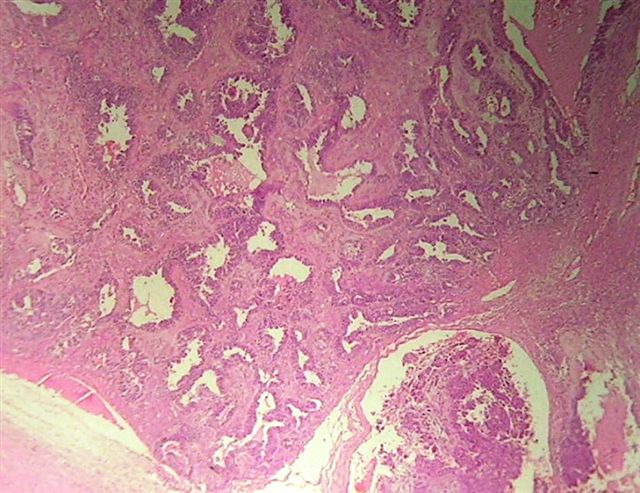

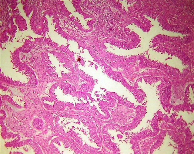
High grade malignant glands

Necrotic foci

High grade epithelial tumor cells and necrosis
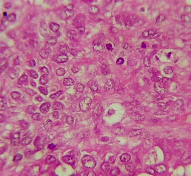
High grade tumor cells with mitotic figures
Images hosted on other servers:


Rhabdomyosarcoma component
Positive stains
Differential diagnosis
