

Ovary
Mucinous tumors
Mucinous cystadenoma and adenofibroma
Author: Gulisa Turashvili, M.D., Ph.D.
Editorial Board Member: C. Blake Gilks, M.D.
Deputy Editor-in-Chief: Jennifer A. Bennett, M.D.

Last author update: 28 September 2021
Last staff update: 24 August 2023
Copyright: 2003-2024, PathologyOutlines.com, Inc.
PubMed Search: Mucinous cystadenoma[TI] OR mucinous adenofibroma[TI] ovary[TIAB] full text[SB]
Table of Contents
Definition / general | Essential features | ICD coding | Epidemiology | Sites | Pathophysiology | Etiology | Clinical features | Diagnosis | Laboratory | Radiology description | Radiology images | Prognostic factors | Case reports | Treatment | Gross description | Gross images | Frozen section description | Frozen section images | Microscopic (histologic) description | Microscopic (histologic) images | Virtual slides | Cytology description | Positive stains | Negative stains | Molecular / cytogenetics description | Sample pathology report | Differential diagnosis | Board review style question #1 | Board review style answer #1 | Board review style question #2 | Board review style answer #2Cite this page: Turashvili G. Mucinous cystadenoma and adenofibroma. PathologyOutlines.com website. https://www.pathologyoutlines.com/topic/ovarytumormucinousbenign.html. Accessed April 26th, 2024.
Definition / general
- Benign mucinous neoplasm composed of cysts and glands lined by gastrointestinal or Müllerian type mucinous epithelium lacking architectural complexity or cytologic atypia
Essential features
- Includes cystadenoma and adenofibroma
- Usually unilateral and composed of variable amounts of cysts and glands lined by bland gastrointestinal or Müllerian type mucinous epithelium and variably cellular stroma
- May be associated with mature teratoma or Brenner tumor
- Excellent prognosis, with rare recurrences associated with cystectomy or rupture
ICD coding
- ICD-O:
- ICD-11:
- 2F32.Y & XH6H73 - other specified benign neoplasm of ovary and mucinous cystadenoma, NOS
- 2F32.Y & XH59X8 - other specified benign neoplasm of ovary and mucinous adenofibroma, NOS
Epidemiology
- Accounts for 80% of primary ovarian mucinous neoplasms
- Mucinous cystadenoma > adenofibroma (Int J Gynecol Pathol 2005;24:4, Cancer 1985;55:1958)
- Median age 50 years (range 13 - 79) (Cancer 1985;55:1958)
Sites
- Usually ovary
- Less commonly, retroperitoneum (Int J Surg Case Rep 2012;3:486)
Pathophysiology
- May arise from mature teratoma (possible germ cell origin) or Brenner tumor (Arch Pathol Lab Med 2008;132:1753)
- KRAS mutations in 68% of cases (Cancer 1997;79:1581)
Etiology
- Unknown
Clinical features
- Abdominal distention with or without palpable mass
- Abdominal or pelvic pain
- Other symptoms related to abdominal / pelvic mass
- Rarely, estrogenic or androgenic manifestations secondary to stromal luteinization (Int J Gynecol Pathol 2005;24:4)
Diagnosis
- Microscopic examination
Laboratory
- Rarely, elevated CA-125 (Cancer 1985;55:1958)
Radiology description
- Ultrasonography:
- Usually large multilocular cystic adnexal mass with numerous thin septations
- Various degrees of echogenicity in different locules
- Low level internal echogenicity due to increased mucin content
- Magnetic resonance imaging:
- Usually large multilocular cyst containing fluid of various viscosity resulting in variable signal intensities on both T1 and T2 sequences (stained glass appearance)
- Reference: Radiographics 2000;20:1445, Radiographics 2019;39:982
Radiology images
Images hosted on other servers:

Palpable lower abdominal mass

Right pelvic pain
Prognostic factors
- Excellent prognosis
- Recurrences may occur after cystectomy or rupture and spillage (J Obstet Gynaecol Res 2006;32:615, Am J Obstet Gynecol 2010;202:142.e1)
Case reports
- 13 year old girl with recurrent ovarian mucinous cystadenoma after cystectomy (Int J Surg Case Rep 2021;83:106006)
- 22 year old woman with metachronous ipsilateral recurrent ovarian mucinous cystadenoma in initial pregnancy and mature teratoma in subsequent pregnancy (Cureus 2019;11:e3818)
- 40 year old woman with an ovarian mixed Brenner tumor and mucinous cystadenoma (J Clin Imaging Sci 2020;10:22)
- 71 year old woman with postmenopausal hyperandrogenism associated with ovarian mucinous cystadenoma (BMJ Case Rep 2021;14:e237505)
Treatment
- Oophorectomy or cystectomy
Gross description
- Usually unilateral (95%)
- Smooth or bosselated surface
- Cystadenoma:
- Uni or multilocular cyst with variably sized smooth walled locules
- Filled with dense, viscous, sticky, gelatinous material
- No solid areas or papillary excrescences
- Mean size 10 cm, rarely > 30 cm
- Adenofibroma:
- Usually smaller
- Predominantly white and solid with variable amounts of small cysts (Int J Gynecol Pathol 2005;24:4, Surg Pathol Clin 2019;12:565)
Gross images
AFIP images
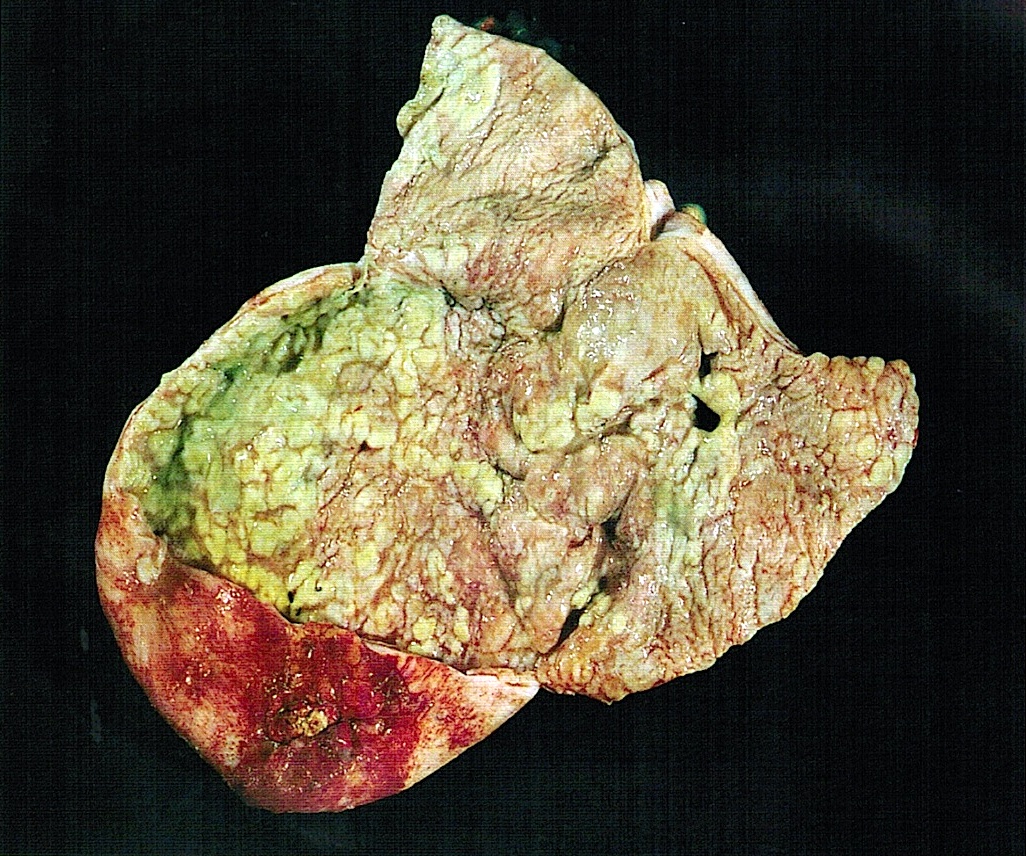
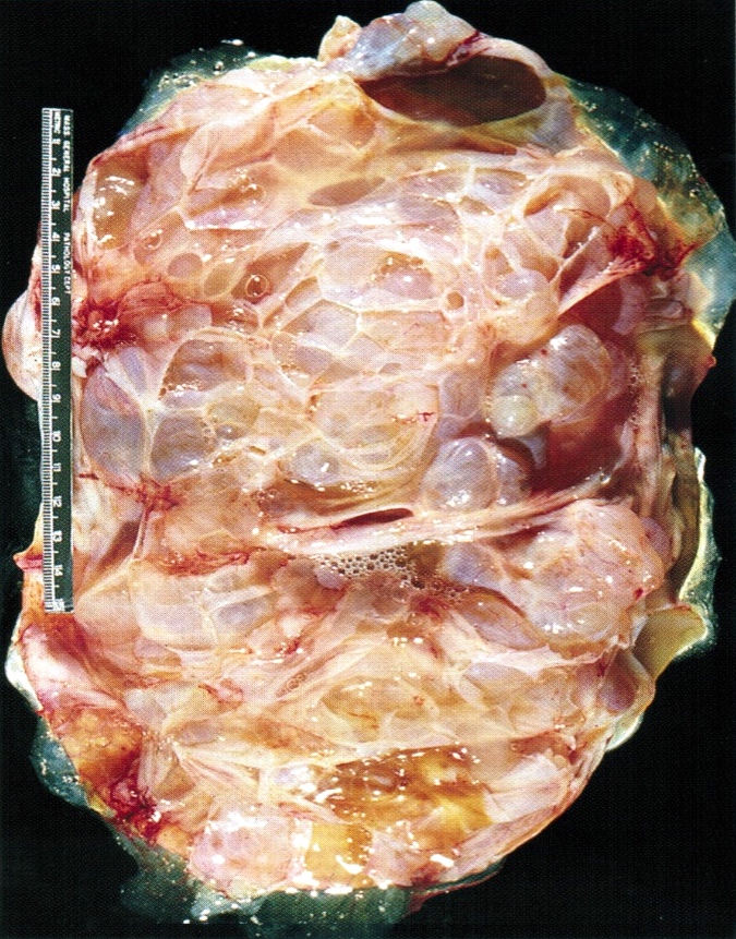
Mucinous cystadenoma
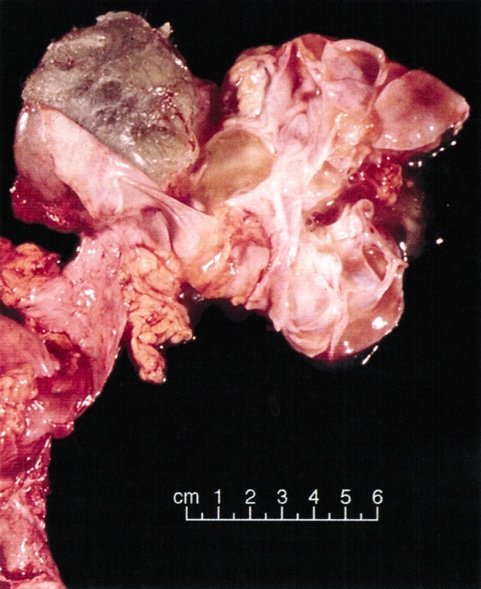
Associated with dermoid cyst
Frozen section description
- Benign cystic neoplasm lined by a single layer of bland mucinous epithelium (cystadenoma) or solid and cystic neoplasm composed of small glands or cysts lined by a single layer of bland mucinous epithelium set in a fibromatous stroma (adenofibroma)
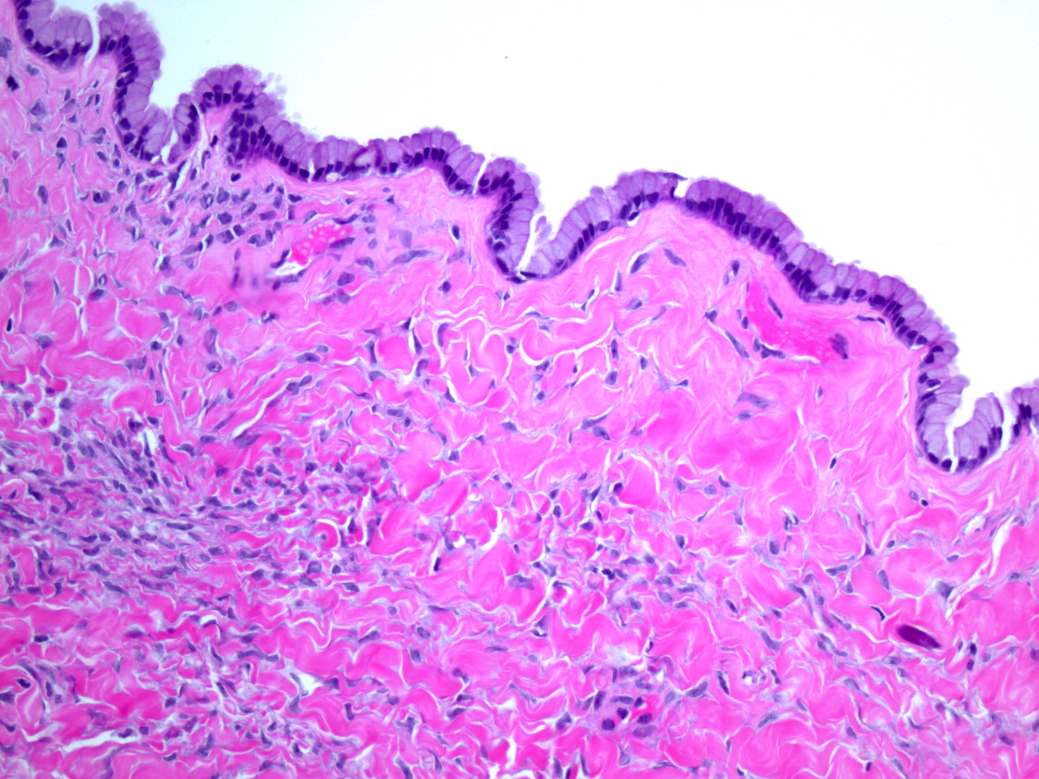
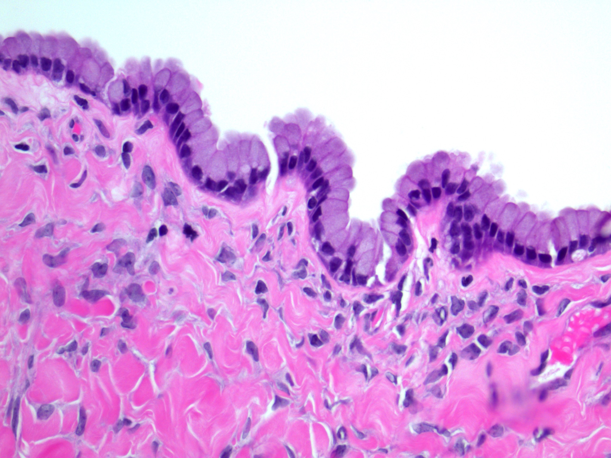
Frozen section images
Contributed by Gulisa Turashvili, M.D., Ph.D.
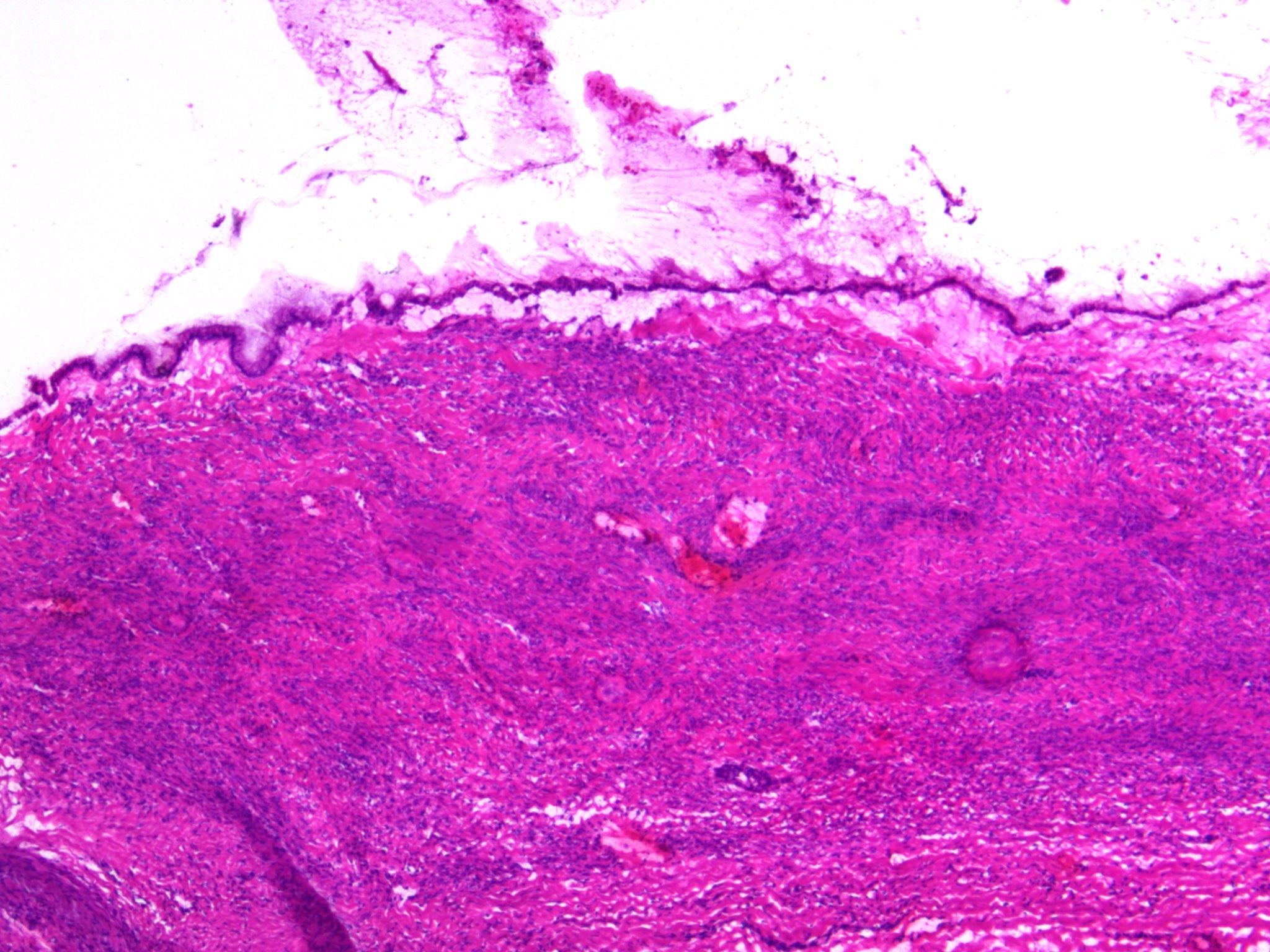
Unilocular cyst
Bland mucinous epithelium
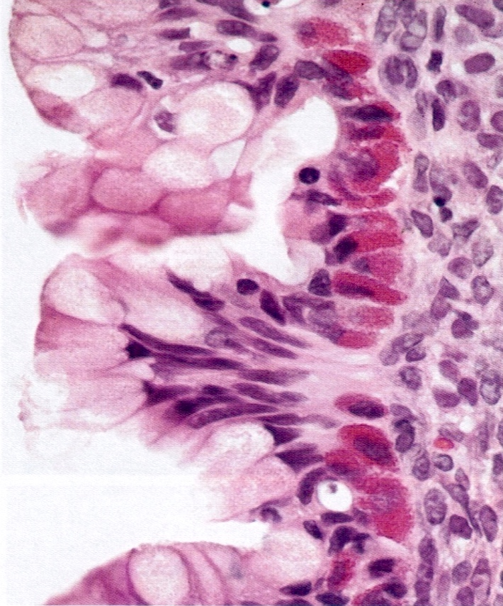
Microscopic (histologic) description
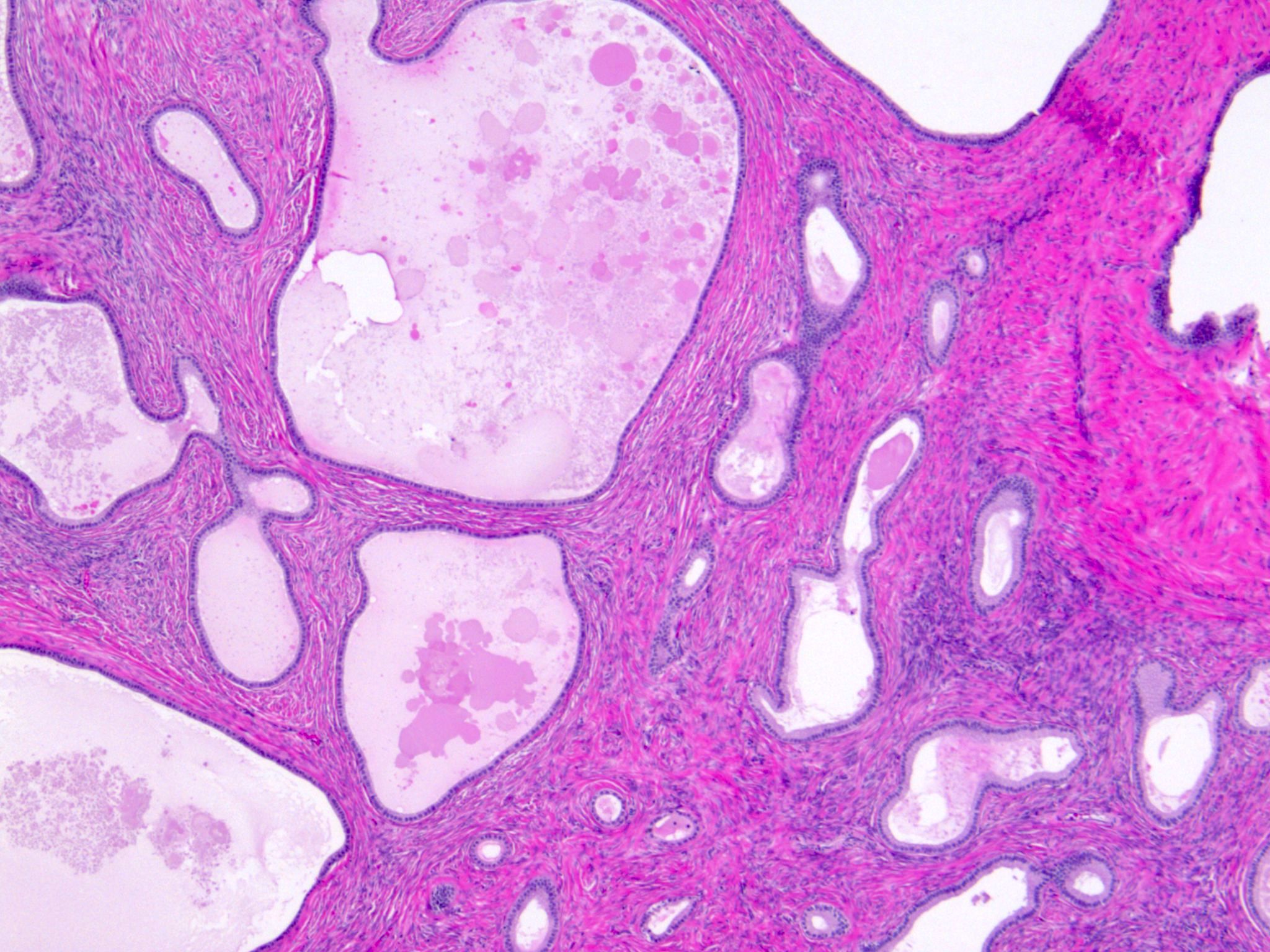
- Mucinous cystadenoma:
- Uni or multilocular cystic neoplasm composed of multiple cysts and glands lined by a single layer of bland mucinous epithelium
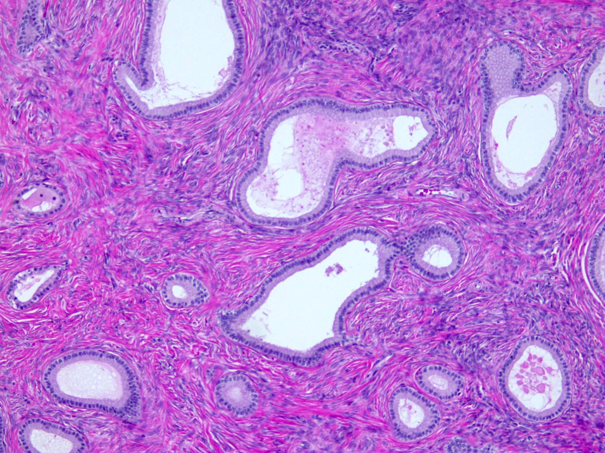
- Mucinous adenofibroma:
- Solid and cystic neoplasm composed of small cysts or glands lined by a single layer of bland mucinous epithelium
- Stroma is fibromatous and variably cellular with or without luteinization
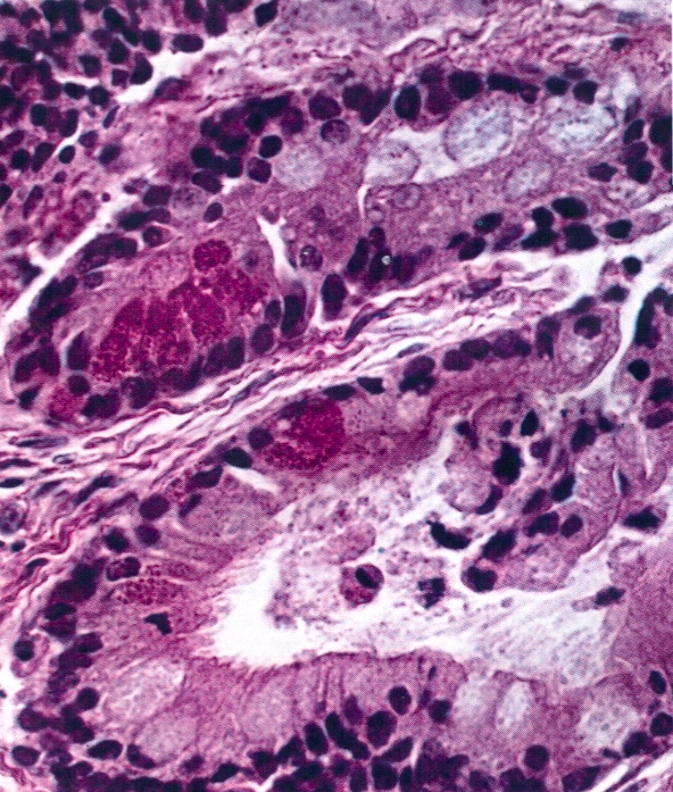
- Focal extravasated mucin or mucin granulomas with numerous histiocytes may be present secondary to cyst / gland rupture
- General features:
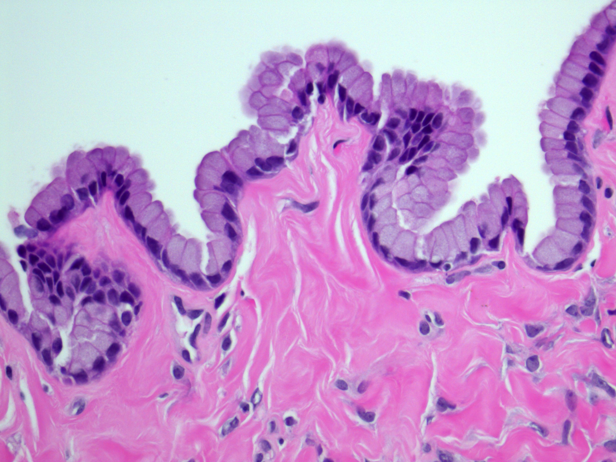
- Simple nonstratified mucinous epithelium resembling Müllerian, intestinal or gastric foveolar type epithelium
- > 80% are intestinal type containing goblet cells
- Epithelium may be undulating but usually no epithelial stratification or tufting
- Columnar, cuboidal to flat nonciliated cells
- Variable amounts of mucinous cytoplasm
- Small basally located nuclei lacking cytological atypia
- Absent or minimal mitotic activity and apoptotic bodies
- Spiculated calcifications (62.5%) (Arch Pathol Lab Med 2008;132:1753)
- Rare features:
- Tubular crypt-like outpouchings or papillary infoldings at the periphery of cysts
- Focal mild cytologic atypia and mitoses
- Goblet cells with or without neuroendocrine cells or Paneth cells
- Periglandular stromal condensation and luteinization (more common in pregnancy) (40%) (Arch Pathol Lab Med 2008;132:1753)
- Sex cord-like differentiation
- Necrosis (12.5%) (Arch Pathol Lab Med 2008;132:1753)
- Pseudoxanthoma cells (47.5%) (Arch Pathol Lab Med 2008;132:1753)
- Muciphages (42.5%) (Arch Pathol Lab Med 2008;132:1753)
- Pseudomyxoma ovarii (stromal dissecting mucin) and giant cell reaction (10%) (Arch Pathol Lab Med 2008;132:1753)
- May be associated with:
- Brenner tumor (18%) (Arch Pathol Lab Med 2008;132:1753)
- Mature teratoma (2 - 11%)
- Mural nodules (Indian J Med Sci 2005;59:499, Cancer 1979;44:1327, J Korean Med Sci 1998;13:680)
- Clear cell carcinoma (J Clin Pathol 2000;53:938, J Pak Med Assoc 2007;57:373, Int J Gynecol Pathol 2009;28:584)
- Large cell neuroendocrine carcinoma (Int J Gynecol Pathol 1996;15:167)
- Endometrioid carcinoma (Ann Diagn Pathol 2003;7:300)
- Sex cord stromal tumors, such as granulosa cell tumor and Sertoli-Leydig cell tumor (Arch Pathol Lab Med 1986;110:528, Int J Gynecol Pathol 2005;24:224)
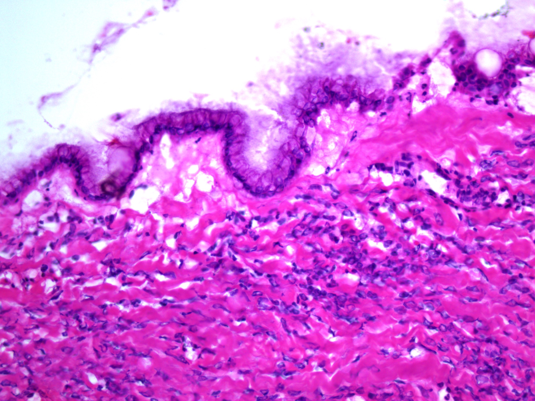
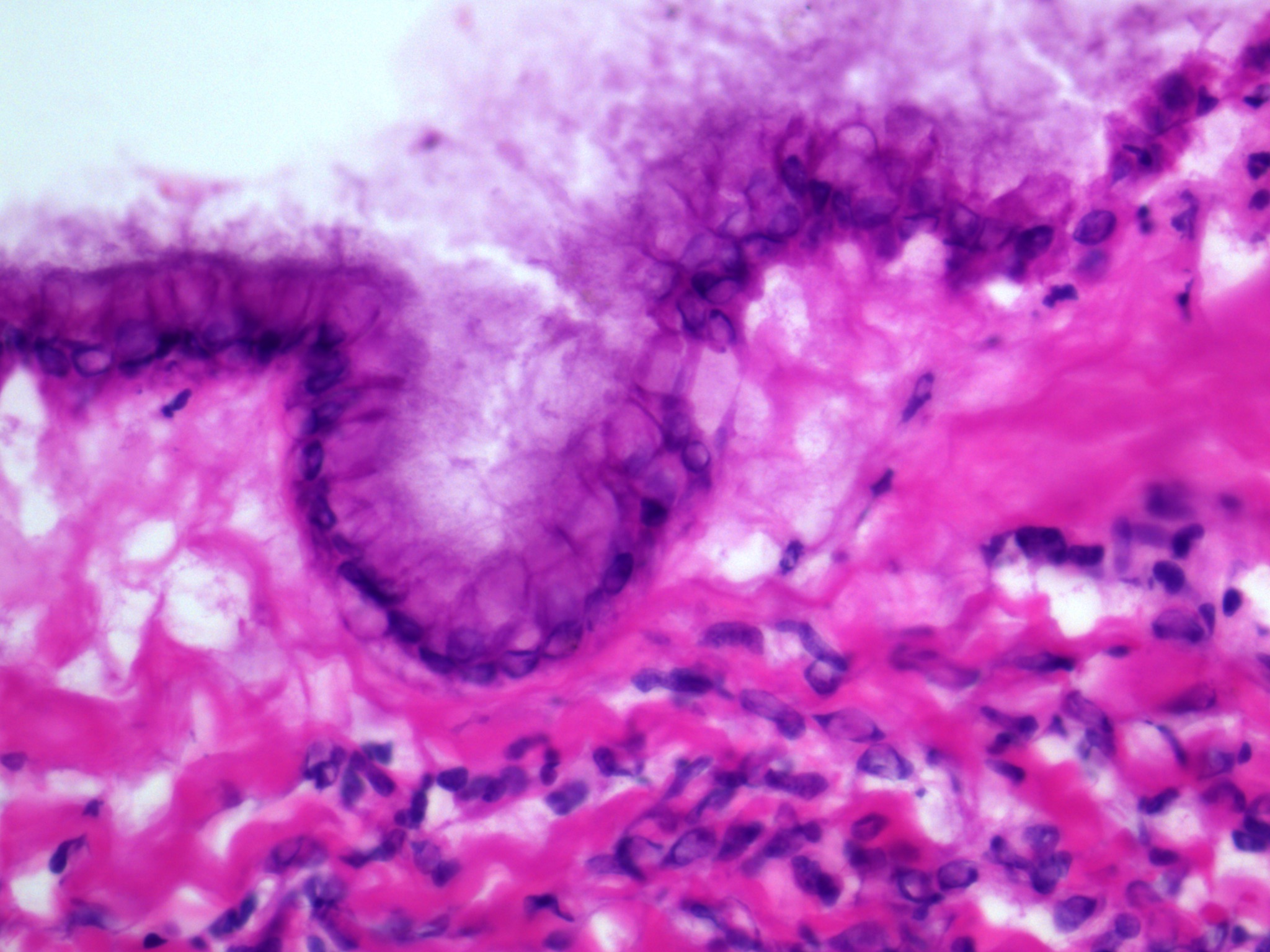
Microscopic (histologic) images
Contributed by Gulisa Turashvili, M.D., Ph.D. and AFIP images

Unilocular cyst
Bland mucinous epithelium
Mucinous adenofibroma
Mucinous cystic tumor
Virtual slides
Images hosted on other servers:

Mucinous cystic neoplasm

Mucinous cystadenoma associated with Brenner tumor

Mucinous cystadenoma with functioning stroma

Mucinous cystadenoma, intestinal type

PAX8

CK7

CK20
Cytology description
- Usually negative cytology or reactive mesothelial cells
Positive stains
- CK7 (Am J Clin Pathol 2002;117:944)
- PAX8: variable, may be focal
- CK20: variable (Am J Clin Pathol 2002;117:944)
- CDX2: variable
Negative stains
Molecular / cytogenetics description
- KRAS mutations in 68% of cases (Cancer 1997;79:1581)
Sample pathology report
- Right fallopian tube and ovary, salpingo-oophorectomy:
- Ovary: mucinous cystadenoma
- Fallopian tube: benign
Differential diagnosis
- Mucinous tumor with focal atypia / proliferation (Hum Pathol 2004;35:949, Hum Pathol 2004;35:918, Am J Surg Pathol 1991;15:227):
- Focal (< 10%) architectural complexity, including papillae and tufting or nuclear pseudostratification and crowding
- Mucinous borderline tumor:
- Architectural complexity with variable degrees of epithelial stratification, tufting and villous or slender filiform papillae in at least 10% of tumor
- Metastatic low grade appendiceal mucinous neoplasm:
- Mucin on ovarian and extraovarian surfaces (pseudomyxoma peritonei)
- Bilateral ovarian involvement
- Detachment of mucinous epithelium from stroma (cleft-like space)
- Hypermucinous (highly differentiated, tall, mucin rich) cells
- Positive for CK20, CDX2 and SATB2; negative for PAX8, usually negative for CK7
- Metastatic adenocarcinoma:
- Variable differentiation with high grade nuclear atypia and mitotic activity (gastrointestinal and pancreatico-hepatobiliary tract)
- Nuclear pseudostratification, apical mitoses and apoptotic bodies (human papillomavirus associated endocervical adenocarcinoma)
- Often bilateral, multinodular, with surface involvement, at least focal infiltrative growth and lack of correlation between architecture and cytology (Anticancer Res 2018;38:5465)
Board review style question #1
Which immunohistochemical markers are useful for differentiating this primary ovarian tumor from a low grade appendiceal mucinous neoplasm involving the ovary?
- CK7, CK20, CDX2, SATB2 and PAX8
- CK7, CK20, CDX2, WT1 and SATB2
- CK7, CK20, PAX8, ER and SATB2
- CK7, CK20, SATB2, ER and PR
- CK7, CK20, SATB2, WT1 and PAX8
Board review style answer #1
A. CK7, CK20, CDX2, SATB2 and PAX8. Low grade appendiceal mucinous neoplasm is usually positive for CK20, CDX2 and SATB2 and negative for CK7 and PAX8.
Comment Here
Reference: Mucinous cystadenoma / adenofibroma
Comment Here
Reference: Mucinous cystadenoma / adenofibroma
Board review style question #2
Which of the following is true for ovarian mucinous cystadenoma?
- Always positive for PAX8
- May be associated with mature teratoma or Brenner tumor
- Most common in postmenopausal women
- Usually bilateral ovarian involvement
- Usually positive for ER and PR
Board review style answer #2
B. May be associated with mature teratoma or Brenner tumor
Comment Here
Reference: Mucinous cystadenoma / adenofibroma
Comment Here
Reference: Mucinous cystadenoma / adenofibroma
