Ovary
Serous tumors
Serous cystadenoma, adenofibroma and surface papilloma
Editorial Board Member: Gulisa Turashvili, M.D., Ph.D.
Deputy Editor-in-Chief: Jennifer A. Bennett, M.D.


Last author update: 11 June 2021
Last staff update: 19 May 2025
Copyright: 2003-2025, PathologyOutlines.com, Inc.
PubMed search: Serous cystadenoma
Table of Contents
Definition / general | Essential features | Terminology | ICD coding | Epidemiology | Sites | Pathophysiology | Etiology | Clinical features | Diagnosis | Laboratory | Radiology description | Prognostic factors | Case reports | Treatment | Gross description | Gross images | Frozen section description | Microscopic (histologic) description | Microscopic (histologic) images | Cytology description | Cytology images | Positive stains | Sample pathology report | Differential diagnosis | Additional references | Practice question #1 | Practice answer #1 | Practice question #2 | Practice answer #2Cite this page: Roe CJ, Hanley K. Serous cystadenoma, adenofibroma and surface papilloma. PathologyOutlines.com website. https://www.pathologyoutlines.com/topic/ovarytumorserousbenign.html. Accessed October 4th, 2025.
Definition / general
- Benign partially or completely cystic lesion measuring > 1 cm in size and composed of cells resembling fallopian tube epithelium or cuboidal nonciliated epithelium resembling ovarian surface epithelium
Essential features
- Benign; > 1 cm in size (< 1 cm signifies a cortical inclusion cyst); composed of cells resembling fallopian tube epithelium
- Presents over a broad age range and are generally asymptomatic
- Usually small, uni to multilocular cysts lined by a single layer of tall, columnar, ciliated cells
- Adenofibromas and cystadenofibromas are composed predominantly of fibrous stroma, with glands and cysts forming a minor component
Terminology
- Includes cystadenoma, cystadenofibroma, adenofibroma, papillary cystadenoma, papillary cystadenofibroma, papillary adenofibroma
- Term used depends on the relative amount of fibrous stroma but distinctions are often arbitrary
ICD coding
- ICD-11: 2F32.3 - serous ovarian cystadenoma
Epidemiology
- Patients present over a broad age range
- Most often found in adult women of reproductive age
Sites
- Ovary, less commonly fallopian tube
Pathophysiology
- DNA copy number changes may be seen in stromal fibromatous cells and epithelial cells (Clin Cancer Res 2011;17:7273, Lab Invest 2004;84:778)
Etiology
- Unknown
Clinical features
- Generally asymptomatic
- Symptoms related to an ovarian mass
- One of the more common ovarian tumors to undergo torsion
Diagnosis
- Cystectomy or oophorectomy
Laboratory
- CA-125 levels may be mildly elevated (rarely marked) (Arch Gynecol Obstet 2007;276:559)
Radiology description
- Typically anechoic with thin, smooth walls and posterior acoustic enhancement; unilocular cysts, thin walls, minimal septations and absence of papillary projections (Radiographics 2000;20:1445)
- Imaging modality: pelvic ultrasound or CT scan
Prognostic factors
- May recur after incomplete excision
Case reports
- 63 year old woman with ruptured benign serous ovarian cystadenoma mimicking ovarian malignancy with peritoneal carcinomatosis (Diagn Interv Imaging 2016;97:1187)
- 64 year old woman with bilateral ovarian fibromas and concomitant unilateral serous cystadenoma (J Obstet Gynaecol 2019;39:1027)
- 65 year old woman with ovarian serous cystadenoma with ectopic adrenal tissue (Int J Surg Case Rep 2017;33:89)
Treatment
- Surgery (cystectomy or oophorectomy)
Gross description
- Cystadenoma:
- Usually 3 - 10 cm (but can be up to 30 cm), oval to round, smooth glistening surface
- Usually watery clear to pale yellow cyst fluid but can be viscous and mucoid
- Rarely papillary excrescences are seen on outer surface
- Cystadenofibroma:
- Varies from solid areas with knobby papillae to firm confluent areas
- Adenofibroma:
- Entirely solid with small cysts
- Reference: Kurman: Blaustein's Pathology of the Female Genital Tract (Springer Reference), 7th Edition, 2019
Gross images
Images hosted on other servers:


Serous cystadenoma

Cystadenofibroma
Frozen section description
- Uni or multiloculated cysts with single layer of cuboidal or columnar epithelium and simple papillary projections, if present
- Bland appearing fibrous stroma in varying amounts
- No invasion, architectural complexity or atypia
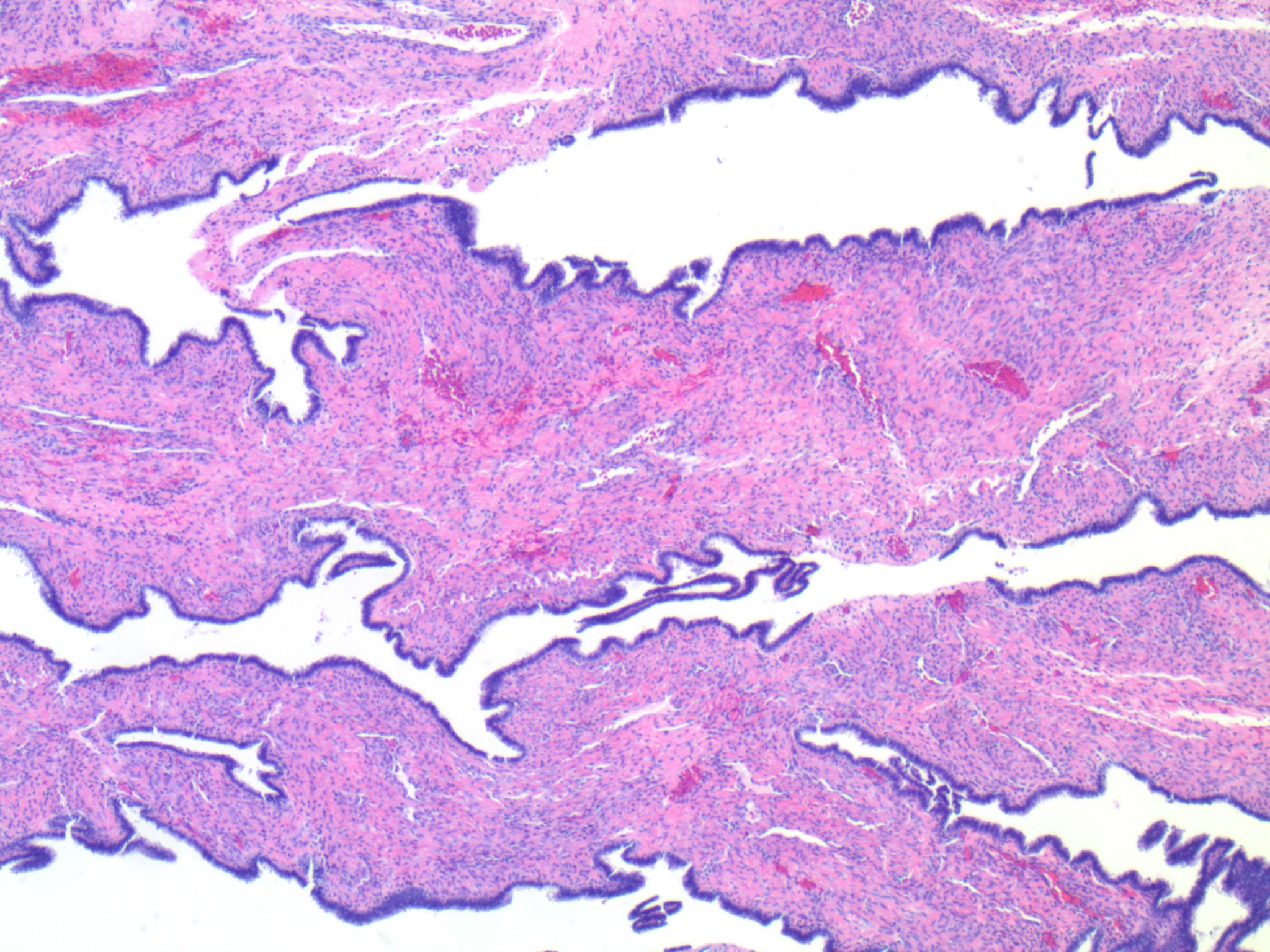
Microscopic (histologic) description
- Usually small, uni to multilocular cysts lined by a single layer of tall, columnar, ciliated cells resembling normal tubal epithelium or cuboidal nonciliated epithelium resembling ovarian surface epithelium
- Stroma contains spindle fibroblasts
- If papillae are present, they are simple
- Adenofibromas and cystadenofibromas are composed predominantly of fibrous stroma, with glands and cysts forming a minor component
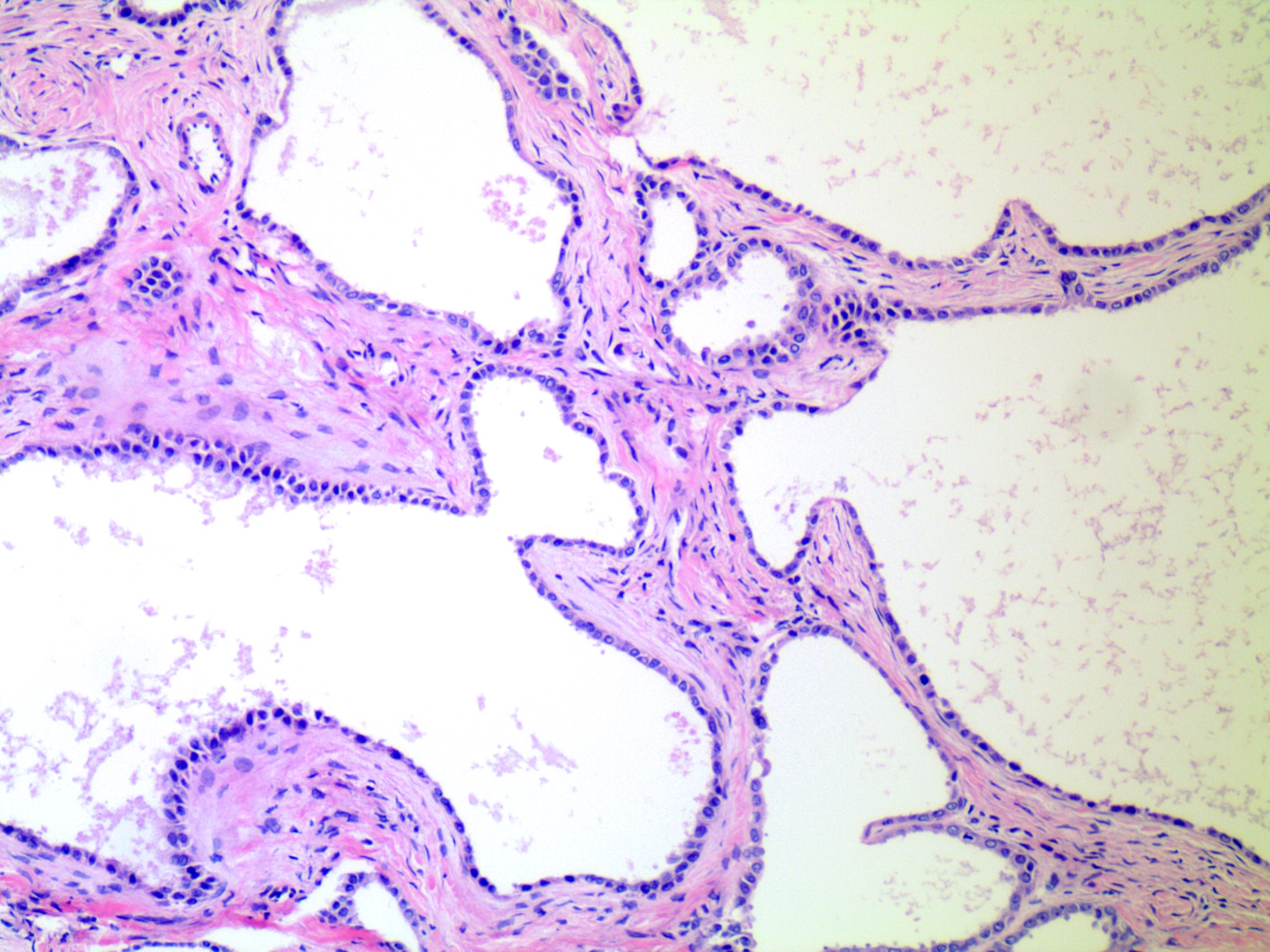
- If < 10% of the total tumor volume shows epithelial proliferation within the cysts that would otherwise qualify as serous borderline tumor, the tumor is designated as serous cystadenoma with focal epithelial proliferation
- Reference: Kurman: Blaustein's Pathology of the Female Genital Tract (Springer Reference), 7th Edition, 2019
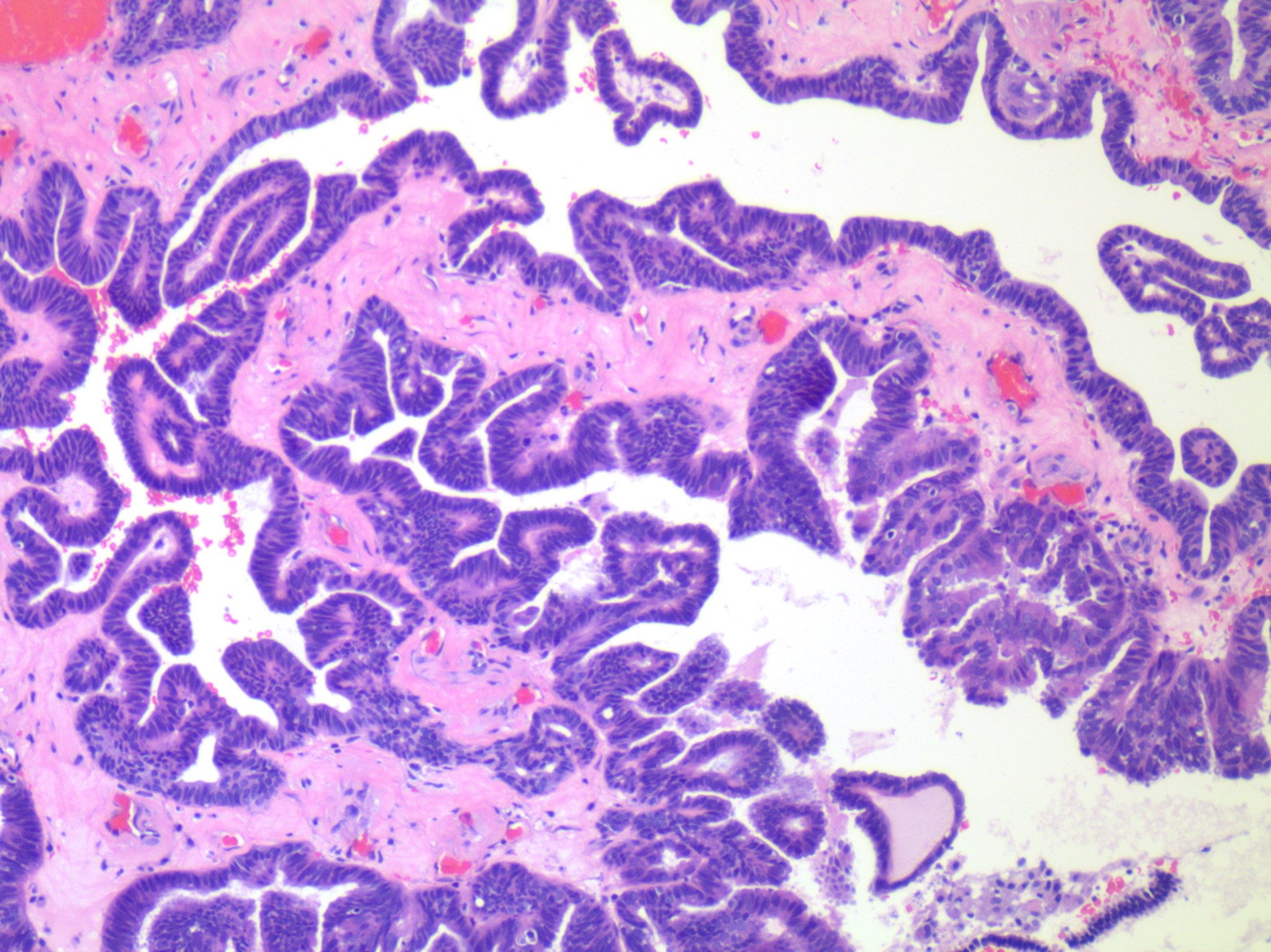
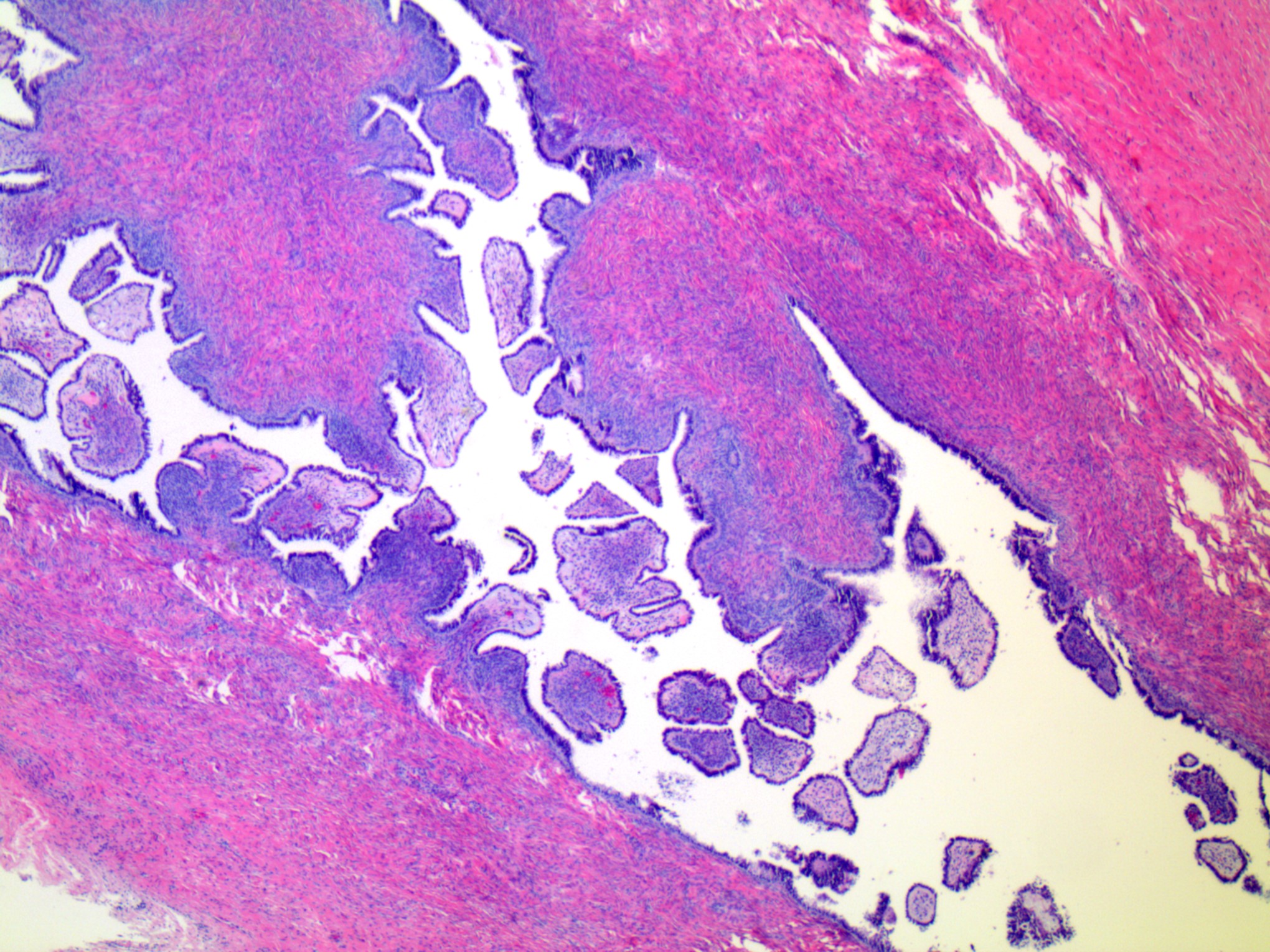
Microscopic (histologic) images
Contributed by Catherine J. Roe, M.D. and Krisztina Hanley, M.D.
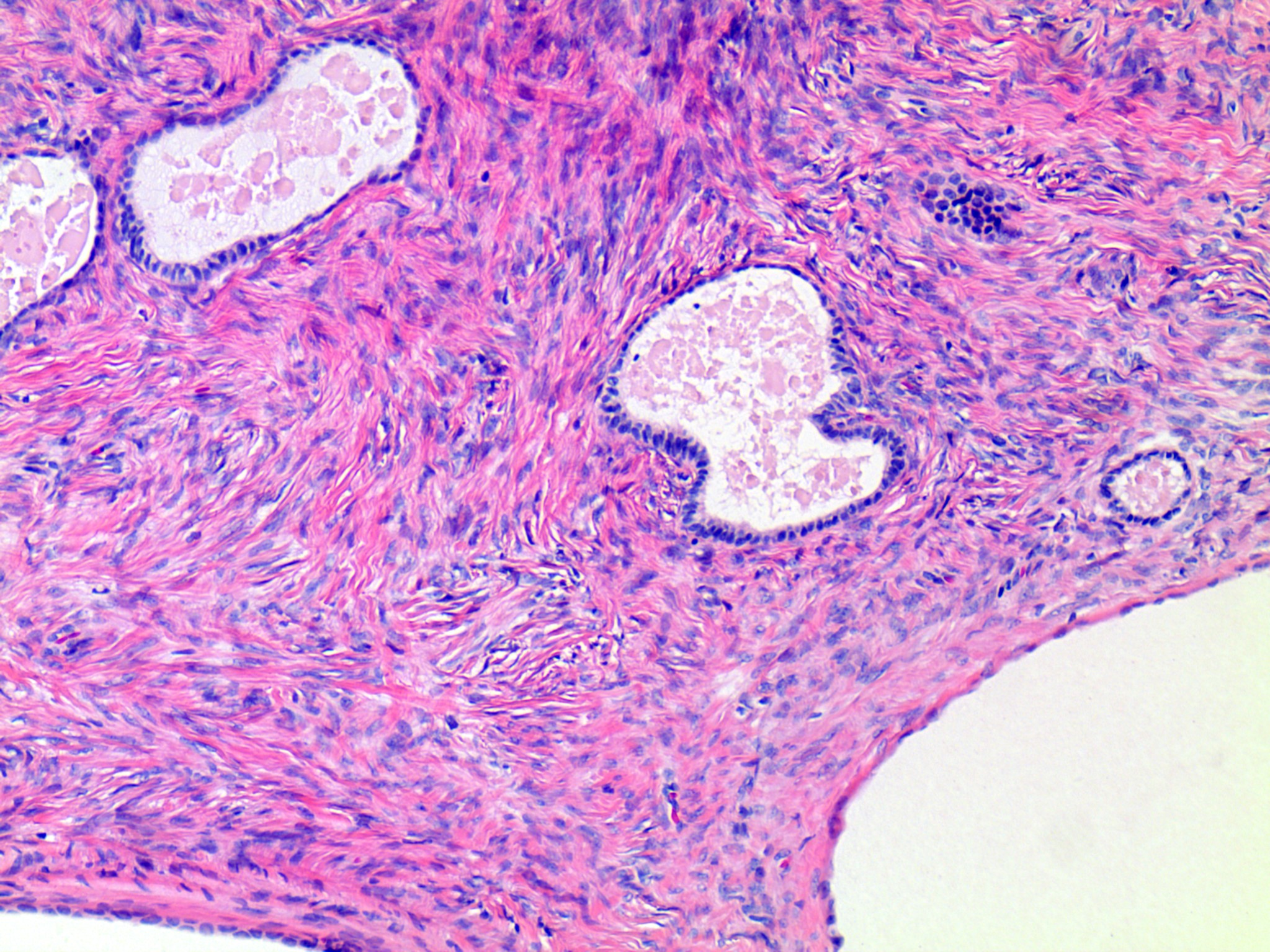

Serous cystadenofibroma
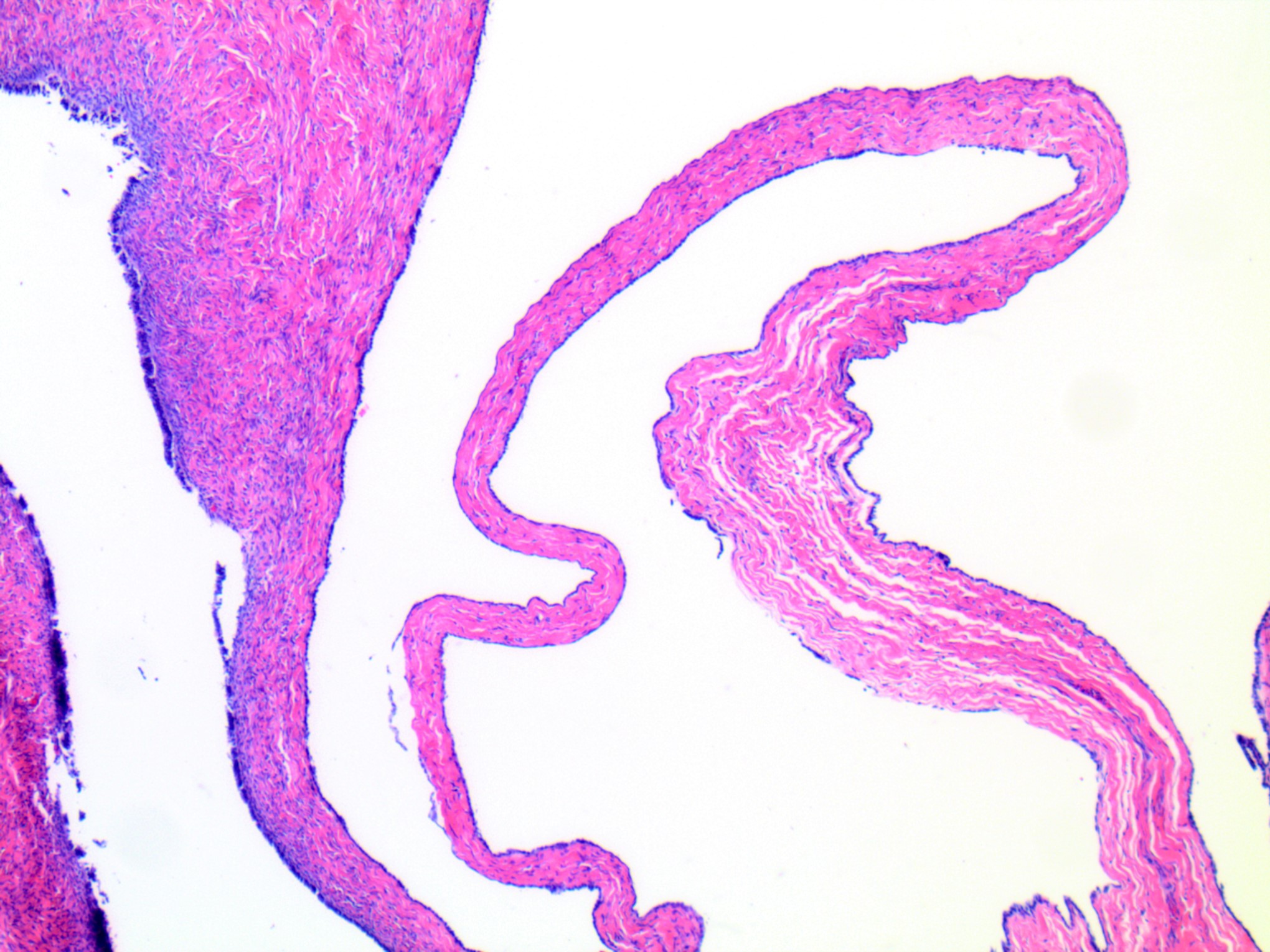
Serous cystadenoma
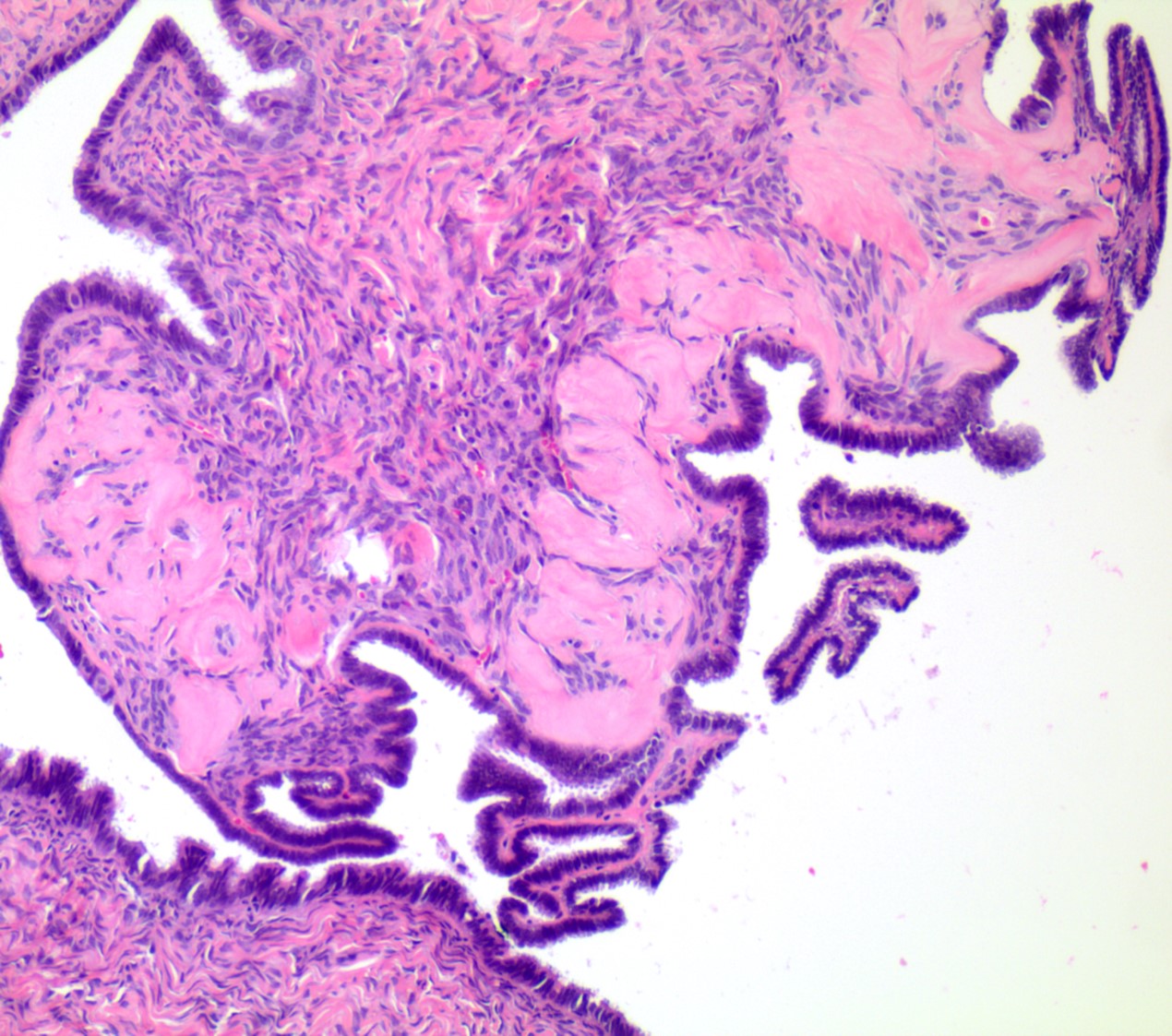
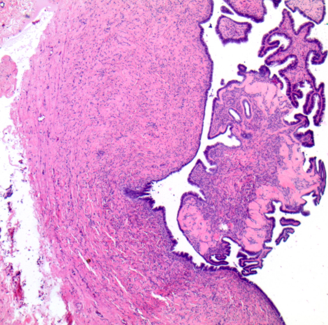
Serous cystadenoma with focal epithelial proliferation
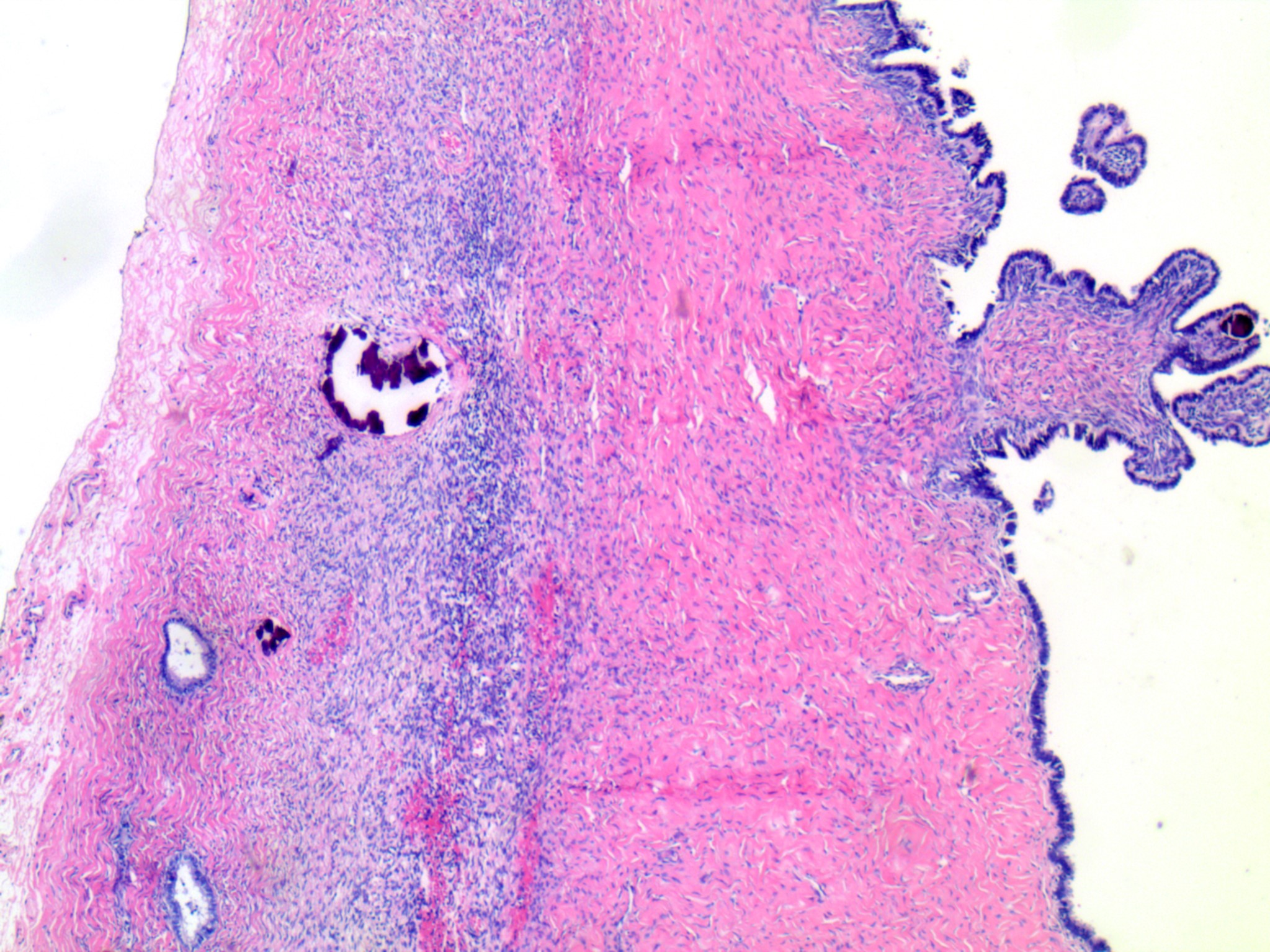
Papillary tufting

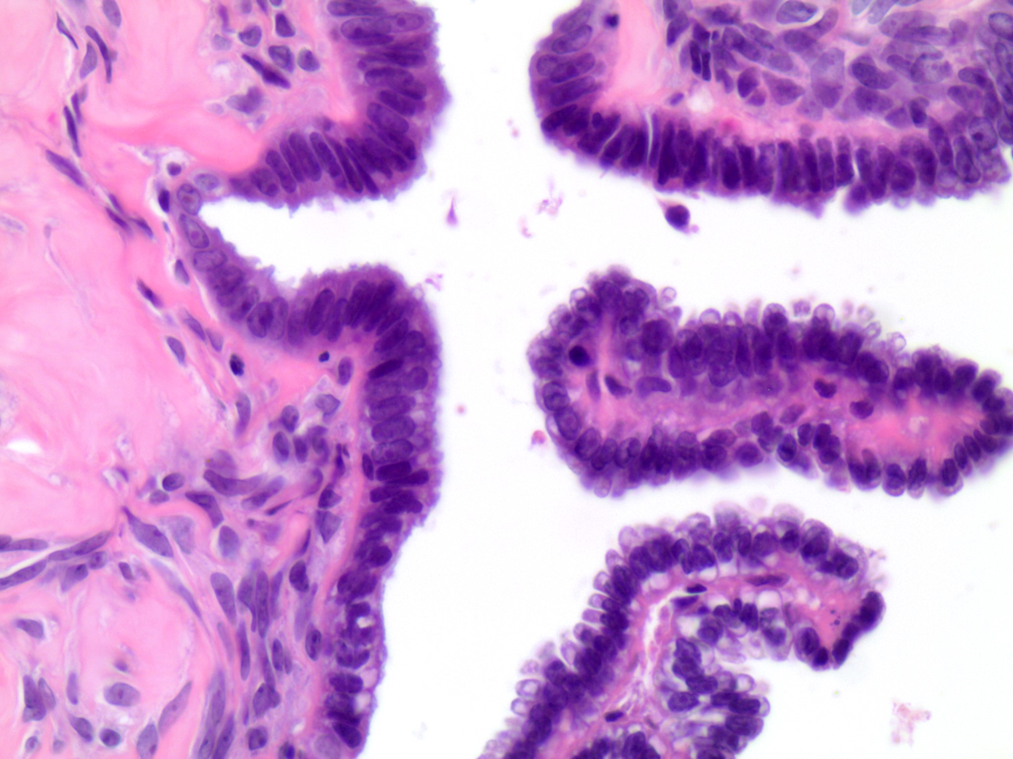
Cytologic features
Focal epithelial proliferation
Cytology description
- Groups, strips or clusters of epithelial cells with small, bland, round to oval nuclei and variable cytoplasm with or without cilia
- Background cyst contents, including histiocytes and proteinaceous debris
- Cannot definitively diagnose on a cytology specimen; histologic examination is required for classification
Cytology images
Images hosted on other servers:

Small epithelial cell cluster

CD68
Positive stains
Sample pathology report
- Right ovary, oophorectomy:
- Serous cystadenofibroma (3.3 cm)
- Left ovary, oophorectomy:
- Serous cystadenoma with focal epithelial proliferation (see comment)
- Comment: The 8.3 cm cystic ovarian mass was extensively sampled. Focal epithelial proliferation (small, noncomplex papillae) is noted, which represents less than 10% of sampled cyst wall. These finding represent a benign serous cystadenoma with focal epithelial proliferation.
Differential diagnosis
- Rete cyst / cystadenoma:
- Located in ovarian hilus, undulating epithelium, smooth muscle wall, cyst lining is a single layer of flat cuboidal cells
- Nests of Leydig cells may be present in the wall
- Paratubal Müllerian cyst (hydatid cyst of Morgagni):
- Paramesonephric cyst attached to fimbria, thin fallopian tube type epithelium with small epithelial plicae projecting into the lumen, may have smooth muscle in the wall
- Peritoneal cyst:
- Lined by mesothelial cells, often associated with ovarian surface adhesions
- Mesonephric cyst:
- Lined by cuboidal cells and usually surrounded by smooth muscle
- Mucinous cystadenoma:
- Lined by a single layer of mucin containing tall columnar epithelium
- Hydrosalpinx:
- Dilated fallopian tube lumen, lined by ciliated epithelium, attenuated or rare plicae, well developed smooth muscle in the wall
- Cortical / epithelial inclusion cyst:
- < 1 cm
- Lined by simple cuboidal to columnar epithelium with ciliated cells, sometimes admixed with nonciliated cells
- Serous borderline tumor:
- Epithelial proliferation with architectural complexity, including branching of irregularly shaped papillae
- Seromucinous cystadenoma:
- Lined by epithelium with 2 or more Müllerian cell types each accounting for at least 10% of the epithelium (Histopathology 2021;78:445)
- Frequently associated with endometriosis (Int J Gynecol Pathol 2021 Feb 11 [Epub ahead of print])
Additional references
Practice question #1
This image is a representative section of a 4.3 cm solid and cystic ovarian mass. What is the diagnosis?
- Fibroma
- Hydrosalpinx
- Mucinous cystadenoma
- Paratubal cyst
- Serous cystadenoma
Practice answer #1
Practice question #2
What feature distinguishes cystadenoma from cortical inclusion cyst?
- Epithelial lining
- Location
- Presence of psammoma bodies
- Relationship to ovarian serosa
- Size
Practice answer #2
