

Ovary
Miscellaneous tumors
Solid pseudopapillary tumor
Author: Kamaljeet Singh, M.D.
Editor-in-Chief: Debra L. Zynger, M.D.

Last author update: 14 December 2018
Last staff update: 7 July 2023
Copyright: 2018-2024, PathologyOutlines.com, Inc.
PubMed Search: Solid pseudopapillary neoplasm ovarian
Table of Contents
Definition / general | Essential features | Terminology | ICD coding | Epidemiology | Sites | Clinical features | Diagnosis | Radiology description | Radiology images | Prognostic factors | Case reports | Treatment | Gross description | Microscopic (histologic) description | Microscopic (histologic) images | Positive stains | Negative stains | Electron microscopy description | Molecular / cytogenetics description | Differential diagnosis | Board review style question #1 | Board review style answer #1 | Board review style question #2 | Board review style answer #2Cite this page: Singh K. Solid pseudopapillary tumor. PathologyOutlines.com website. https://www.pathologyoutlines.com/topic/ovarytumorspn.html. Accessed April 19th, 2024.
Definition / general
- Ovarian tumor that is morphologically, immunohistochemically and genetically identical to pancreatic solid pseudopapillary neoplasm
- Categorized as miscellaneous tumor of ovary in WHO classification (Kurman: WHO Classification of Tumours of the Female Reproductive Organs, 4th Edition, 2014)
Essential features
- Rare tumor of reproductive age group; 10 reported cases
- Possible origin from genital ridge related cells, presumed epithelial / neuroendocrine nature
- Solid nests with pseudopapillary architecture, cysts with colloid-like material, papillae with myxoid stroma and tumor cell nuclei oriented away from the papillae
- Abnormal nuclear β catenin immunohistochemical staining and E-cadherin (negative NCH-38 clone)
- Limited cases with followup, presumed to be of low malignant potential, like pancreatic solid pseudopapillary neoplasm
Terminology
- Primary ovarian solid pseudopapillary neoplasm
- Extrapancreatic solid pseudopapillary neoplasm
- Miscellaneous ovarian tumors (Kurman: WHO Classification of Tumours of the Female Reproductive Organs, 4th Edition, 2014)
ICD coding
- ICD-10: C56.9 - Malignant neoplasm of unspecified ovary
Epidemiology
- Rare tumor
- Females of reproductive age, range 17 - 57 years
- 10 reported cases: 6 from U.S., 2 from China, 1 each from India and Japan (Am J Surg Pathol 2010;34:1514, Hum Pathol 2012;43:1339, Ann Diagn Pathol 2012;16:498, Int J Gynecol Pathol 2018;37:110, Int J Clin Exp Pathol 2015;8:8645, Int J Gynecol Pathol 2011;30:539, Indian J Pathol Microbiol 2016;59:348, Pathol Int 2014;64:460)
Sites
- Ovary
Clinical features
- Abdominal mass, fullness, swelling or pain (6 cases)
- Pelvic pain (2 cases)
- Incidental radiologic finding (2 cases)
- Postmenopausal bleeding (1 case)
Diagnosis
- Exclude metastasis of solid pseudopapillary neoplasm from extraovarian site
Radiology description
- Ultrasound: solid and cystic complex adnexal mass with high density in cyst (Int J Clin Exp Pathol 2015;8:8645)
- Multiloculated heterogeneous ovarian cystic mass (Hum Pathol 2012;43:1339)
Radiology images
Images hosted on other servers:

Solid pseudopapillary neoplasm
Prognostic factors
- Unknown due to limited number of cases
- Only patient who died of disease was stage IV
- Tumor showed high mitotic rate (62/50 high powered fields), necrosis and lymphovascular invasion (Ann Diagn Pathol 2012;16:498)
- 6 patients had no evidence of disease at followup, including a stage IIIC case at 18 months followup
Case reports
- 17, 21 and 57 year old women; first reported cases (Am J Surg Pathol 2010;34:1514)
- 22 and 47 year old women with metastasis from pancreas to ovary (Mol Clin Oncol 2016;4:845)
- 47 year old woman with overlap of microcystic stromal tumor and SPN (Int J Clin Exp Pathol 2015;8:11792)
- 48 year old woman with clinically aggressive tumor (Hum Pathol 2012;43:1339)
- 49 year old woman with CTNNB1 point mutation (Int J Gynecol Pathol 2018;37:110)
Treatment
- Surgery is mainstay of treatment
Gross description
- Circumscribed ovarian / adnexal mass with solid and cystic hemorrhagic cut surface
Microscopic (histologic) description
- Well circumscribed tumor
- Tumor cell sheets, nests and bands surrounded by fibrous septa
- Thin delicate capillary sized vessels forming myxohyaline pseudopapillary cores; nuclei located away from the papillary core
- Moderate amount of pale eosinophilic cytoplasm with central / eccentric round to oval nuclei with uniform chromatin
- Inconspicuous mitoses, usually no necrosis, no significant nuclear atypia
- Abundant foamy cytoplasm, paranuclear vacuoles, nuclear grooves and intra / extracellular PAS+ diastase resistant eosinophilic globules can be present
- Microcysts with colloid-like material
- Infarction can be present
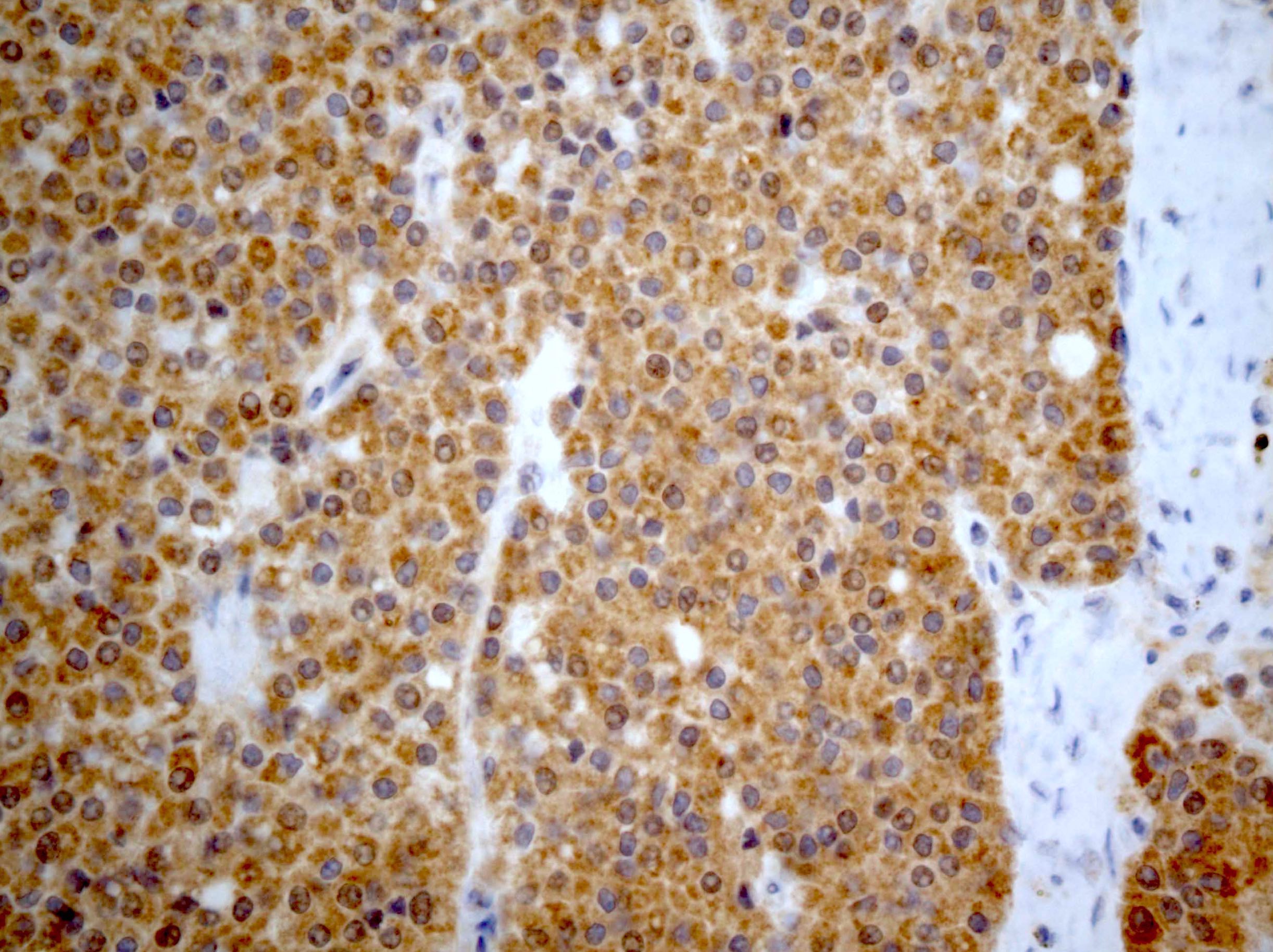
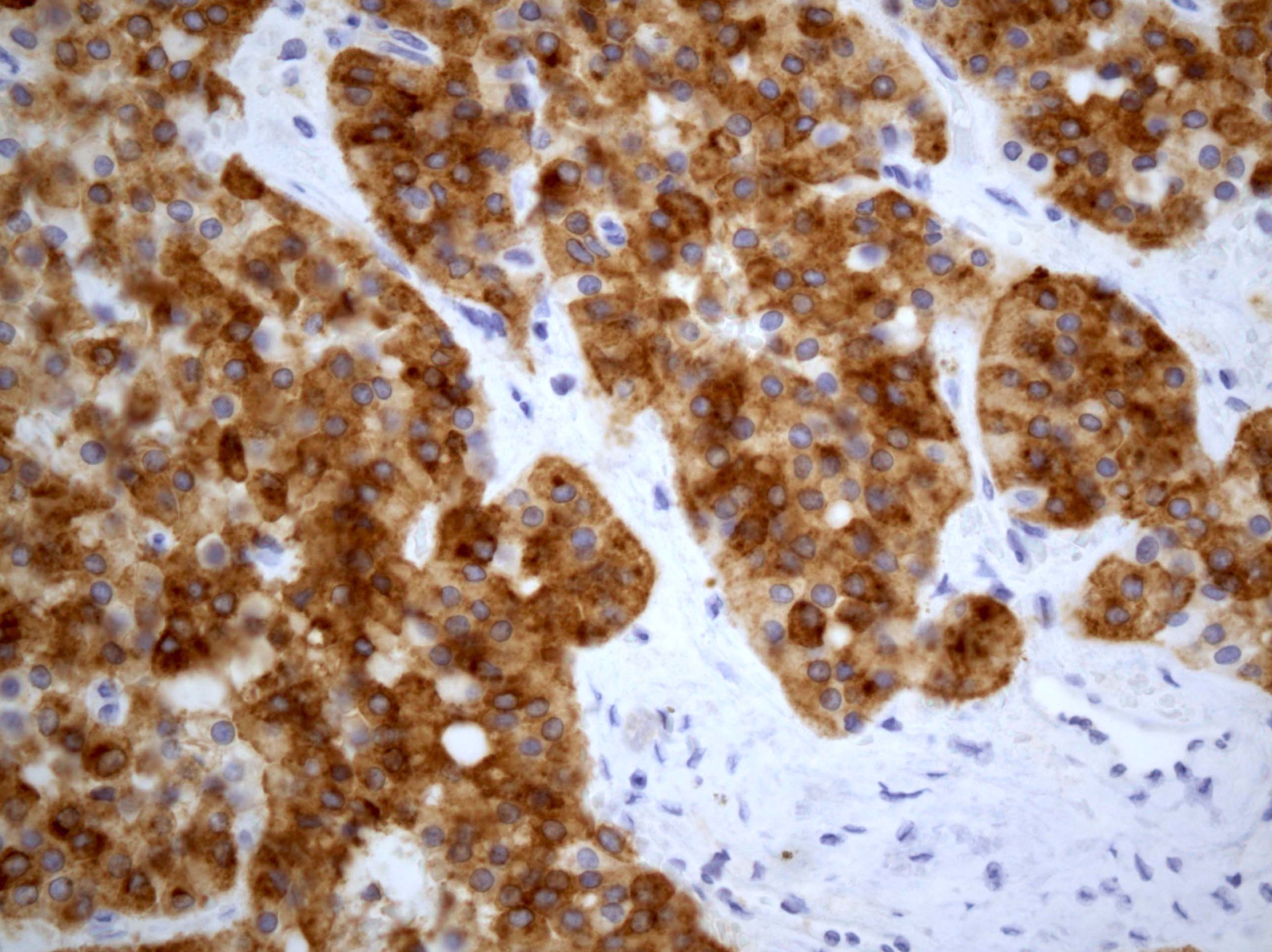
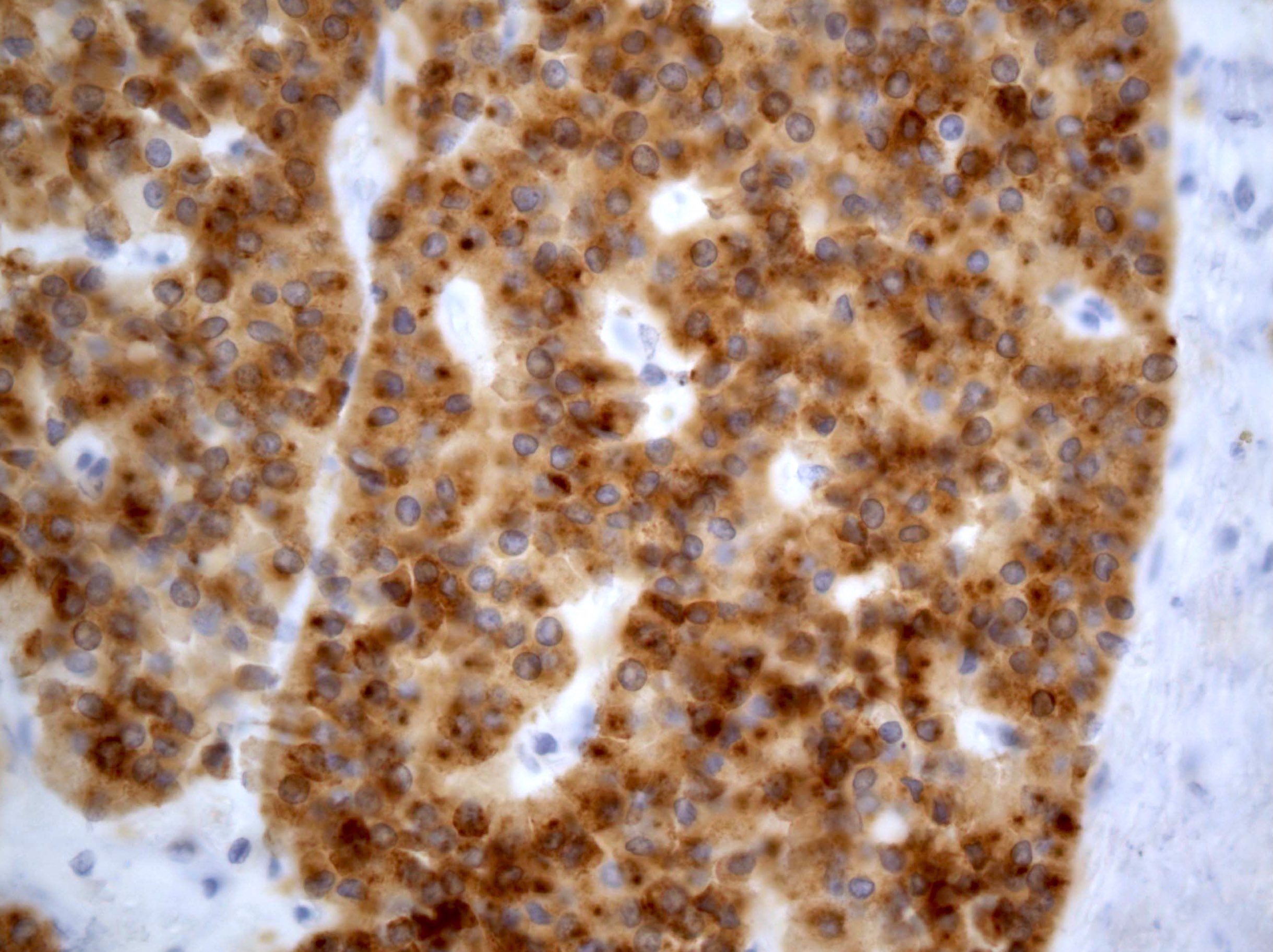
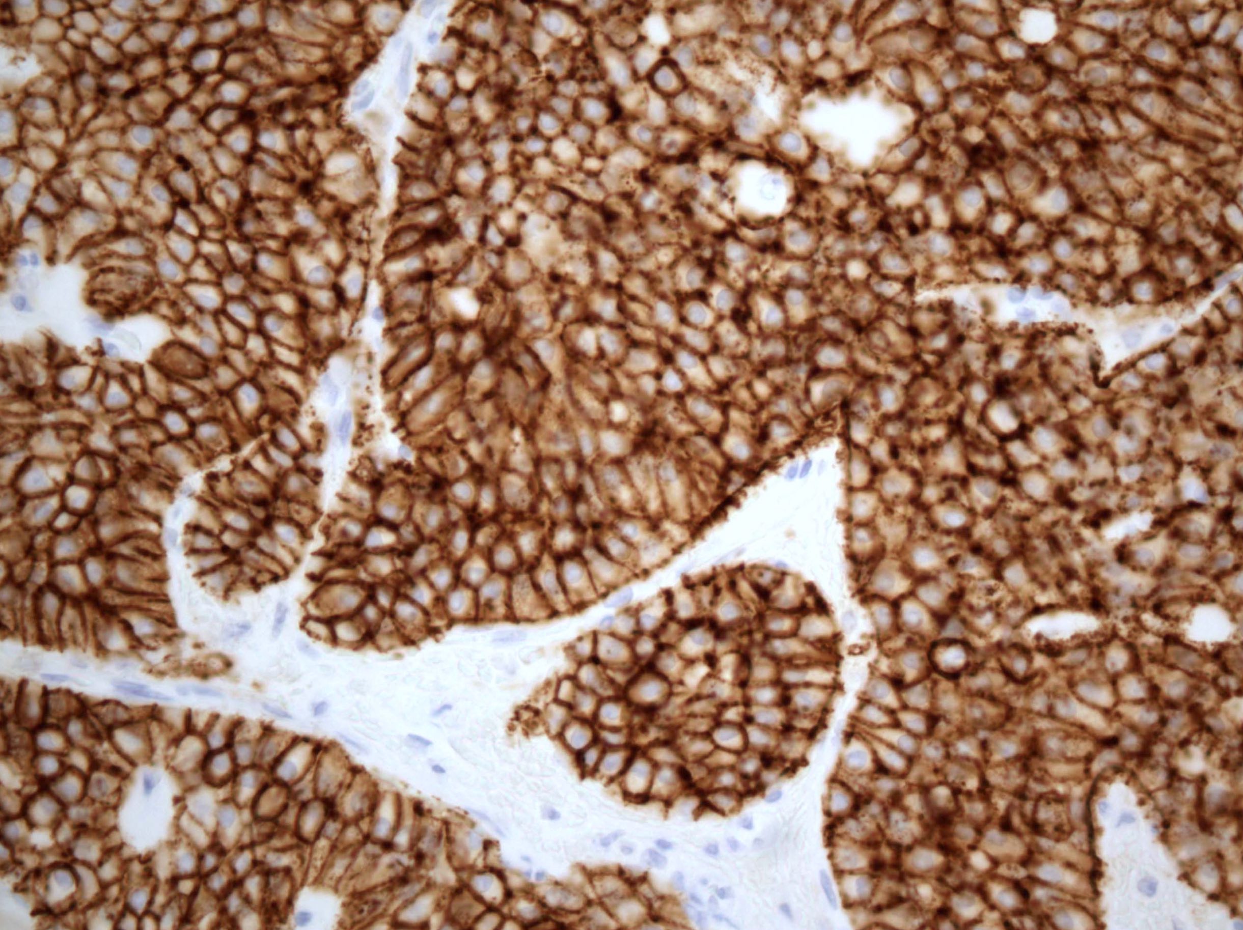
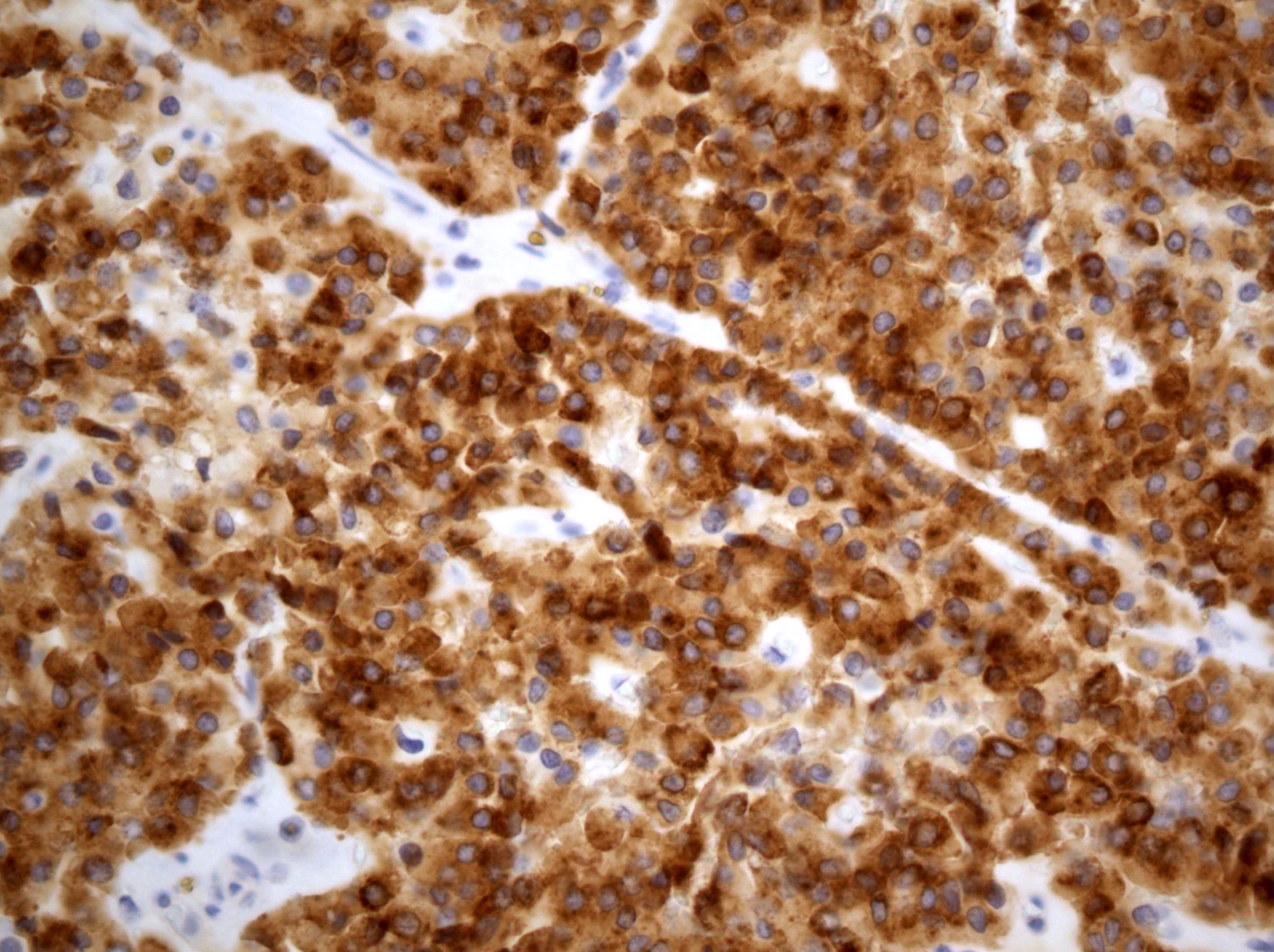
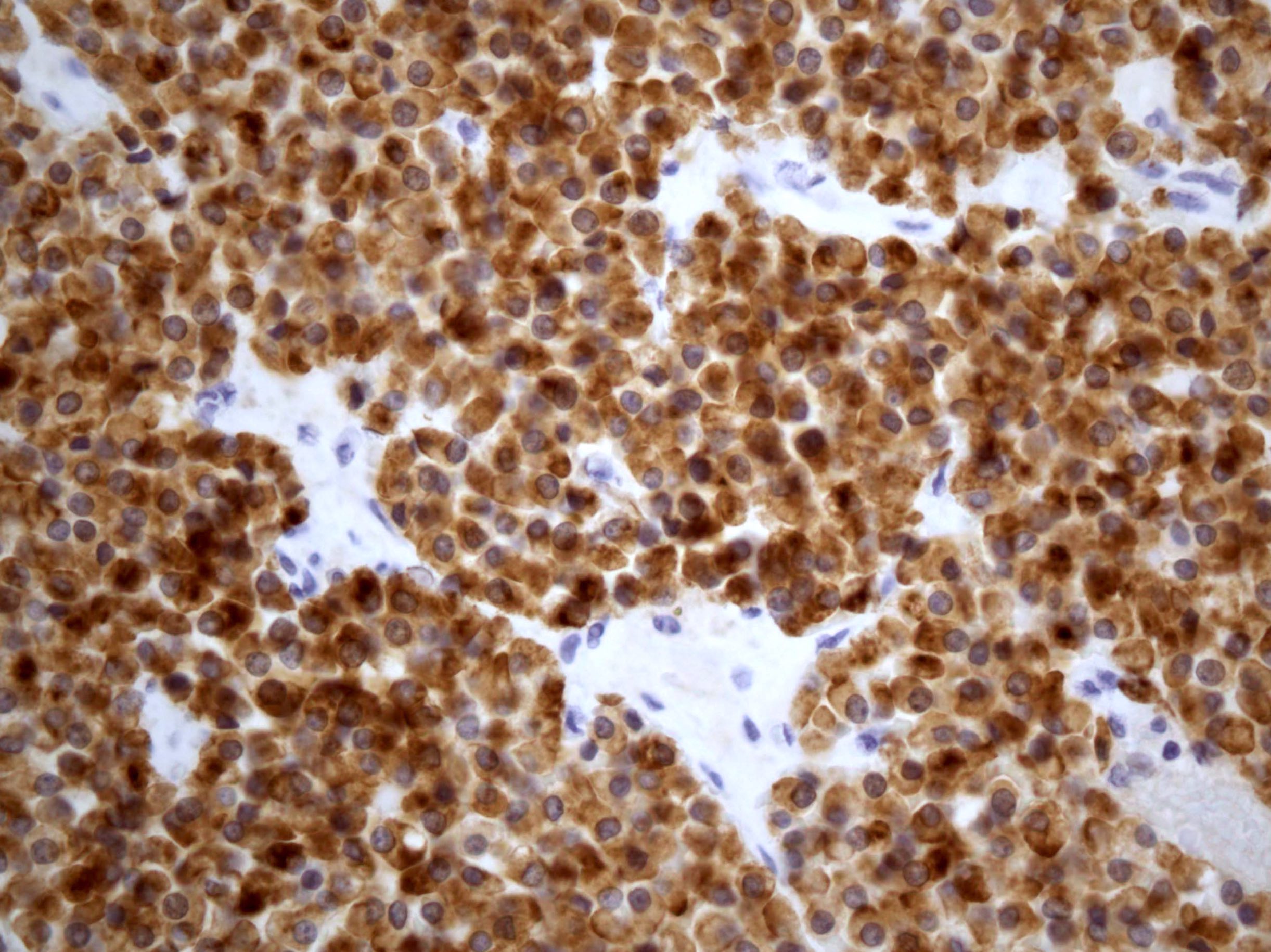
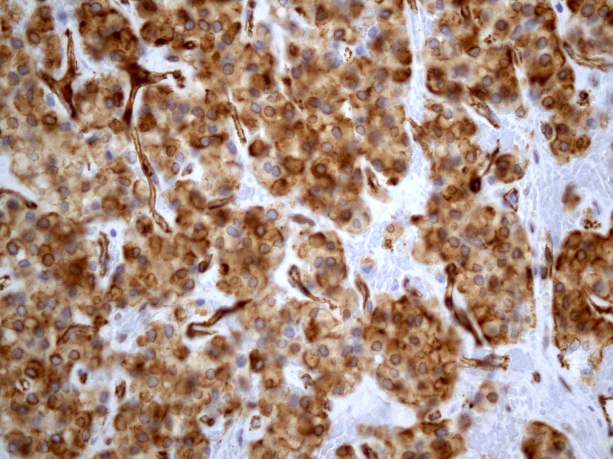
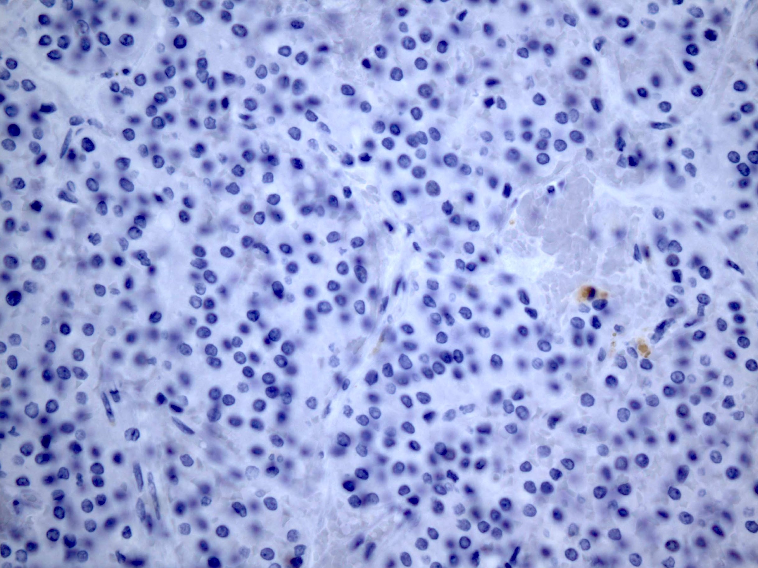
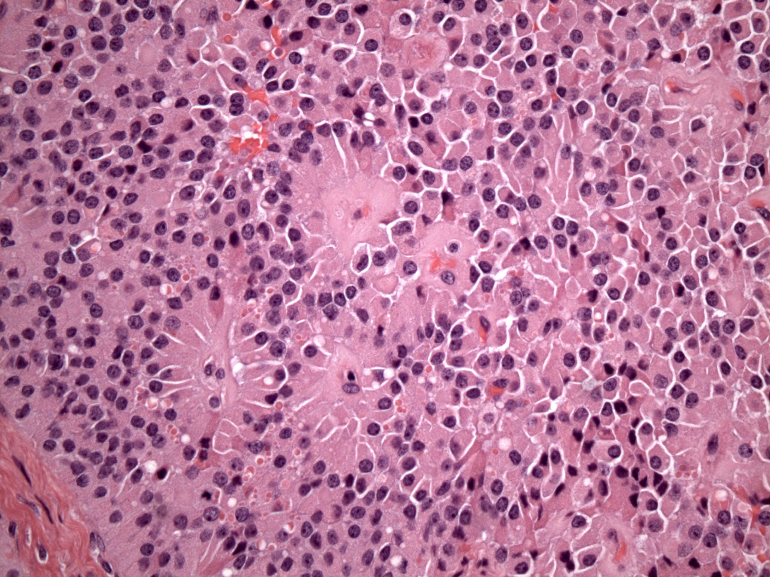
Microscopic (histologic) images
Contributed by Kamaljeet Singh, M.D.
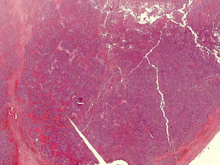
Circumscribed mass
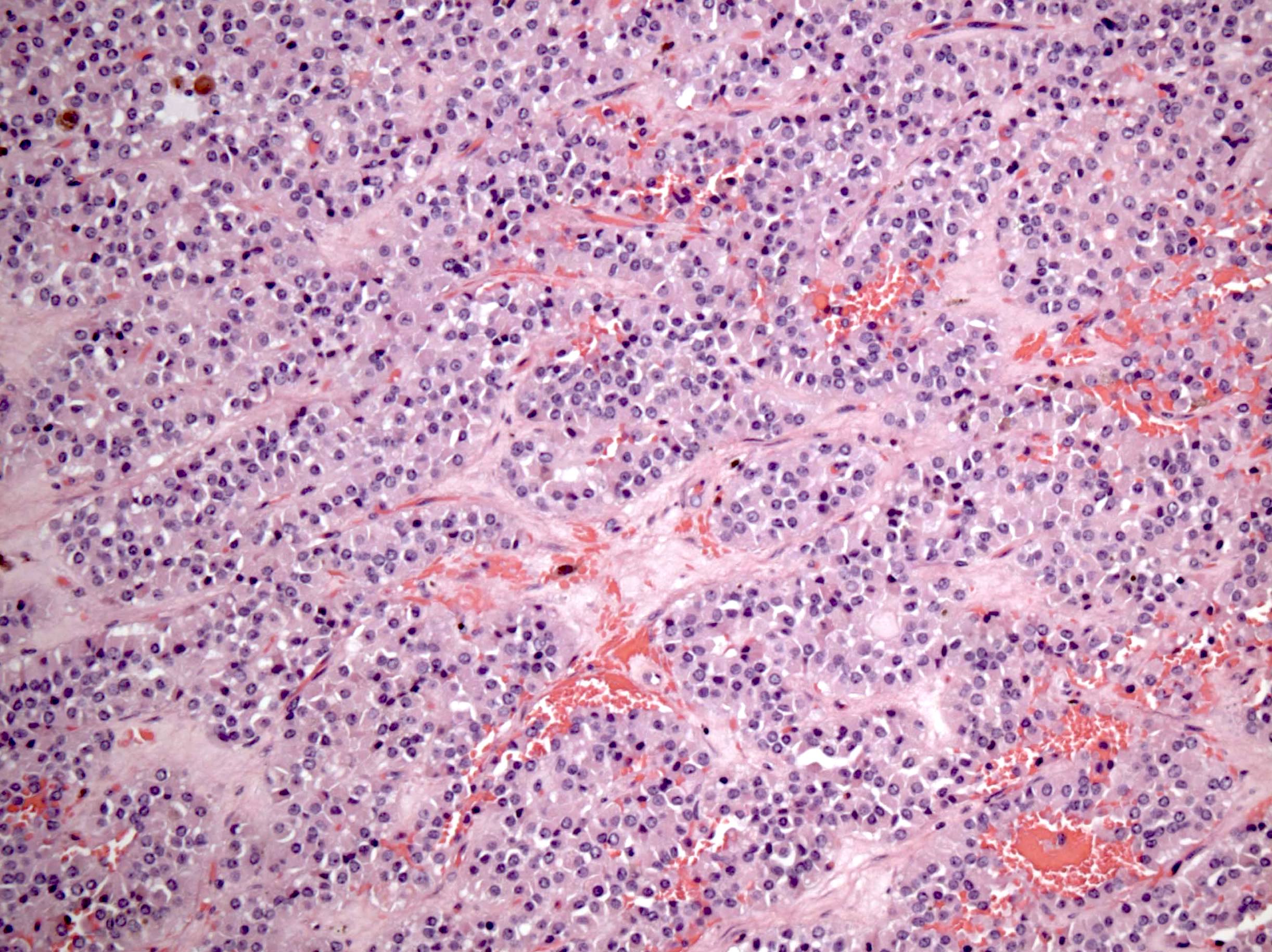
Nests with fibrous septa

Pseudopapillae

Intracytoplasmic vacuoles
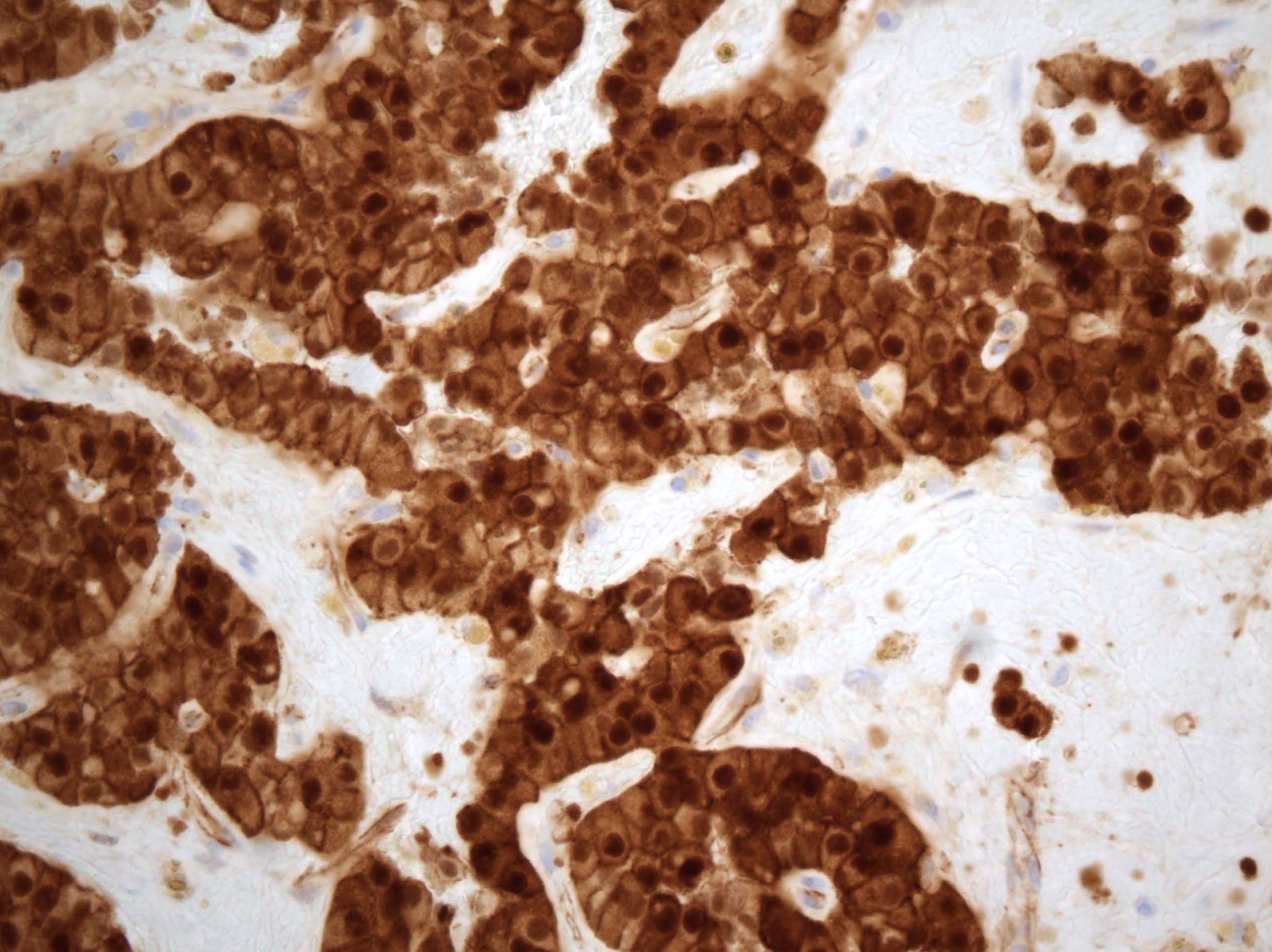
β catenin
c-kit
CAM5.2
CD10
CD99
Synaptophysin
Vimentin
WT1
E-cadherin
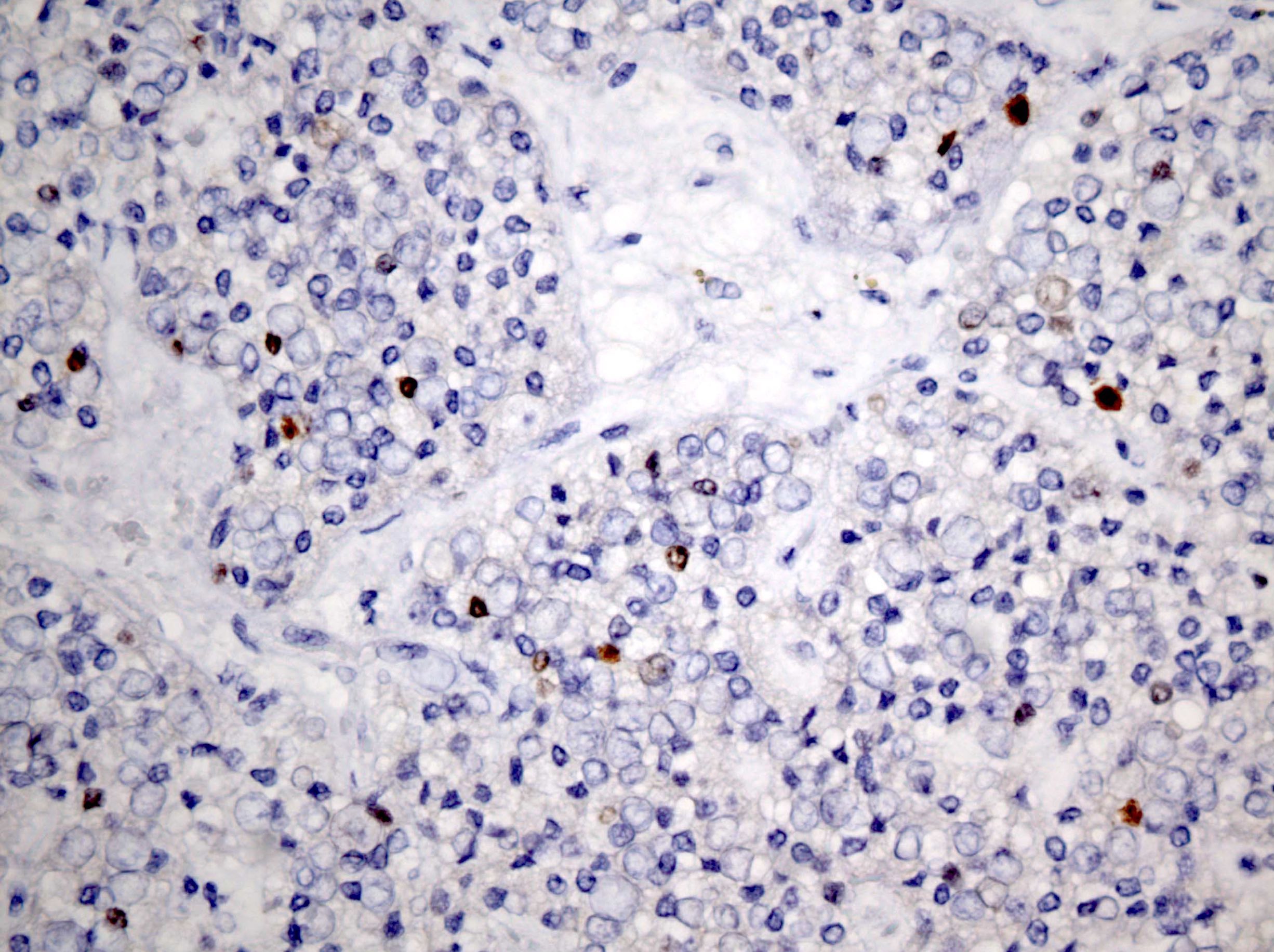
Ki67
Positive stains
Negative stains
- E-cadherin: no staining with NCH-38 clone
- Chromogranin: usually negative
- Calretinin, inhibin, S100, desmin, TTF1, GFAP and HMB45
Electron microscopy description
- Tumor cells with numerous pleomorphic mitochondria interspersed among short strands of rough endoplasmic reticulum (Ann Diagn Pathol 2012;16:498)
Molecular / cytogenetics description
- c.110C > T point mutation in CTNNB1 exon 3 (Pathol Int 2014;64:460)
- c.98C > G point mutation in CTNNB1 exon 3 (Int J Gynecol Pathol 2018;37:110)
Differential diagnosis
- Granulosa cell tumor:
- Cuboidal to polygonal cells with Call-Exner bodies arranged in macrofollicular, trabecular, solid and insular patterns
- Inhibin+ and calretinin+
- Steroid cell tumor:
- Sheets and nests of large vacuolated cells with clear or eosinophilic cytoplasm; minimal to no nuclear atypia
- Inhibin+ and calretinin+
- Metastatic pancreatic solid pseudopapillary neoplasm (Mol Clin Oncol 2016;4:845):
- Similar morphologically
- Clinical correlation and pancreatic / abdominal imaging required
- Metastatic neuroendocrine tumor:
- Cells arranged in nests, sheets and ribbons separated by thin walled vessels
- Salt and pepper chromatin and moderate granular cytoplasm
- Keratin+, chromogranin+ and synaptophysin+
- Struma ovarii:
- Mature thyroid tissue with variable sized follicles lined by cuboidal cells
- TTF1+ and thyroglobulin+
- Paraganglioma / pheochromocytoma:
- Zellballen pattern with supporting vessels and spindle sustentacular cells
- S100+ sustentacular cells, chromogranin+ and synaptophysin+
- Melanoma:
- Gastrointestinal stromal tumor:
- Ependymoma:
Board review style question #1
The most characteristic immunohistochemical staining pattern of solid pseudopapillary neoplasm of the ovary is
- c-kit-
- Consistent keratin expression
- Consistent positive expression of both chromogranin and synaptophysin
- Nuclear β catenin+ and E-cadherin-
- S100+ and HMB45+
Board review style answer #1
Board review style question #2
The image below is taken from an H&E stained section of a 3.0 cm circumscribed hemorrhagic left ovarian lesion of a 48 year old woman. Plasma inhibin levels were normal. Tumor cells expressed CAM5.2, vimentin and synaptophysin and were negative for S100, inhibin, FOXL2, E-cadherin and chromogranin. Genetic analysis showed c.98C > G point mutation in CTNNB1 exon 3. The diagnosis is
- Ependymoma
- Granulosa cell tumor
- Microcystic stromal tumor
- Solid pseudopapillary neoplasm
- Strumal carcinoid
Board review style answer #2
