
Pancreas
Exocrine carcinomas
Adenosquamous carcinoma
Authors: Orhun Çiğ Taşkın, M.D., Volkan Adsay, M.D.
Editorial Board Members: Raul S. Gonzalez, M.D., Wei Chen, M.D., Ph.D.
Deputy Editor-in-Chief: Debra L. Zynger, M.D.


Last author update: 3 May 2023
Last staff update: 3 May 2023
Copyright: 2002-2024, PathologyOutlines.com, Inc.
PubMed Search: Adenosquamous carcinoma pancreas
Table of Contents
Definition / general | Essential features | ICD coding | Epidemiology | Sites | Etiology | Diagnosis | Radiology description | Radiology images | Prognostic factors | Case reports | Treatment | Gross description | Microscopic (histologic) description | Microscopic (histologic) images | Cytology description | Cytology images | Positive stains | Negative stains | Molecular / cytogenetics description | Sample pathology report | Differential diagnosis | Board review style question #1 | Board review style answer #1 | Board review style question #2 | Board review style answer #2Cite this page: Taşkın OÇ, Adsay NV. Adenosquamous carcinoma. PathologyOutlines.com website. https://www.pathologyoutlines.com/topic/pancreasadenosquamous.html. Accessed April 25th, 2024.
Definition / general
- Malignant tumor that consists of both squamous cell carcinoma and ductal adenocarcinoma components (Digestion 2005;72:104, Cancer 2003;99:372, Int J Pancreatol 1999;26:85, Arch Surg 1999;134:599)
Essential features
- Malignant tumor consisting of both squamous cell carcinoma and ductal adenocarcinoma components (Digestion 2005;72:104, Cancer 2003;99:372, Int J Pancreatol 1999;26:85, Arch Surg 1999;134:599)
- Closely related to and admixed with pancreatic ductal adenocarcinoma (PDAC); some require 30% squamous differentiation for the diagnosis of adenosquamous carcinoma
- Cases with < 30% of squamous component also behave like adenosquamous carcinoma and can be acknowledged as PDAC with focal (< 30%) squamous differentiation
- Metastatic tumor can be solely composed of the glandular component (Mod Pathol 2001;14:443)
- Often has necrotic and cystic components (Arch Surg 1999;134:599)
- Behaves even worse than ordinary PDAC
ICD coding
- ICD-O: 8560/3 - adenosquamous carcinoma
Epidemiology
- Accounts for about ~2% of exocrine pancreatic malignancies
- M:F = 2:1
- Mean age of 65 years (Arch Surg 1999;134:599)
Sites
- Body and tail of the pancreas is more often affected than the head (J Surg Res 2012;174:12)
Etiology
- Ionizing radiation of the pancreas may predispose to the occurrence of adenosquamous carcinoma (Mod Pathol 2001;14:443)
Diagnosis
- Generally discovered with radiology and confirmed on fine needle aspiration biopsy (FNAB) or resection
Radiology description
- Similar to pancreatic ductal adenocarcinoma but many cases present with more round, lobulated lesion with extensive central necrosis
- Portal venous system may contain tumor thrombus (Abdom Radiol (NY) 2016;41:508)
Radiology images
Contributed by Orhun Çığ Taşkın, M.D.

Cystic and necrotic changes
Prognostic factors
- Similar to PDAC, stage is the most important prognostic factor
Case reports
- 63 year old man and 69 year old woman with adenosquamous carcinoma of the pancreas (Annu Rev Pathol 2020;15:97)
- 66 year old woman with adenosquamous carcinoma of the pancreas (Clin Case Rep 2022;10:e6181)
- 80 year old Japanese woman with adenosquamous carcinoma coexisting with intraductal papillary mucinous neoplasm of the pancreas (J Med Case Rep 2023;17:72)
Treatment
- Similar to pancreatic ductal adenocarcinoma
- If the tumor is operable, complete surgical resection, followed by adjuvant chemotherapy; increasingly neoadjuvant therapy is utilized just as in PDAC (Hum Pathol 2010;41:113, J Gastrointest Oncol 2015;6:115)
- Even more aggressive than ordinary PDAC with a median survival of < 1 year (Gastrointest Cancer Res 2013;6:75)
- Based on the immunohistochemical results, 11% of patients are assumed to be potential candidates for therapy with antibodies against PD-1 / PDL1 (Pancreatology 2021;21:920)
Gross description
- Large, firm mass of the pancreas, with ill defined borders, often with necrotic component, with or without cystic areas
- Some cases are more demarcated
- Reference: World J Gastrointest Oncol 2015;7:132
Microscopic (histologic) description
- Adenocarcinoma component can bare all the characteristics of classic pancreatic ductal adenocarcinoma
- Squamous component can show keratinization with intercellular bridges or sheets of squamous cells with keratohyaline granules or pearls
- Some are adenoacanthoma-like, with bland mature squamous elements, whereas others are more basal-like (with high N:C ratio and basophilic appearance)
- Adenocarcinoma and squamous components can vary greatly in the amount and distribution
- Sarcomatous component can also be encountered (Anticancer Res 2019;39:4575)
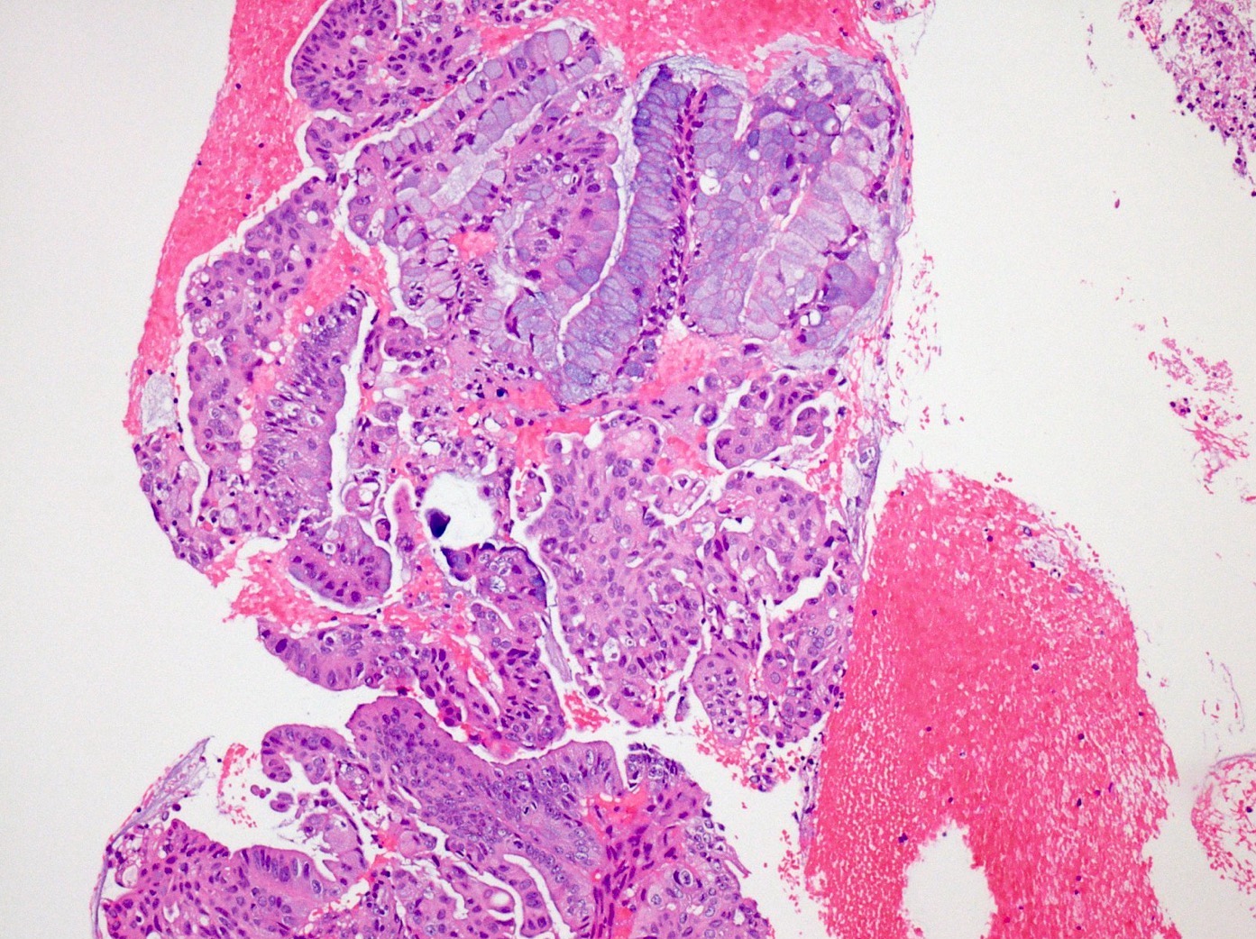
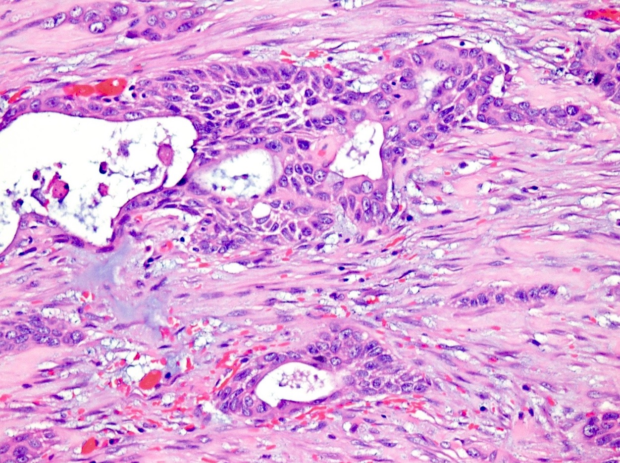
Microscopic (histologic) images
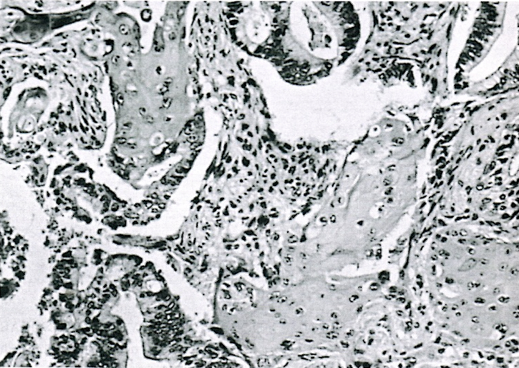
Contributed by Orhun Çığ Taşkın, M.D., Raul S. Gonzalez, M.D. and AFIP

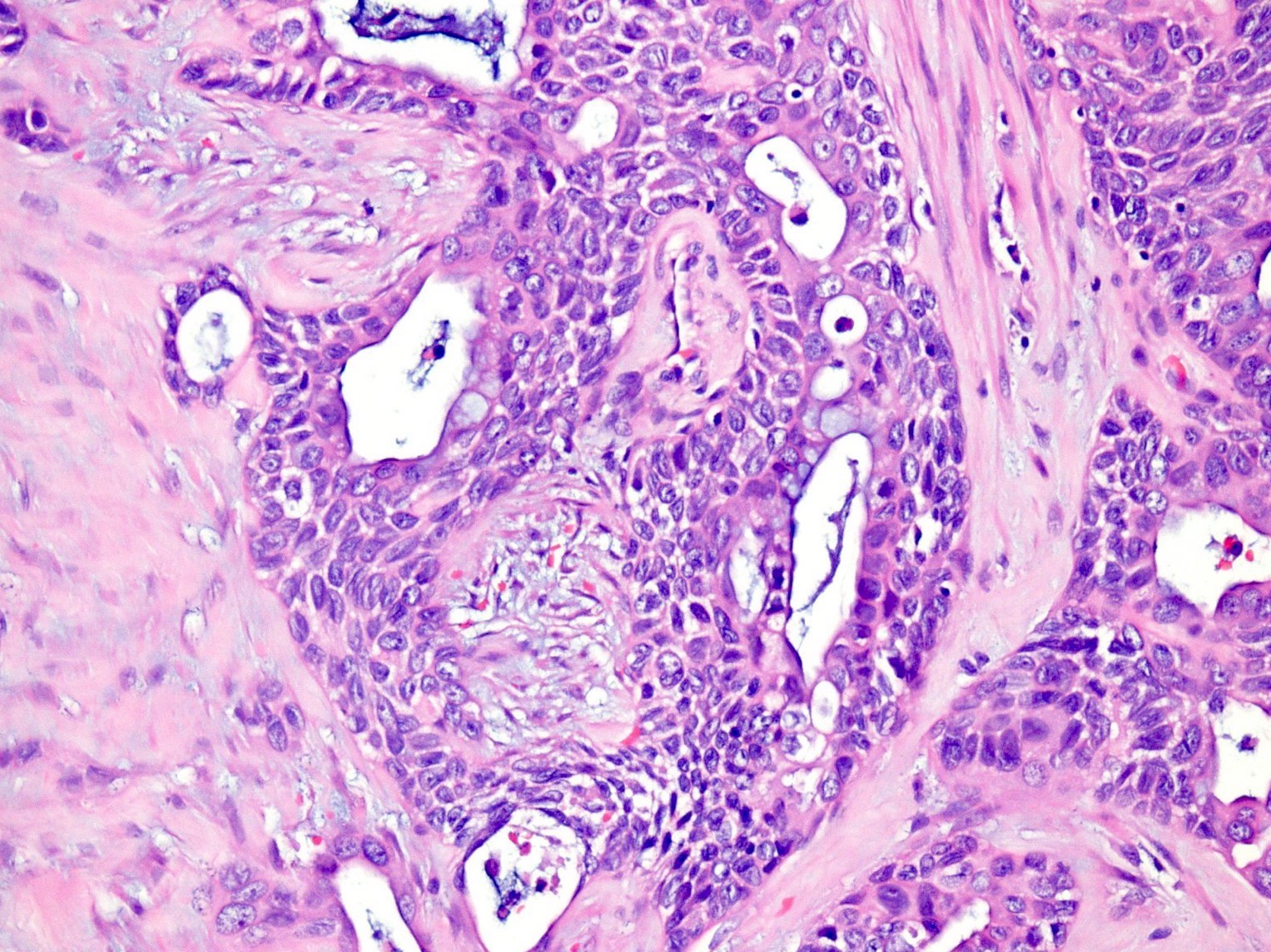
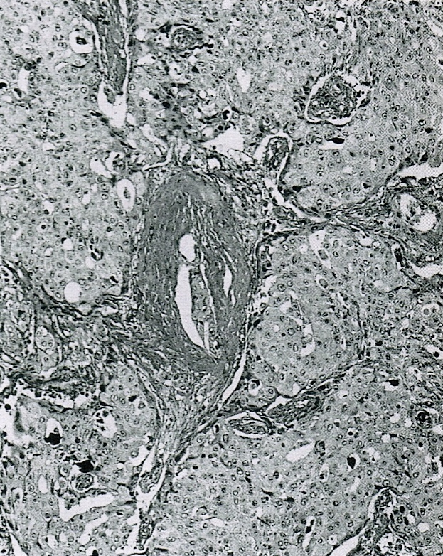
Glandular and squamous components
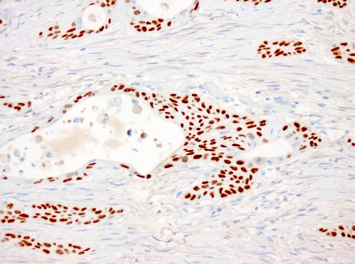
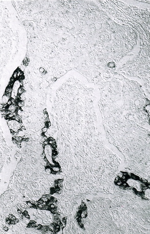
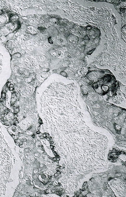
p63
Adenocarcinoma and squamous carcinoma
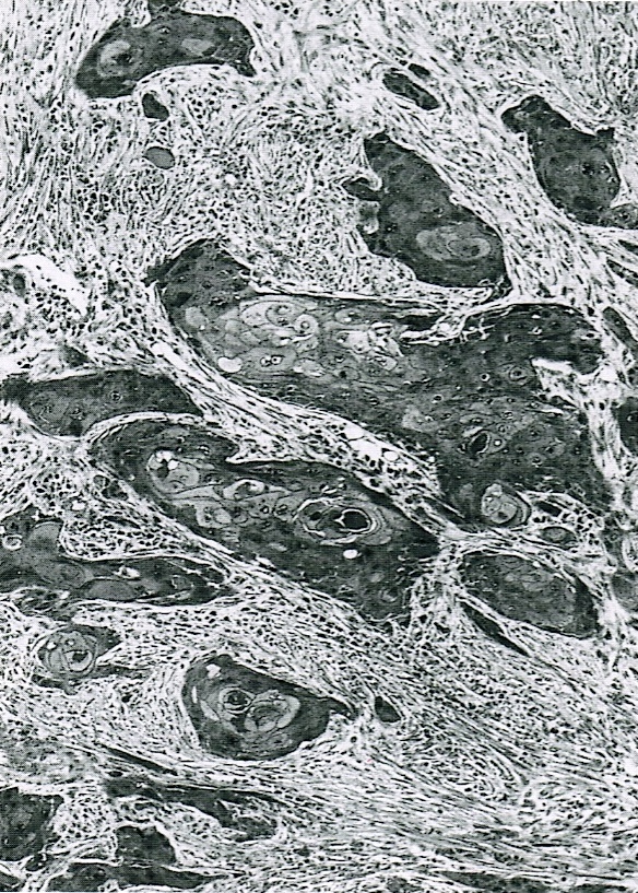
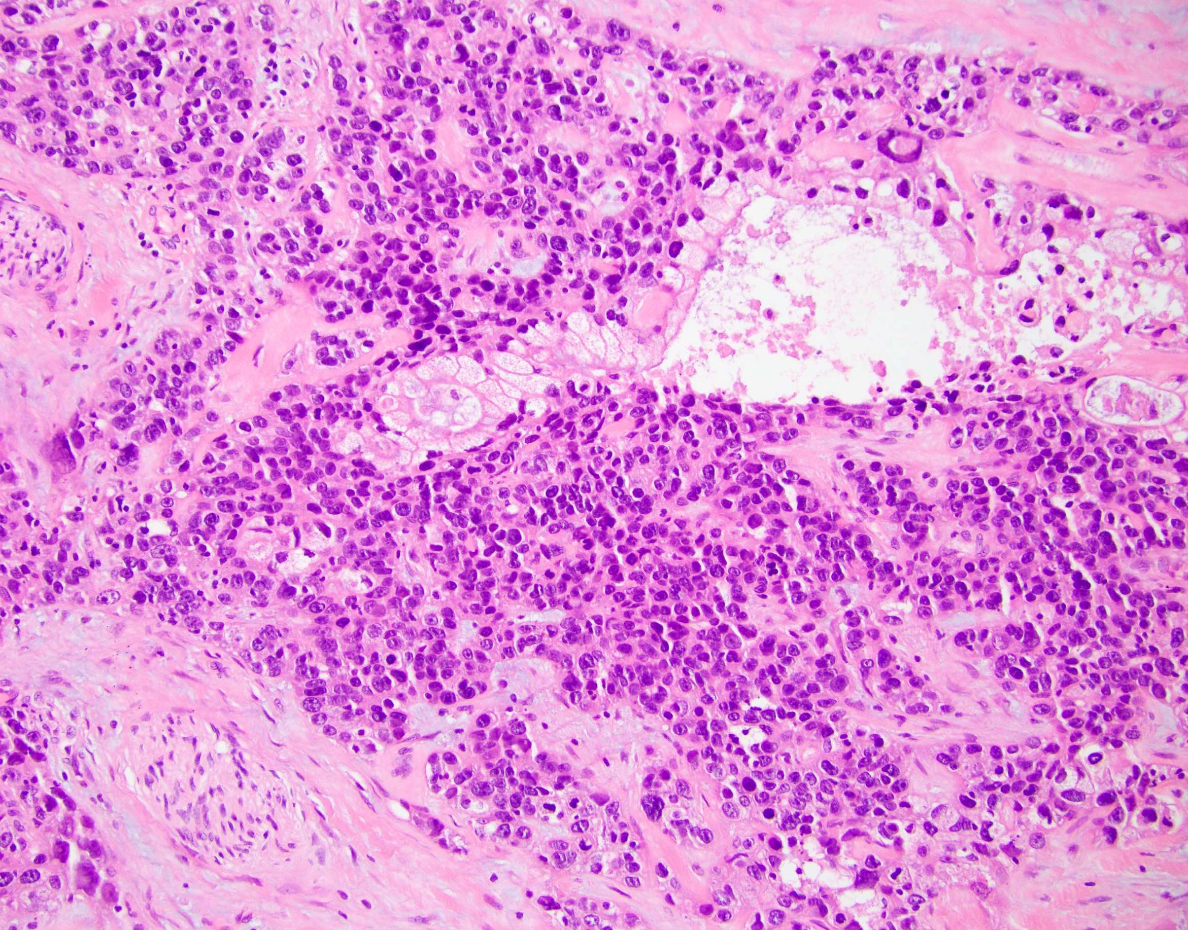
Squamous components
Mucoepidermoid pattern
CAM5.2
CK13
Cytology description
- Squamous component may be undersampled but malignant squamous cells are highly significant for the diagnosis (Acta Cytol 2013;57:139)
- Glandular and squamous components can both be distinguished
- Dense globules, silhouettes of squamous cells, anucleate squames, atypical cytoplasm and enlarged pyknotic nuclei are present in the prominent necrotic background
- Sheets and clusters of atypical cells with nuclei of variable sizes and shapes (Cancer 2003;99:372)
Cytology images
Contributed by Orhun Çığ Taşkın, M.D.
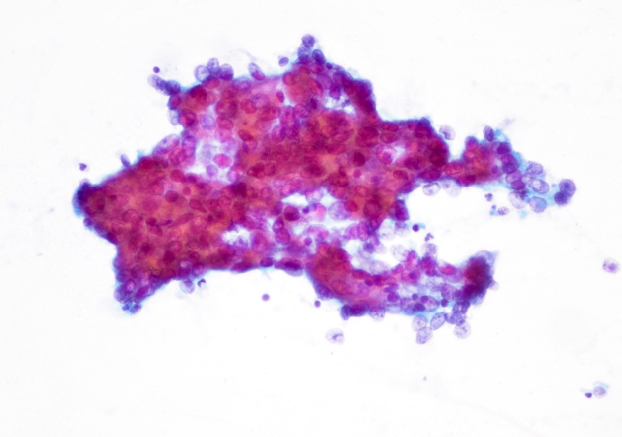
Pap stain
Cell block

Cell block, p40
Positive stains
- Immunoreactivity for AE1 / AE3 is identified
- p63, p40, high molecular weight cytokeratin specifically highlights the squamous component (Mod Pathol 2005;18:1193, Mod Pathol 2009;22:651)
Negative stains
- Loss of SMAD4 / DPC4 protein expression
Molecular / cytogenetics description
- KRAS mutations occur in almost all cases (Mod Pathol 2001;14:443, Mod Pathol 2009;22:651)
- TP53 mutations and 3p loss are commonly encountered
- Recently discovered basal-like molecular subtype of PDAC is believed to also represent adenosquamous carcinoma (Nature 2016;531:47)
- Both components are believed to originate from the same progenitor cell (J Pathol 2017;243:155)
- Susceptibility genes including MAP3K1, PDE4DIP and BCR are frequently mutated in the germlines of patients (Cancer Biomark 2020;27:389)
Sample pathology report
- Pancreas, resection:
- Adenosquamous carcinoma (see synoptic report)
Differential diagnosis
- Pure squamous cell carcinoma:
- Extremely rare; most cases contain a small amount of glandular component, which is enough to call the tumor adenosquamous carcinoma
- Metastasis of adenosquamous carcinoma of the lung:
- Needs to be carefully excluded (Virchows Arch 2004;444:527)
Board review style question #1
A 65 year old man presented with back pain and severe weight loss. Radiologic images showed a 4 cm relatively round, lobulated mass with extensive central necrosis in the pancreatic body. He underwent resection. Histologic sections demonstrate conventional ductal adenocarcinoma admixed with sheets of squamous cells with keratohyaline granules and intercellular bridges. Which of the following immunohistochemical stains would be positive in this case?
- Beta catenin nuclear expression
- Chromogranin A
- p63
- Synaptophysin
Board review style answer #1
C. p63. The histology described in this case is consistent with pancreatic adenosquamous carcinoma. The squamous components should be positive with p63 immunohistochemistry. Neuroendocrine neoplasms are positive for chromogranin and synaptophysin. Beta catenin nuclear expression is observed in solid pseudopapillary neoplasm.
Comment Here
Reference: Adenosquamous carcinoma
Comment Here
Reference: Adenosquamous carcinoma
Board review style question #2
Which of the following is true for adenosquamous carcinoma of the pancreas?
- It has a better prognosis than ordinary pancreatic ductal adenocarcinoma
- It is the most common type of pancreatic malignancy
- It usually affects pediatric patients
- p63 specifically highlights the squamous component
Board review style answer #2
D. p63 specifically highlights the squamous component. The squamous components of the tumor should be positive with p63 immunohistochemistry. Answer A is incorrect as adenosquamous carcinomas behave worse than pancreatic ductal adenocarcinomas. Answer B is incorrect as pancreatic ductal adenocarcinoma is the most common pancreatic malignancy. Answer C is incorrect as adenosquamous carcinomas occur in elder patients (mean age of 65 years).
Comment Here
Reference: Adenosquamous carcinoma
Comment Here
Reference: Adenosquamous carcinoma
