

Pleura & peritoneum
Peritoneum
Mesothelioma (peritoneum)-epithelioid
Authors: Aysha Mubeen, M.D., Raul S. Gonzalez, M.D.
Deputy Editor-in-Chief: Debra L. Zynger, M.D.


Last author update: 12 January 2021
Last staff update: 8 September 2022
Copyright: 2003-2024, PathologyOutlines.com, Inc.
PubMed Search: Peritoneum epithelioid mesothelioma


Table of Contents
Definition / general | Essential features | Epidemiology | Pathophysiology | Clinical features | Radiology images | Prognostic factors | Case reports | Treatment | Clinical images | Gross description | Microscopic (histologic) description | Microscopic (histologic) images | Virtual slides | Cytology description | Cytology images | Positive stains | Negative stains | Electron microscopy description | Sample pathology report | Differential diagnosis | Additional references | Board review style question #1 | Board review style answer #1Cite this page: Mubeen A, Gonzalez RS. Mesothelioma (peritoneum)-epithelioid. PathologyOutlines.com website. https://www.pathologyoutlines.com/topic/pleuraperitmesothelioma.html. Accessed April 17th, 2024.
Definition / general
- Mesothelioma is a neoplasm arising from mesothelial cells that line serous cavities, such as the pleura and peritoneum
- Pleural mesothelioma is much more common than peritoneal mesothelioma
- Epithelioid mesothelioma is the most frequent histologic type of malignant mesothelioma; sarcomatoid and biphasic subtypes are less common
- 20 - 33% of malignant mesothelioma arises in the peritoneum (Semin Oncol 2002;29:51)
Essential features
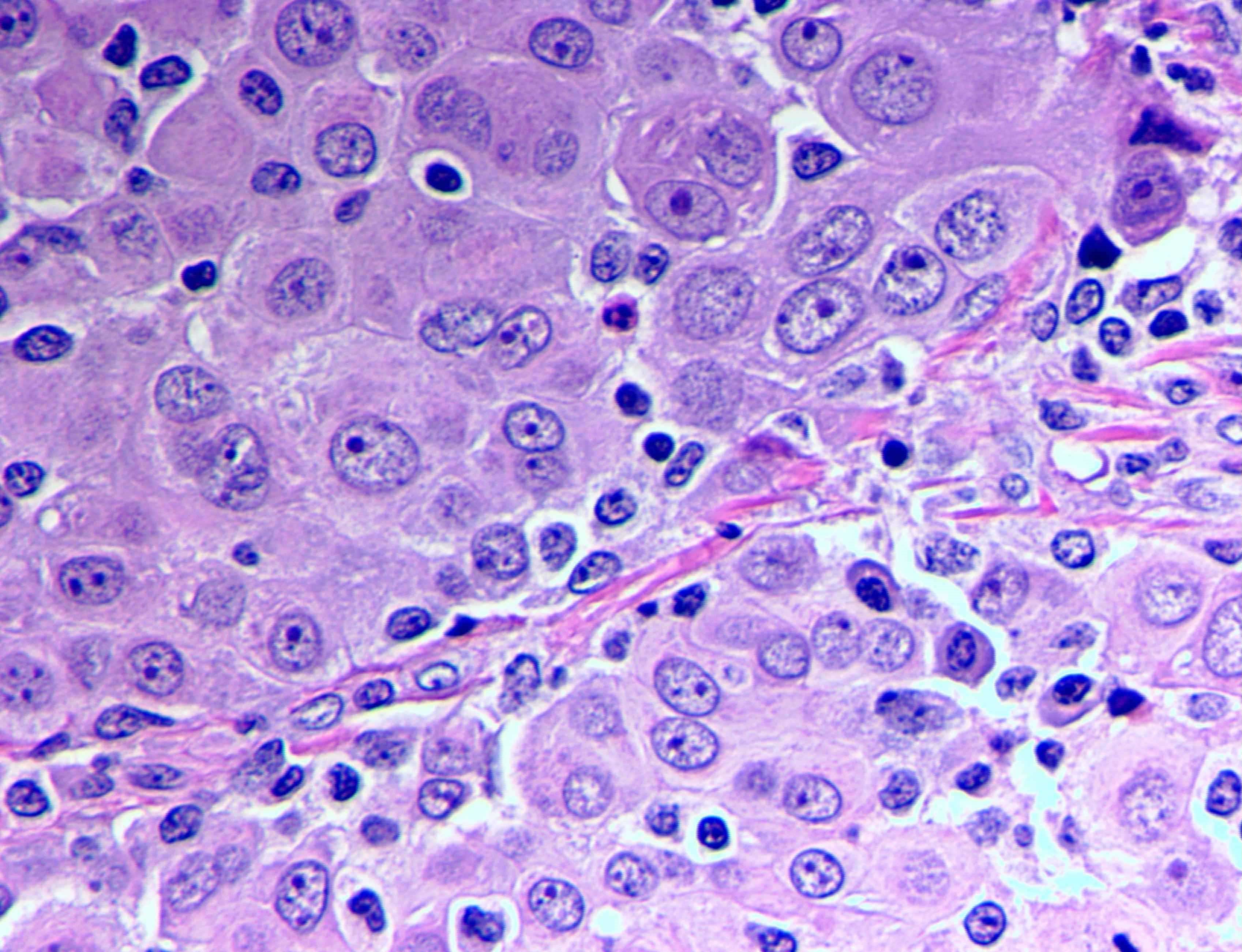
- Hallmark of epithelioid mesothelioma is the epithelioid cells which are polygonal cells with moderate to abundant eosinophilic cytoplasm, vesicular round nuclei and prominent nucleolus; often mimic nonneoplastic, reactive mesothelial cells (Arch Pathol Lab Med 2018;142:89)
- The most common histologic patterns of epithelioid mesothelioma are tubulopapillary, adenomatoid, solid well differentiated, solid poorly differentiated and acinar (Arch Pathol Lab Med 2012;136:241)
- Myxoid variant of peritoneal epithelioid mesothelioma is extremely rare with only 5 reported cases
Epidemiology
- Often due to asbestos exposure, whether in the pleura, peritoneum and pericardium; cumulative asbestos exposure is directly proportional to risk of cancer (J Med Case Rep 2008;2:121)
Pathophysiology
- Asbestos fibers lead to chronic inflammation, which causes the release of free radicals
- Latent period between asbestos exposure and disease averages 20 - 30 years (Cancer Treat Rev 2012;38:605)
Clinical features
- No distinctive symptoms, causing difficulties in diagnosis and treatment
- When symptomatic, usually present with abdominal pain, ascites and abdominal distention
Radiology images
Images hosted on other servers:

CT with dilated loops of bowel
Prognostic factors
- Prognosis is poor
- Survival rate of myxoid variant appears to be better than epithelioid mesothelioma in general (Virchows Arch 2005;447:828)
- Suggested favorable prognostic factors are small nuclear size and low Ki67 labeling index (World J Gastroenterol 2009;15:4856, Med Mol Morphol 2010;43:53)
Case reports
- 34 year old woman with epithelioid mesothelioma after radiation for cervical cancer (Mol Clin Oncol 2018;8:302)
- 44 year old woman with extensive myxoid change in well differentiated papillary mesothelioma (Ann Diagn Pathol 2002;6:164)
- 59 year old man with abdominal bloating and vague abdominal pain (Case #441)
- 60 year old woman with myxoid variant of epithelioid malignant mesothelioma (Cesk Patol 2014;50:149)
- 76 year old woman with small bowel obstruction secondary to carcinomatosis caused by primary peritoneal mesothelioma (Am J Case Rep 2015;16:496)
Treatment
- Systemic chemotherapy
- Cytoreductive and palliative surgery
Clinical images
Images hosted on other servers:

Fig D: bilateral
intratubal masses
Gross description
- Diffuse thickening or multiple nodules on the peritoneum
- Myxoid variant is gelatinous
Microscopic (histologic) description
- Invasive epithelioid cells are arranged in different patterns (which form the basis of the different subtypes of epithelioid mesothelioma)
- Form tubules and papillae with / without psammoma bodies (tubulopapillary variant), gland-like structures (acinar variant) and are in solid sheets, nests or cords (solid variant)
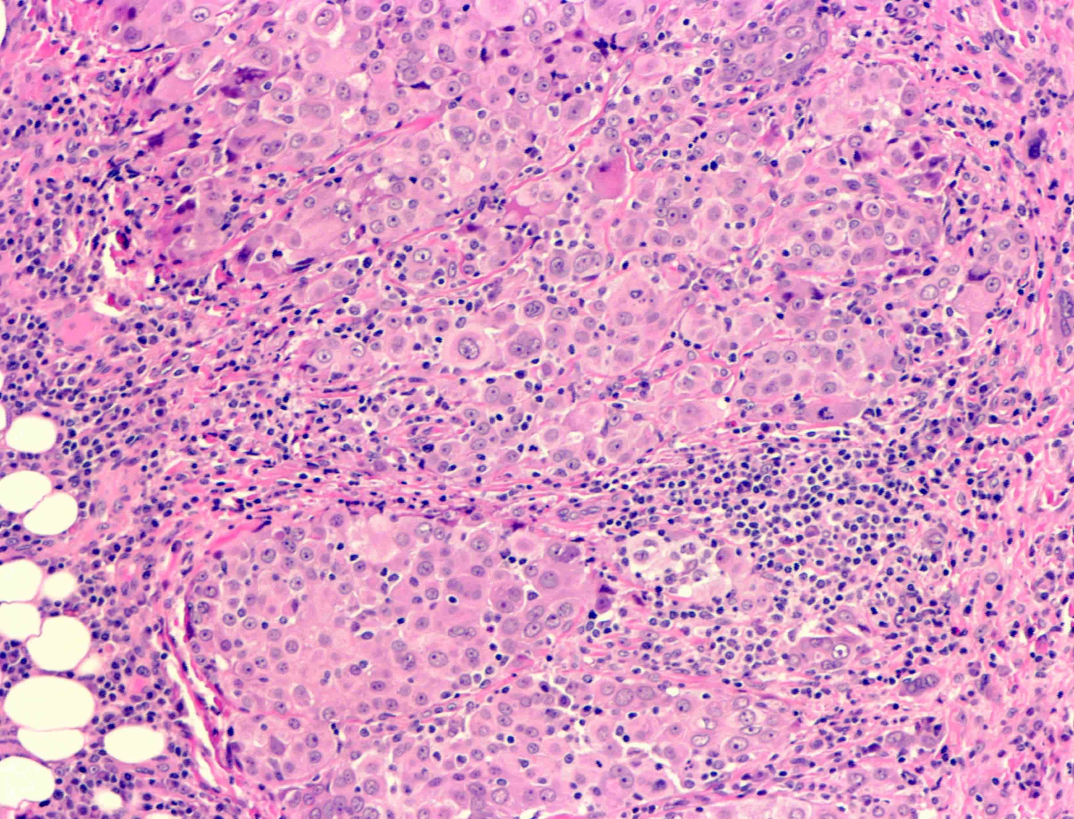
- Myxoid variant:
- Dyscohesive medium to large epithelioid cells with a moderate to abundant amount of eosinophilic cytoplasm dispersed in a myxoid background
- Some cells can have intracytoplasmic clear vacuoles
- Nuclei with coarse chromatin and prominent nucleoli
- Mitotic figures are usually inconspicuous
- Difficulty to differentiate from other myxoid lesions of the peritoneum (e.g. adenocarcinoma) thus panel of immunohistochemical markers is generally required
Microscopic (histologic) images
Contributed by Aysha Mubeen, M.D.
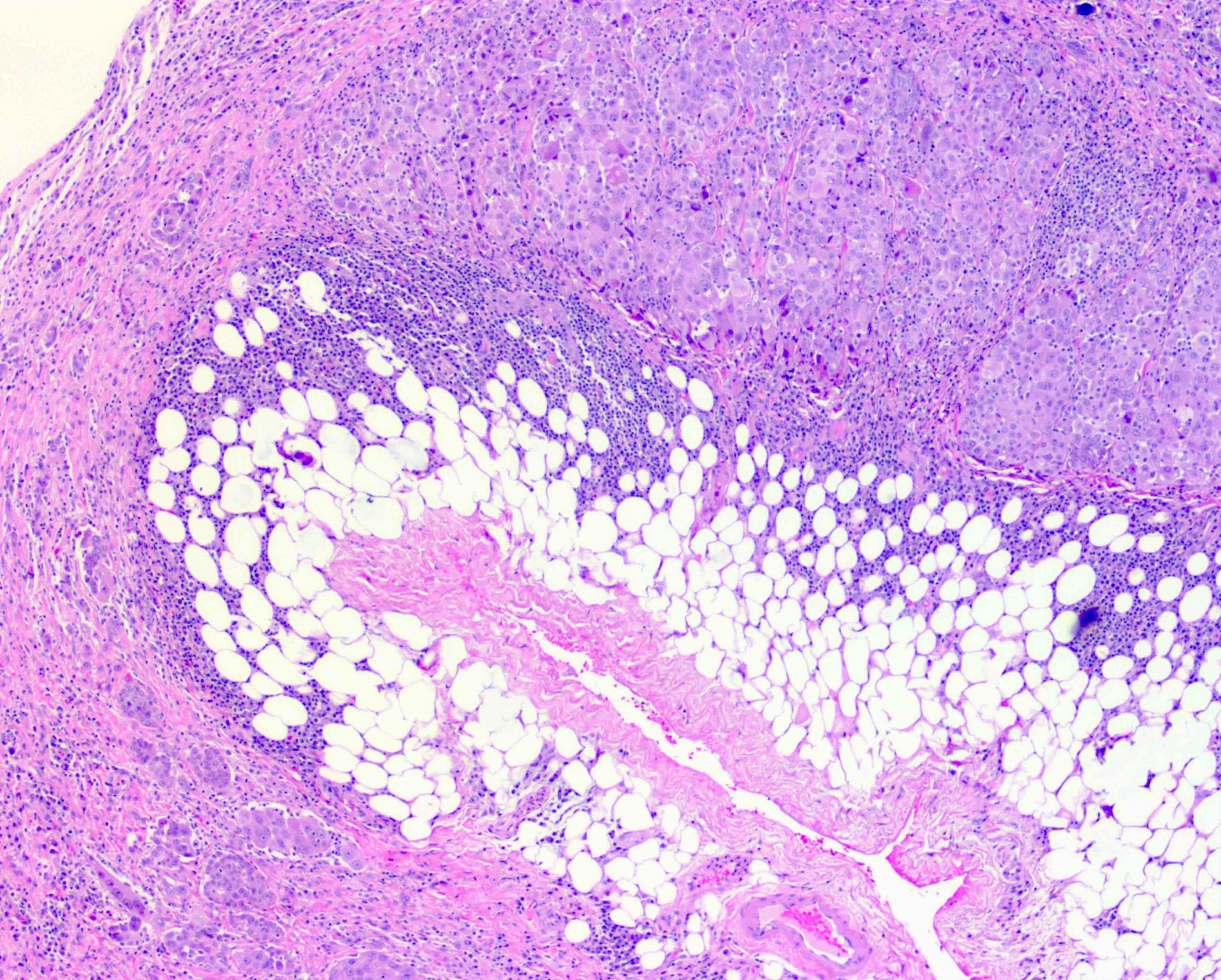
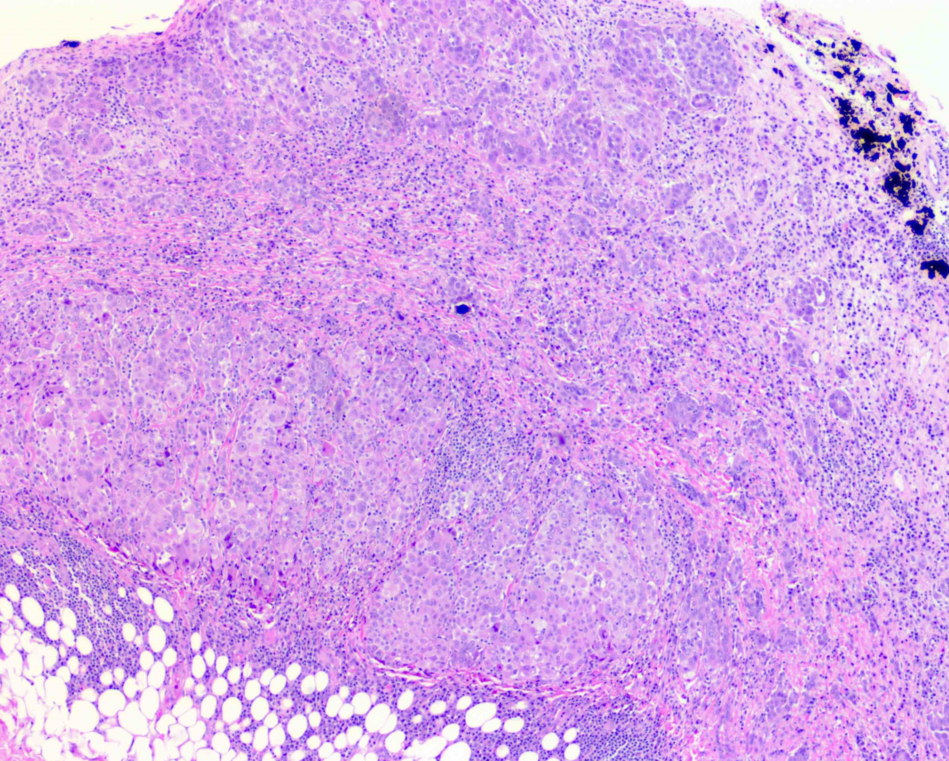
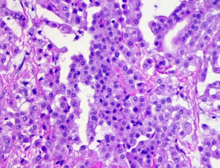
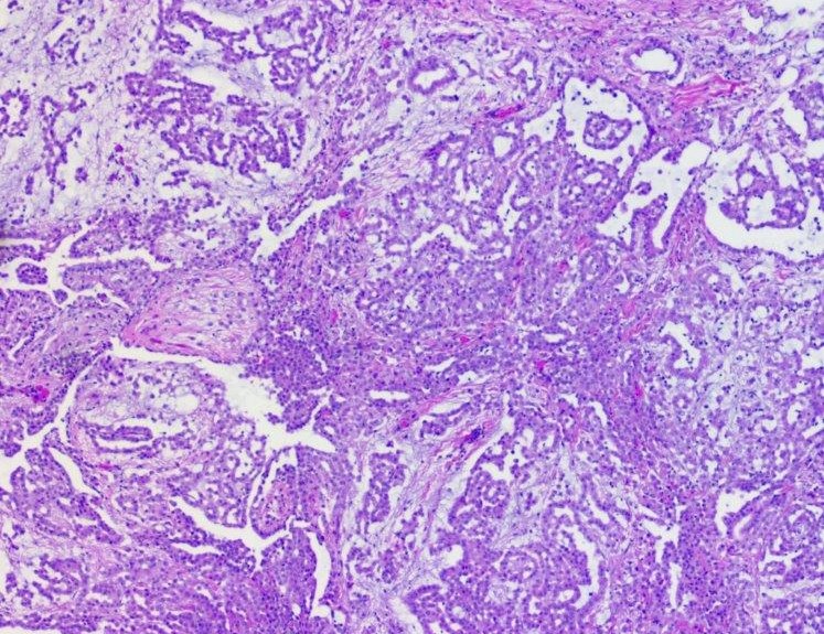
Epithelioid mesothelioma (pleural)
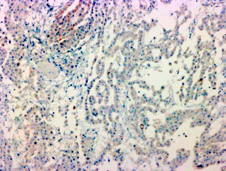
MOC31
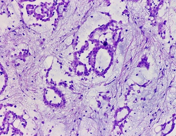
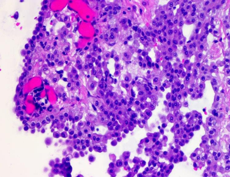
Myxoid variant of peritoneal mesothelioma
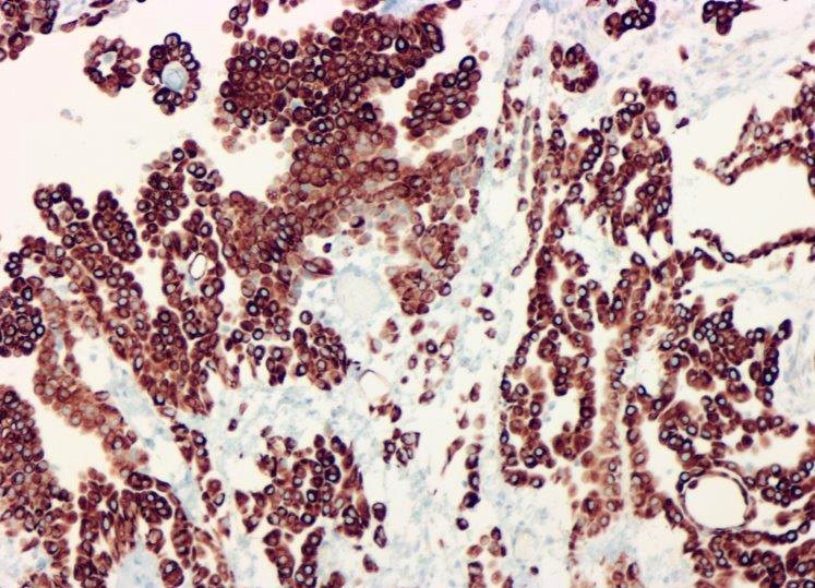
Calretinin
Virtual slides
Images hosted on other servers:


Epithelioid mesothelioma: 70 year old man with pleural effusion
Cytology description
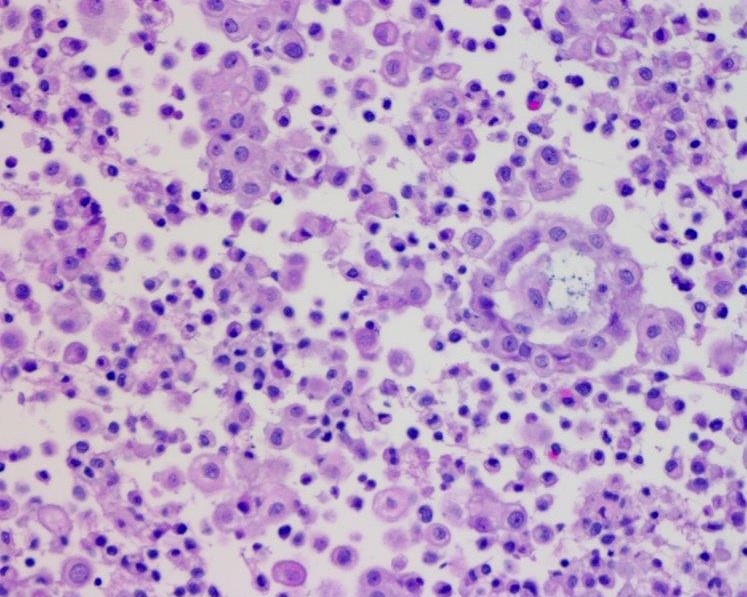
- Clusters of epithelioid cells (morulae) with knobby contour
- Abundant cytoplasm, round nuclei and prominent nucleoli
- Mild atypia
Cytology images
Contributed by Aysha Mubeen, M.D.
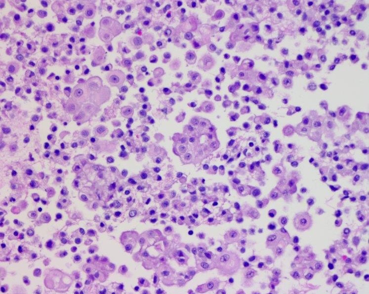

Cell block, myxoid variant
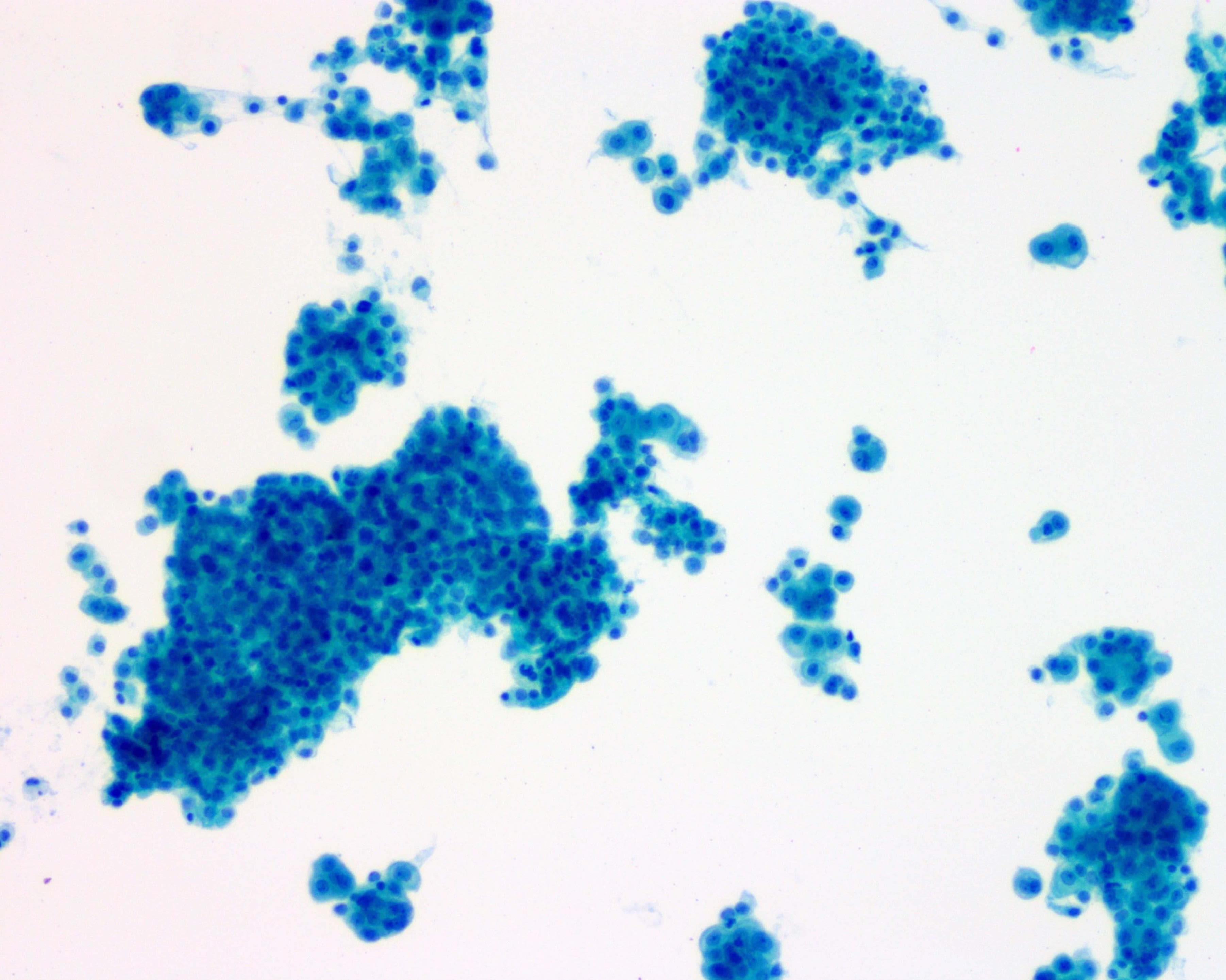
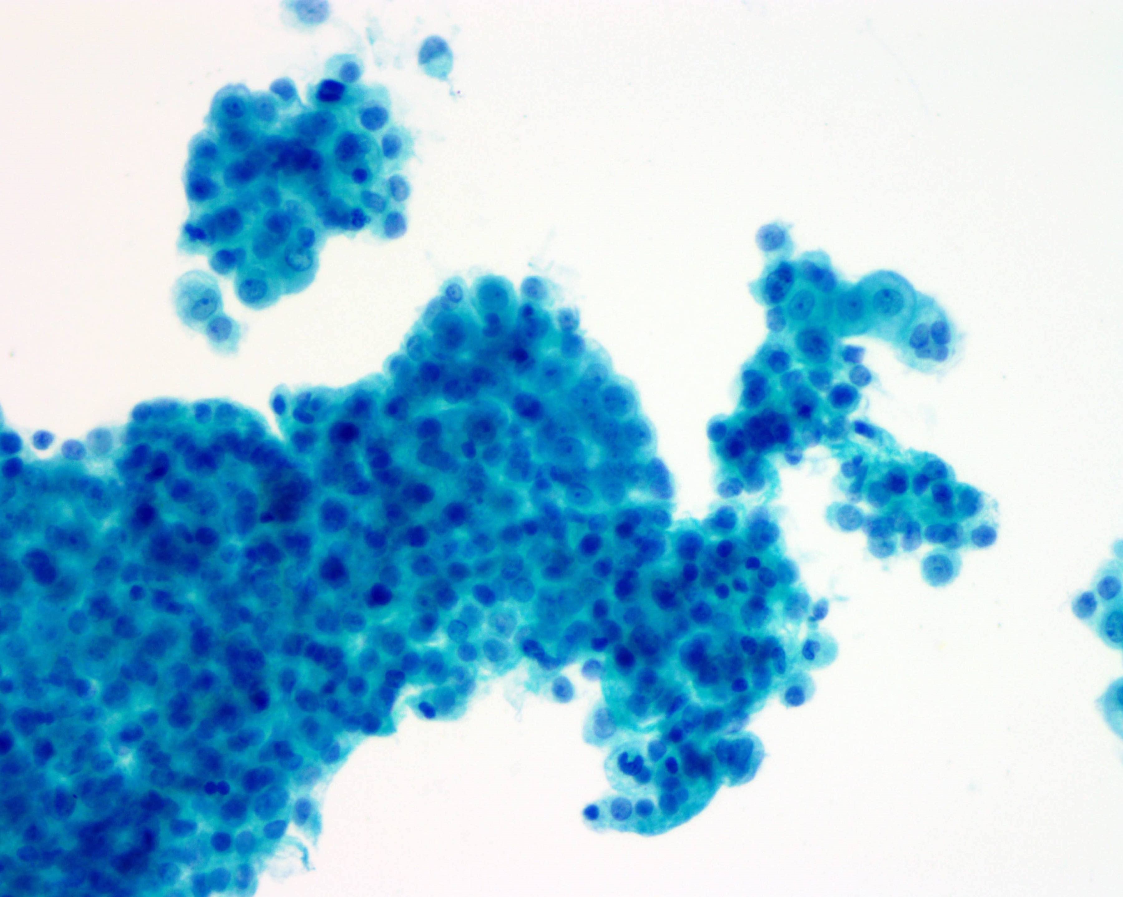
ThinPrep, myxoid variant
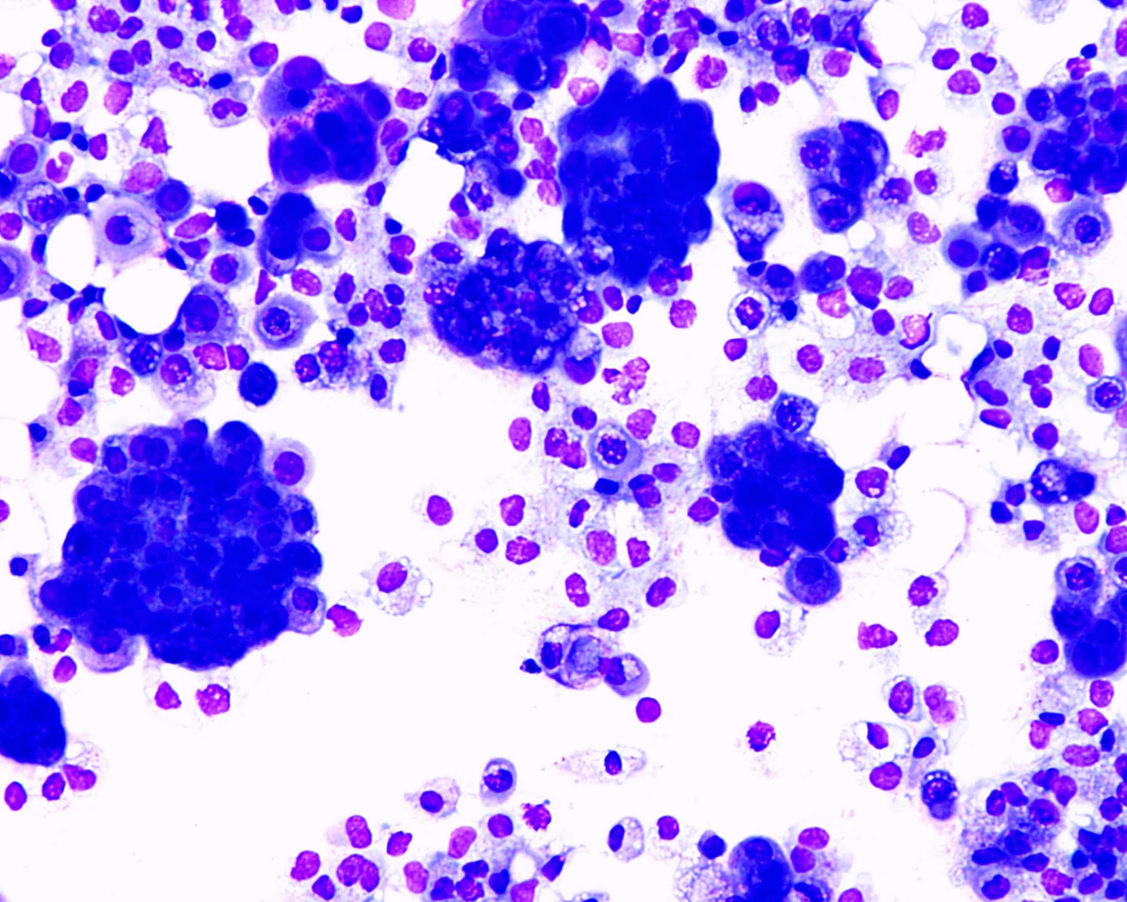
DiffQuik, myxoid variant
Electron microscopy description
- Very long, thin apical microvilli and the absence of glycocalyx (compared to adenocarcinoma, which has shorter villi)
Sample pathology report
- Peritoneum, resection:
- Multifocal epithelioid mesothelioma (largest focus 4.5 cm) (see comment)
- Margins of resection unremarkable.
- Comment: There is not currently an AJCC TNM cancer staging system for peritoneal mesothelioma. Immunohistochemical stains for calretinin and D2-40 are positive in the tumor.
Differential diagnosis
- Mucinous adenocarcinoma
- Papillary serous carcinoma involving the peritoneum (Am J Surg Pathol 2007;31:1139)
- Pleura - mesothelioma versus adenocarcinoma
- Pseudomyxoma peritonei
Additional references
Board review style question #1
Which of the following stains is positive in the myxoid variant of epithelioid mesothelioma and helps to differentiate it from mucinous adenocarcinoma?
- B72.3
- Claudin4
- D2-40
- MOC31
Board review style answer #1
