
Salivary glands
Primary salivary gland neoplasms
Benign
Lymphadenoma
Author: Adriana Handra-Luca, M.D., Ph.D.

Last author update: 1 September 2012
Last staff update: 1 December 2021
Copyright: 2003-2024, PathologyOutlines.com, Inc.
PubMed Search: Lymphadenoma[TI] salivary
Table of Contents
Definition / general | Clinical features | Case reports | Treatment | Gross description | Gross images | Microscopic (histologic) description | Microscopic (histologic) images | Cytology description | Cytology images | Positive stains | Negative stains | Molecular / cytogenetics description | Differential diagnosisCite this page: Handra-Luca A. Lymphadenoma. PathologyOutlines.com website. https://www.pathologyoutlines.com/topic/salivaryglandssebaceousadenoma.html. Accessed April 24th, 2024.
Definition / general
- Rare benign tumor with nests and islands of bland epithelium composed in part of sebaceous elements, with lymphoid stroma
- Called sebaceous lymphadenoma if lymphoid stroma is prominent
- Also called benign lymphoepithelial cyst with sebaceous differentiation
Clinical features
- 0.1% of salivary gland neoplasms, < 0.5% of salivary adenomas
- Over 90% occur in or near the parotid gland or in minor salivary glands
- May arise from salivary duct inclusions within parotid lymph node, similar to Warthin tumor (Am J Clin Pathol 1980;74:683)
- Fine needle aspiration cytology identifies a benign process but usually not diagnosed prior to excision (Acta Otorhinolaryngol Ital 2007;27:144)
Case reports
- 53 year old woman with progressively enlarging, painless parotid mass (University of Pittsburgh: Parotid Gland Mass [Accessed 19 March 2018])
- 57 year old man with neck mass (Arch Pathol Lab Med 2005;129:e171)
- 67 year old woman with parotid mass (Acta Otorhinolaryngol Ital 2007;27:147)
- 68 year old woman with synchromous squamous cell carcinoma (World J Surg Oncol 2003;1:30)
- 86 year old man with stable parotid mass (Case #103)
- Benign tumor with transition to sebaceous adenocarcinoma (Eur Arch Otorhinolaryngol 2006;263:940)
Treatment
- Excision is curative, no / rare recurrences, rare malignant transformation (Eur Arch Otorhinolaryngol 2006;263:940)
Gross description
- Solid or cystic, well circumscribed, tan-yellow mass, up to 3 cm, with variable encapsulation
Gross images
Case #103
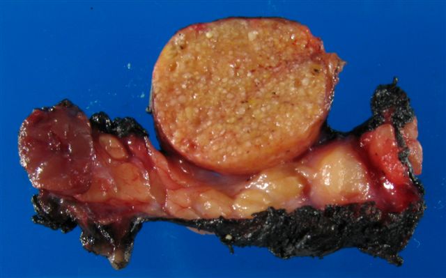
Large nodule is
sebaceous adenoma,
small nodule on left
is oncocytoma
Images hosted on other servers:


Parotid gland masses
Microscopic (histologic) description
- Benign, encapsulated, solid and cystic (Mod Pathol 2012;25:26)
- Nests and islands of benign squamous cells, often lining a cyst; epithelial nests have focal sebaceous differentiation
- Background is prominent lymphoid infiltrate, often with germinal centers
- May be associated foreign body reaction, histiocytes, oncocytic change
- Rarely malignant change to sebaceous carcinoma
- May be combined / hybrid / synchronous tumors with Warthin tumor, acinic cell carcinoma, adenoid cystic carcinoma, squamous cell carcinoma (Eur J Cancer B Oral Oncol 1996;32B:251, Pathol Res Pract 1993;189:577, J Oral Surg 1979;37:826)
Microscopic (histologic) images
Case #103
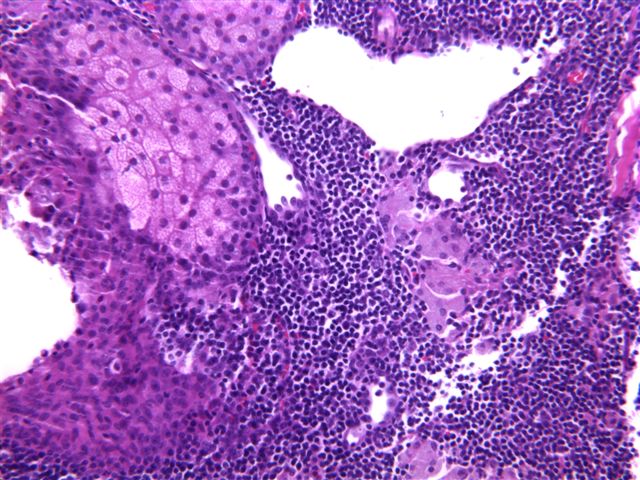

Large nodule
Images hosted on other servers:


Benign parotid mass with sebaceous features and lymphoid stroma

Parotid gland mass


With synchronous squamous cell carcinoma
Cytology description
- Mixed population of large and small lymphocytes, plasma cells and occasional tingible body macrophages
- 3 dimensional, cohesive aggregates of epithelial cells, often with cytoplasmic vacuoles characteristic of sebaceous differentiation, surrounded by layers of basaloid cells (Acta Cytol 2004;48:551)
Cytology images
Case #103
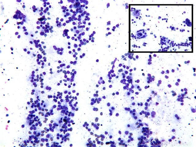
Diff-Quik touch prep
Molecular / cytogenetics description
- May have trisomy 9 (Oncol Rep 1994;1:561)
Differential diagnosis
- Low grade mucoepidermoid carcinoma: epithelial islands, ducts and cysts tend to be haphazardly distributed with variable shapes and sizes; usually infiltration of connective tissue or parenchyma; cells have some atypia, cells are mucin+
- Normal sebaceous glands: present in 10% of parotid glands but no mass
- Warthin tumor: prominent cysts and lymphoid stroma, cysts have bilayered oncocytic epithelium
