

Skin nontumor
Vesiculobullous and acantholytic reaction patterns
Epidermolysis bullosa acquisita
Author: Nat Pernick, M.D.

Last author update: 1 July 2011
Last staff update: 11 November 2020
Copyright: 2002-2024, PathologyOutlines.com, Inc.
PubMed Search: Epidermolysis bullosa acquisita
Table of Contents
Definition / general | Etiology | Clinical features | Treatment | Microscopic (histologic) description | Immunofluorescence images | Positive stains | Videos | Differential diagnosisCite this page: Pernick N. Epidermolysis bullosa acquisita. PathologyOutlines.com website. https://www.pathologyoutlines.com/topic/skinnontumoreba.html. Accessed April 20th, 2024.
Definition / general
- Rare, noncongenital, autoimmune, chronic listering disease of skin and mucus membranes (eMedicine)
Etiology
- Usually IgG autoantibodies against NC1 (noncollagenous domain of type VII collagen), major component of anchoring fibrils that connect basement membrane to dermal structures; also antibodies to central triple helical (collagenous) domain of type VII collagen and IgA antibodies instead of IgG
Clinical features
- Occurs at any age, usually affects elderly
- Blisters, scars and milia at trauma prone areas
- Some patients have generalized inflammatory skin blister phenotype
- Chronic disorder with partial remissions and exacerbations
- Causes significant morbidity but death due to disease is rare
Treatment
- Corticosteroids and immunosuppressants but relatively resistant to treatment
Microscopic (histologic) description
- Subepidermal blister with mixed inflammatory cell dermal infiltrate
- Often has bullous pemphigoid-like features (Acta Derm Venereol 2011;91:307)
Immunofluorescence images
Images hosted on other servers:
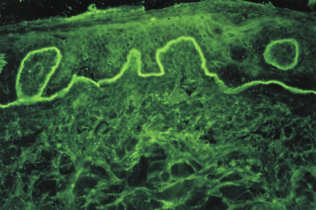
Direct immunofluorescence on perilesional skin: linear band of IgG along dermal-epidermal junction

Indirect immunofluorescence on salt-split normal human skin substrate using serum from affected patient: IgG autoantibodies on dermal side of basement membrane
Positive stains
- Direct immunofluorescence on perilesional skin shows linear band of IgG along dermal-epidermal junction
- Indirect immunofluorescence on salt-split normal human skin substrate using serum from affected patient shows IgG autoantibodies on dermal side of basement membrane
Videos
Epidermolysis bullosa acquisita
Differential diagnosis
- Bullous pemphigoid: mild dermal infiltrate including eosinophils; reactivity on epidermal side in NaCl split skin
