Table of Contents
Definition / general | Epidemiology | Sites | Etiology | Clinical features | Diagnosis | Laboratory | Prognostic factors | Case reports | Treatment | Clinical images | Microscopic (histologic) description | Microscopic (histologic) images | Videos | Differential diagnosis | Additional referencesCite this page: Carlquist EM, Gardner JM, Stuart LN. Impetigo contagiosa. PathologyOutlines.com website. https://www.pathologyoutlines.com/topic/skinnontumorimpetigocontagiosa.html. Accessed September 21st, 2023.
Definition / general
- Contagious superficial infection of skin
- 2 types of impetigo: impetigo contagiosa (nonbullous impetigo) and bullous impetigo
Epidemiology
- Nonbullous impetigo comprises > 70% of all cases of impetigo (An Bras Dermatol 2014;89:293)
- Primarily an endemic disease in school aged children but may occur in epidemics
Sites
- Occurs in areas of exposed skin, especially limbs and face (An Bras Dermatol 2014;89:293)
- May occur in normal skin or in areas of previous dermatoses (secondary impetigo) (An Bras Dermatol 2014;89:293, Am Fam Physician 2014;90:229)
Etiology
- In U.S., most common cause is currently Staphylococcus aureus
- Previously, most common cause was group A streptococci, either alone or in association with S. aureus (Pediatr Dermatol 1987;4:185)
- Infection with resistant strains, including methicillin resistant S. aureus (MRSA) is continually increasing (Pediatr Dermatol 2012;29:243)
Clinical features
- Early lesions: easily ruptured vesicopustules on an erythematous base
- Later lesions: ruptured lesions are replaced by thick, adherent, distinct golden yellow (honey colored) crust
- Satellite lesions due to self inoculation are common
Diagnosis
- Primarily a clinical diagnosis
Laboratory
- Swab for culture of lesions may rarely be helpful
Prognostic factors
- May resolve spontaneously after several weeks without scarring (Pediatr Dermatol 2012;29:243), but more severe cases require treatment
- Most common complication is poststreptococcal glomerulonephritis, occurs in up to 5% of those infected (Int J Dermatol 2003;42:251)
- There are conflicting opinions as to whether treatment of impetigo decreases risk of poststreptococcal glomerulonephritis (Calif Med 1966;105:113, Cochrane Database Syst Rev 2012;1:CD003261)
Case reports
- Newborn boy with impetigo contagiosa (BMJ Case Rep 2014 May 7)
- 9 year old boy with impetigo contagiosum and atopic dermatitis (Int Arch Allergy Immunol 2008;146:169)
Treatment
- Observation is reasonable in uncomplicated infections (Pediatr Dermatol 2012;29:243)
- Over the counter topical agents include bacitracin and neomycin (Pediatr Dermatol 2012;29:243)
- Topical disinfectants, including sodium hypochlorite baths, help decrease colonization (Pediatr Dermatol 2012;29:243)
- Topical antibacterials may minimize resistance and adverse effects from systemic agents (Pediatr Dermatol 2012;29:243); include mupirocin and retapamulin
- Oral antibacterials are indicated for infection of deeper structures including subcutaneous tissues and muscle fascia (cellulitis), fever, lymphadenopathy, pharyngitis, infections near oral cavity, scalp infections, > 5 lesions (An Bras Dermatol 2014;89:293)
Microscopic (histologic) description
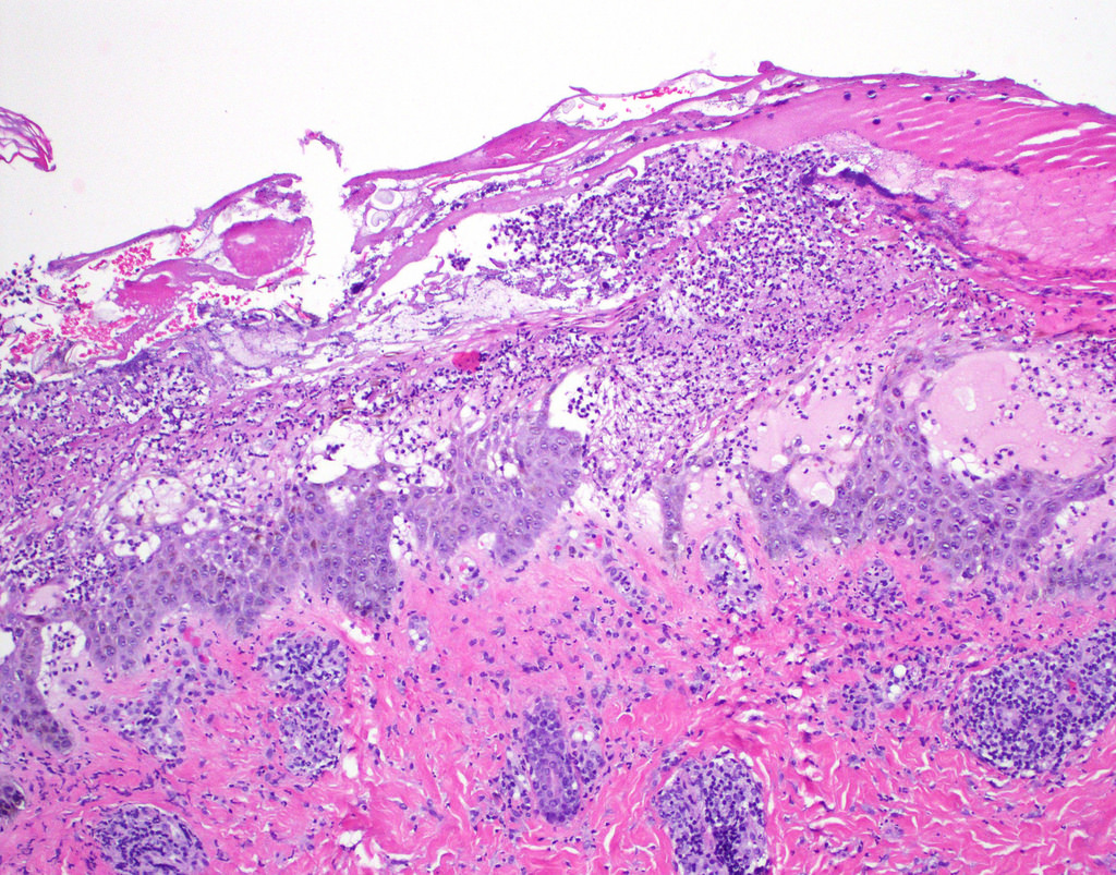
- Spongiosis of epidermis giving rise to vesicopustules in upper layers of epidermis
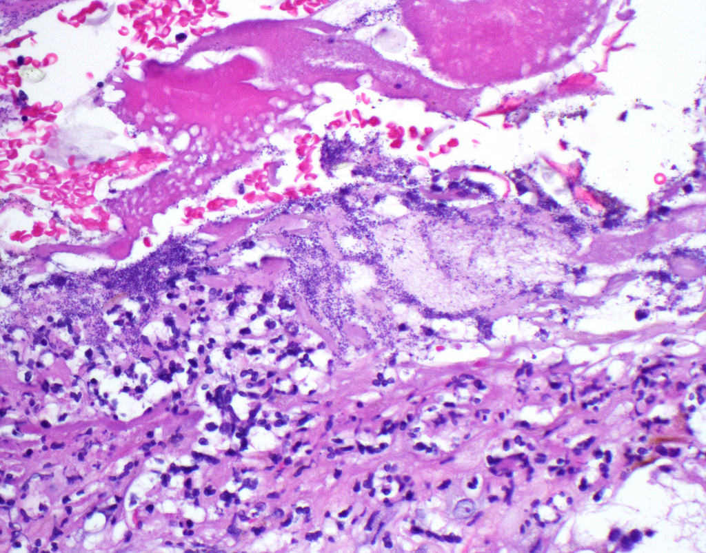
- As lesions progress, epidermis may become eroded and covered with a thick layer of serous crust with neutrophilic debris and Gram positive cocci
- Superficial dermis displays a moderately dense mixed inflammatory infiltrate
Videos
Impetigo contagiosa
Differential diagnosis
- Ecthyma: may be a variant of impetigo but lesions are more punched out, sharply demarcated and result in scarring; also due to superficial bacterial infection usually with Streptococcus pyogenes
- Secondary impetiginization: similar histologic changes to impetigo may be associated with various other skin conditions; represents secondary infection with cutaneous Gram positive cocci
Additional references