

Skin nontumor
Infestations
Onchocerciasis
Deputy Editor-in-Chief: Debra L. Zynger, M.D.

Last author update: 1 May 2018
Last staff update: 19 July 2021
Copyright: 2003-2024, PathologyOutlines.com, Inc.
PubMed Search: Onchocerciasis [title] Infestations AND (Humans[Mesh])
Table of Contents
Definition / general | Essential features | Terminology | Epidemiology | Sites | Etiology | Clinical features | Diagnosis | Case reports | Treatment | Clinical images | Microscopic (histologic) description | Microscopic (histologic) images | Differential diagnosis | Board review style question #1 | Board review style answer #1Cite this page: Wang J, Nagarajan P. Onchocerciasis. PathologyOutlines.com website. https://www.pathologyoutlines.com/topic/skinnontumoronchocerciasis.html. Accessed April 19th, 2024.
Definition / general
- Chronic dermatitis accompanied by progressive keratitis, uveitis and loss of sight caused by the filarial nematode, Onchocerca volvulus which is transmitted to humans through the bite of a blackfly (simulium species) (WHO: Onchocerciasis (river blindness) - disease information [Accessed 9 April 2018])
- Larval worms (microfilariae) migrate in the skin and the eye and lead to irreversible blindness and skin diseases
Essential features
- Caused by the filarial nematode Onchocerca volvulus, through the bite of a blackfly
- Dying larvae evoke focal inflammation resulting initially in dermal microabscesses followed by granuloma formation
- Pigmentation and atrophy of skin at advanced stage
- Treatment is a single dose of ivermectin (150 mcg / kg) every month for 3 - 6 months until patient becomes asymptomatic
Terminology
- Also called river blindness
Epidemiology
- 90% in sub-Saharan Africa
- Also found in Yemen as well as Central and Southern America
- 37 million people infected worldwide in 2006 (PLoS Med 2006;3:e260)
Sites
- Eye and skin
Etiology
- Antigens from dying parasites can activate innate immune responses (Br J Dermatol 2003;149:782, J Helminthol 2015;89:375)
Clinical features
- Onchocercal nodules are located close to bony prominences outside the inguinal and cervical regions (Postgrad Med J 2010;86:578 )
- Acute and chronic onchodermatitis: scattered pruritic papules, vesicles or pustules distributed over the shoulders, waist or buttocks (Int J Dermatol 2004;43:170)
- In advanced disease, the lesions can be spottily depigmented "leopard skin", scaly and atrophic "lizard skin" or thickened and hyperkeratotic "elephant skin"
- May also have lymphedema of the groin or "hanging groin" and skin atrophy (Br J Dermatol 1993;129:260)
Diagnosis
- Based on a history of exposure, clinical manifestations and supportive laboratory evidence of infection
- Skin snips are the gold standard to investigate the presence of microfilariae (Enferm Infecc Microbiol Clin 2017 Dec 20 [Epub ahead of print])
Case reports
- 5 year old girl with a subcutaneous nodule on forehead and histopathologic analysis of the nodule revealed the presence of Onchocerca volvulus worm (Pediatr Dev Pathol 2015;18:164)
- 7 year old girl with a conjunctival nodule (Neth J Med 2015;73:437)
- 9 year old girl with papular, indurated and itching skin lesions located on the limbs and positive anti-filarial antibodies in serum (Klin Padiatr 2006;218:41)
- 53 year old man presented with a 4 month history of intense, migratory urticaria (Int J Dermatol 2005;44:125)
- 60 year old woman developed leopard skin-like changes, rashes and pruritus on the left leg and onchocercal microfilariae were identified by a skin snip (Pan Afr Med J 2015;22:298)
Treatment
- Ivermectin (150 mcg / kg) administered orally as a single dose and repeated every 3 to 6 months until the patient is asymptomatic (Lancet 2002;360:203)
Clinical images
Images hosted on other servers:
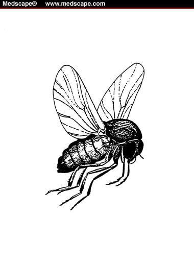
Blackfly (Simulium damnosum)
Microscopic (histologic) description
- At early stage, microfilariae concentrate in the papillary dermis with clusters of inflammatory cells surrounding vessels and adnexa
- Focal microabscesses and granuloma is evoked by dying larvae
- At advanced stages, secondary acanthosis, parakeratosis, pigment incontinence and melanophagocytosis appear
- Worms may be calcified or degenerated with dermal scaring and atrophy
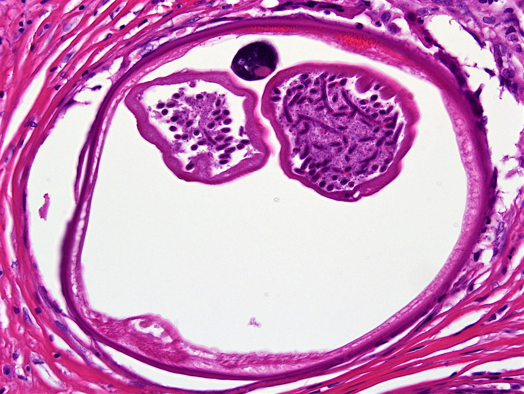
- Onchocercoma (onchocercal nodule) is a subcutaneous ball of worms embedded in inflammatory granulation tissue
- In cross - section, onchocerca typically has a cuticle, subjacent thin layer of muscle
- Within the lumen are paired uteri containing microfilaria (J Am Acad Dermatol 2015;73:947)
Microscopic (histologic) images
Contributed by Eddie Fridman, M.D.
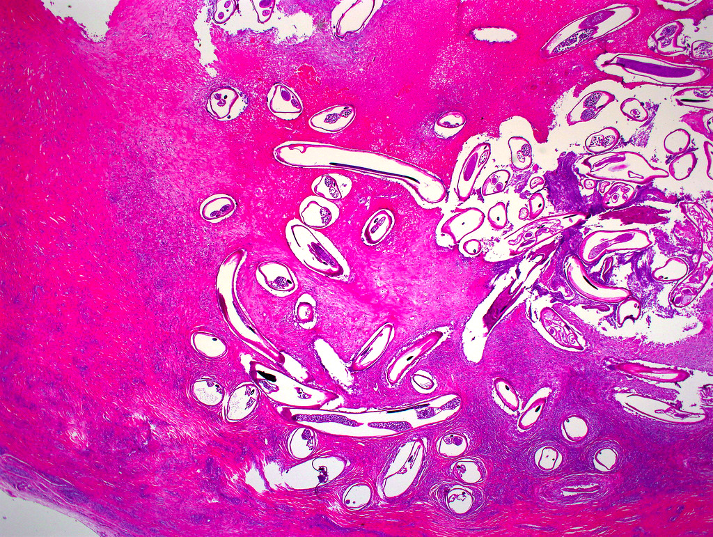
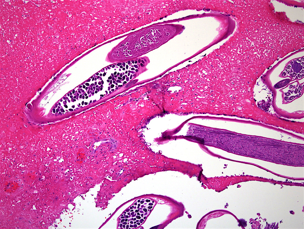
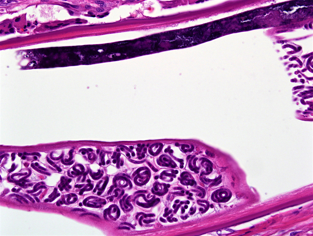
36 year old Ethiopian man with no previous history and a clinical diagnosis of inclusion cyst / pilomatrixoma of left thigh
Images hosted on other servers:


Figs C and D: Microfilariae of Onchocerca volvulus in tissue


Figs B and D: Adults of Onchocerca volvulus
Differential diagnosis
- Other helminthic diseases such as:
- Cestodes: cutaneous cysticercosis is characterized by a cyst containing purulent material containing parasites with prominent cuticle, scolices or hooklets (Indian Dermatol Online J 2012;3:135, Indian Pediatr 2015;52:715)
- Dirofilariasis: can also produce dermal / subcutaneous abscesses but they do not contain gravid uteri (Cutis 2015;95:131, Trop Doct 2009;39:189)
- However, the larval cuticle is characterized by ridges
- Hookworms: cutaneous involvement by hookworm infestation typically manifests as larva migrans (Eur J Clin Microbiol Infect Dis 2012;31:915)
- Schistosomiasis: high degree of clinical suspicion is necessary (An Bras Dermatol 2016;91:109)
- Histologic features include dermal mixed inflammatory infiltrate with variable numbers of eosinophils
- In early lesions, schistosomal eggs (with a lateral spine) may be identified, while later lesions are predominantly granulomatous and fibrotic
- Sparganosis:
sparganum larva has a thick eosinophilic cuticle with microvilli, two layers of smooth muscle, and a row of tegumental cells
- Larval parenchyma is edematous with basophilic calcareous bodies
Board review style question #1
- Which description of Onchocerciasis is not correct?
- A chronic dermatitis caused by the filarial nematode, Onchocerca volvulus which is transmitted to humans through the bite of a blackfly (simulium species).
- Dying larvae can evoke focal microabscesses followed by granuloma formation.
- Skin snips are the gold standard to diagnose Onchocerciasis.
- The disease occurs most commonly in South America.
- Treatment is a single dose of Ivermectin (150 mcg/kg) every 3 months.
Board review style answer #1
