

Skin nontumor
Infectious disorders
Tuberculosis
Last author update: 1 July 2011
Last staff update: 16 April 2021
Copyright: 2002-2024, PathologyOutlines.com, Inc.
PubMed Search: Tuberculosis [title] skin

Table of Contents
Definition / general | Epidemiology | Clinical features | Case reports | Clinical images | Microscopic (histologic) description | Microscopic (histologic) images | Positive stainsCite this page: Hamodat M. Tuberculosis. PathologyOutlines.com website. https://www.pathologyoutlines.com/topic/skinnontumortb.html. Accessed April 19th, 2024.
Definition / general
- Cutaneous disease is uncommon in U.S
Epidemiology
- Primary (inoculation) tuberculosis of the skin is the cutaneous analogue of the pulmonary ghon focus
- Primary cutaneous tuberculosis may be associated with tattooing, ritual circumcision, nose piercing, inoculation of homeopathic solutions or injury by contaminated objects to laboratory workers, surgeons or prosectors; there may be no obvious source of infection
- Erythema induratum of Bazin: also called nodular vasculitis if no coexisting tuberculosis; rare, more common in past; presents as recurrent tender subcutaneous nodules on calves of women with tuberculin hypersensitivity
- Lichen scrofulosorum: lichenoid eruption of minute papules in children and adolescents with tuberculosis (Indian J Dermatol Venereol Leprol 2010;76:494)
- Lupus vulgaris: most common form of re-infection tuberculosis, occurring predominantly in young adults; refers to painful cutaneous TB skin lesions with a nodular appearance, usually near the nose, eyelids, lips, cheeks and ears; untreated lesions may develop into disfiguring skin ulcers ("lupus" historically meant an ulcerative skin disorder), and are associated with squamous cell carcinoma
- Tuberculids: heterogeneous group of cutaneous lesions due to TB infection elsewhere in the body
Clinical features
- Red indurated papule appears 1 - 3 weeks after penetrating injury; papule ulcerates and forms a so-called "tuberculoid chancer"
Case reports
- Cases occuring after mesotherapy cosmetic treatment (Biomedica 2010;30:321)
Clinical images
Images hosted on other servers:

Adult woman with
erythrocyanotic
circulation and
ulcerated lesions
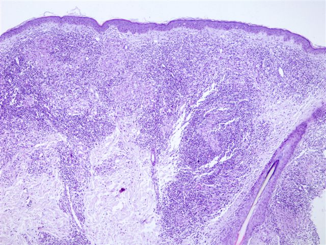
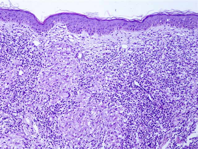
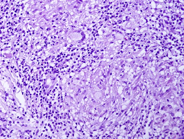
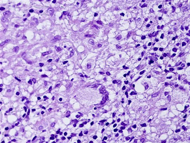
Microscopic (histologic) description
- Early lesions have a mixed dermal infiltrate of neutrophils, lymphocytes and plasma cells; followed by superficial necrosis and ulceration
- Weeks later, tuberculoid granulomas form, with variable caseation necrosis
- Acid-fast bacilli are usually easy to find in the early lesions, but are rare once granulomas develop
- Erythema induratum of Bazin: granulomatous vasculitis affecting subcutaneous large vessels (may need multiple serial sections to identify); also granulomatous inflammation of lobules of subcutis; lesions may contain acid-fast bacilli by PCR
Microscopic (histologic) images
Contributed by Angel Fernandez-Flores, M.D., Ph.D.
Positive stains
- Acid fast stains and PCR are positive
