

Skin melanocytic tumor
Melanoma
Sentinel node biopsy
Author: Christopher S. Hale, M.D.

Last author update: 1 May 2013
Last staff update: 3 November 2020
Copyright: 2002-2024, PathologyOutlines.com, Inc.
PubMed Search: Sentinel node biopsy melanoma
Table of Contents
Definition / general | Recommendations | Diagrams / tables | Prognostic factors | Clinical images | Microscopic (histologic) images | Positive stains | Molecular / cytogenetics description | Videos | Differential diagnosisCite this page: Hale CS. Sentinel node biopsy. PathologyOutlines.com website. https://www.pathologyoutlines.com/topic/skintumormelanocyticsentinelmelanoma.html. Accessed April 18th, 2024.
Definition / general
- Sentinel lymph node is defined as first extracutaneous target of lymphogenous tumor cell spread and potential source of subsequent lymph node metastases and distant metastases (eMedicine)
- Interval sentinel nodes: nodes receiving direct lymphatic drainage from primary site but lying between tumor and a recognized node field (Ann Surg Oncol 2011;18:3292)
- Lymphatic mapping and sentinel lymph node biopsy are widely used for staging
Recommendations
- Recommended for patients with intermediate thickness melanomas (Breslow thickness, 1 to 4 mm) of any anatomic site (J Clin Oncol 2012;30:2912)
- Tumor in sentinel node by H&E predicts recurrence (Mod Pathol 2007;20:427), usually leads to dissection of lymph nodes in affected nodal basin (Ann Surg Oncol 2008;15:1566), which reduces recurrence (Curr Treat Options Oncol 2008;9:243)
- Measuring antimony (originating from antimony sulfide colloid) can confirm sentinel nature of node (Mod Pathol 2004;17:1191)
Diagrams / tables
Images hosted on other servers:

Intraoperative lymphatic mapping and sentinel node
Prognostic factors
- Predictors of positive sentinel nodes: Breslow thickness < 1 mm (Int J Surg 2008;6:205); lymphatic invasion using D2-40, ulceration (Arch Dermatol 2008;144:462), younger age (Ann Surg Oncol 2008;15:630), low density of tumor infiltrating lymphocytes (TILs, J Clin Oncol 2012;30:2678), absence of regression (Ann Surg Oncol 2011;18:3593)
- Predictors of positive non sentinel nodes in sentinel node positive patients (occurs in 20 - 35%): amount of tumor in sentinel node, Breslow thickness of primary melanoma and density of dendritic WBCs in sentinel node paracortex (Mod Pathol 2004;17:747); also perinodal intralymphatic tumor (Ann Surg Oncol 2008;15:1723), depth of invasion in sentinel node (Ann Surg Oncol 2008;15:1202)
- Intraoperative touch imprints may be accurate (Anticancer Res 2008;28:465)
- Patients with triple negative (H&E, S100 / HMB45, RT-PCR) nodes have a markedly improved survival (Mod Pathol 2008;21:438)
- Micrometastasis: single metastasis < 2 mm are NOT associated with metastases in non sentinel nodes (J Surg Oncol 2008;98:46)
Selected protocols for sentinel nodes:
- 3 levels at 250 micrometer intervals, each level has 1 section stained with H&E, S100 and HMB45 (Am J Surg Pathol 2005;29:305) OR
- 2 H&E sections, S100 and HMB45 (Am J Surg Pathol 2003;27:1197)
- 4 sequential sections of both halves of each sentinel node examined - first and fourth sections stained with H&E, second section stained for S100 and third section for HMB45 (Semin Diagn Pathol 2008;25:100)
- Complete step sectioning of nodes with high gamma counts (Ann Surg Oncol 2008;15:1492)
Clinical images
Images hosted on other servers:

Lymphatic mapping
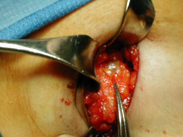
Intraoperative left
axillary node
seen after uptake
with blue dye
Microscopic (histologic) images
Images hosted on other servers:


D2-40+ lymphatic in tumor


Increased 11q13 (cyclin D1) by FISH

Tattoo pigment (not tumor)
Positive stains
- S100 and NKI-C3 are most sensitive stains for nodal metastases but are nonspecific; MART1 is most specific (Am J Surg Pathol 2001;25:1039)
- S100 and MART1 together are recommended (Hum Pathol 2004;35:217)
- Pattern of reticulin staining may help distinguish positive node from nodal nevus (Am J Dermatopathol 2013;35:452)
Molecular / cytogenetics description
- RT-PCR: prognostic significance of melanocytic mRNA in histologically negative nodes is controversial (significant - APMIS 2008;116:199, not significant - Mod Pathol 2007;20:427)
- RT-PCR detects significant sentinel node metastases that are missed by histopathology but overestimates number of patients with clinically significant metastases (Melanoma Res 2003;13:313)
- FISH detects chromosomal aberrations in 83% of nodal metastatic melanoma vs. 6% of nodal nevi (Am J Surg Pathol 2010;34:231)
Videos
Wide excision and sentinel node mapping
Differential diagnosis
- Antigen presenting interdigitating dendritic cells
- Benign nevus cells: usually in capsule, also parenchyma, no atypia, MART1+, S100+, HMB45- and Ki67- (Am J Surg Pathol 2002;26:1351, Am J Surg Pathol 2003;27:673)
- Histiocytes: no atypia
- Tattoo pigment, see Dermatol Online J 2005;11:14
