

Skin melanocytic tumor
Melanoma
Superficial spreading melanoma (low CSD melanoma)
Author: Bethany R. Rohr, M.D.
Deputy Editor-in-Chief: Jonathan D. Ho, M.B.B.S., D.Sc.

Last author update: 4 October 2023
Last staff update: 26 February 2024
Copyright: 2021-2024, PathologyOutlines.com, Inc.
PubMed Search: Superficial spreading melanoma
Table of Contents
Definition / general | Essential features | Terminology | ICD coding | Epidemiology | Sites | Pathophysiology | Etiology | Clinical features | Diagnosis | Prognostic factors | Case reports | Treatment | Clinical images | Microscopic (histologic) description | Microscopic (histologic) images | Positive stains | Negative stains | Videos | Sample pathology report | Differential diagnosis | Additional references | Board review style question #1 | Board review style answer #1 | Board review style question #2 | Board review style answer #2 | Board review style question #3 | Board review style answer #3Cite this page: Rohr BR. Superficial spreading melanoma (low CSD melanoma). PathologyOutlines.com website. https://www.pathologyoutlines.com/topic/skintumormelanocyticsuperficialspreadingmelanomalowCSD.html. Accessed April 18th, 2024.
Definition / general
- Superficial spreading melanoma (SSM) is the most common subtype of melanoma in the Western world (Arch Pathol Lab Med 2020;144:500)
Essential features
- Develops on sun exposed sites with low cumulative sun damage (CSD) (Arch Pathol Lab Med 2020;144:500)
- Has a radial growth phase (Arch Pathol Lab Med 2020;144:500)
- Most common mutation in superficial spreading melanoma is BRAFV600E (Arch Pathol Lab Med 2020;144:500)
Terminology
- Pagetoid melanoma (Arch Pathol Lab Med 2020;144:500)
ICD coding
Epidemiology
- Superficial spreading melanoma accounts for up to 56% of cutaneous melanomas (Br J Dermatol 2021;185:700)
- Low cumulative sun damage (Arch Pathol Lab Med 2020;144:500)
- Intermittent sun exposure throughout childhood
- Tanning bed use
- Increased total number of melanocytic nevi (Arch Pathol Lab Med 2020;144:500)
- Dysplastic nevi (Arch Pathol Lab Med 2020;144:500)
- Increased size of nevi (Arch Pathol Lab Med 2020;144:500)
Sites
- Back in men (Arch Pathol Lab Med 2020;144:500)
- Legs, calves in women (Arch Pathol Lab Med 2020;144:500)
Pathophysiology
- Mutation patterns overlap with other melanoma subtypes
- Low CSD melanoma (Arch Pathol Lab Med 2020;144:500)
- Mutations (Arch Pathol Lab Med 2020;144:500)
- BRAF most common
- V600E most common amongst BRAF
- NRAS
- TERT
- Biallelic inactivation of CDKN2A
- PTEN, TP53 in advanced primary melanomas
- BRAF most common
Etiology
- Low cumulative sun damage (Arch Pathol Lab Med 2020;144:500)
- Intermittent sun exposure and sunburns
Clinical features
- Irregular red, brown or black macule, patch, papule or plaque (Arch Pathol Lab Med 2020;144:500)
- ABCDE: asymmetry, border irregularity, color variation, diameter > 6 mm, other evolution history
Diagnosis
- Excisional biopsy
- Histopathologic diagnosis is gold standard
- Sentinel lymph node biopsy (SLNB)
- For primary tumors with Breslow depth of 0.8 mm or deeper
- Consider SLNB if < 0.8 mm with ulceration
- Breslow depth: measured from the top of the granular layer or base of ulceration to the deepest invasive melanoma cell
- For primary tumors with Breslow depth of 0.8 mm or deeper
- References: Ital J Dermatol Venerol 2021;156:300, NCCN: NCCN Guidelines - Melanoma: Cutaneous [Accessed 3 October 2023]
Prognostic factors
- Adverse prognosis indicators (Ital J Dermatol Venerol 2021;156:300)
- Thicker (Breslow) depth: most important
- Presence of ulceration
- Dermal mitoses
- Deeper anatomic (Clark) level of invasion (I-V)
- Presence of lymphovascular invasion
- Presence of neurotropism
- Increased risk of local recurrence
- Presence of sentinel lymph node biopsy (SLNB)
- Positive excision margins
- Report proximity of melanoma in situ or invasive melanoma to excision margins when able
Case reports
- 32 year old man with primary superficial spreading melanoma recurring with desmoplastic melanoma features (Virchows Arch 2022;480:945)
- 47 year old woman with superficial spreading melanoma developing within a nevus spilus (J Osteopath Med 2023;123:223)
- 78 year old man with a history of superficial spreading melanoma developing a malignant peripheral nerve sheath tumor (World J Clin Cases 2021;9:6457)
Treatment
- Treatment includes excision with or without sentinel lymph node biopsy; additional treatment based on stage (NIH: Melanoma Treatment (PDQ®) - Health Professional Version [Accessed 7 July 2023], NCCN: NCCN Guidelines - Melanoma: Cutaneous [Accessed 3 October 2023], American Cancer Society: Immunotherapy for Melanoma Skin Cancer [Accessed 3 October 2023])
- Wide local excision 1 - 2 cm
- SLNB for primary tumors with Breslow depth of 0.8 mm or deeper
- Consider SLNB if < 0.8 mm with ulceration
- No clinical or radiographic evidence of nodal disease
- Targeted or immunotherapy / checkpoint inhibitors
- Targeted therapies include BRAF and MEK inhibitors
- Immune checkpoint inhibitors include PD1, PDL1, CTLA4 and LAG3 inhibitors
- Talimogene laherparepvec (T-VEC) injections
- Interleukin 2
- Chemotherapy
- Radiation therapy
- Regular skin examination with a dermatologist
Clinical images
Images hosted on other servers:


Irregularly pigmented plaques


Dermoscopy
Microscopic (histologic) description
- Radial growth phase with or without vertical growth phase (Arch Pathol Lab Med 2020;144:500)
- Large epithelioid melanocytes
- Prominent junctional and intraepidermal component
- Irregular and enlarged nests of melanocytes along dermal - epidermal junction
- Junctional melanocyte confluence
- Dermal component is composed of similar appearing melanocytes that fail to mature and disperse with descent; dermal mitoses are noted
- Pagetoid scatter
- Presence of melanocytes above the basal layer
- If invasive
- Failure of dermal melanocytes to mature (i.e., become smaller) and disperse (i.e., smaller nests, single unit melanocytes) with descent into the dermis
- With or without dermal mitotic figures
- With or without melanin pigmentation
- Absent to moderate underlying solar elastosis
- Lacks severe solar elastosis
- Precursor or associated melanocytic nevus may be present (Arch Dermatol 2003;139:1620)
- Most frequent melanoma subtype to arise with a nevus
- Pathologic stage classification AJCC guidelines, eighth edition (2018) (Ital J Dermatol Venerol 2021;156:300)
- Tumor (Breslow) depth is the strongest predictor of clinical outcome, used for staging
- Measure vertically from the top of granular layer of the epidermis to the deepest invasive melanoma cells
- If ulcerated, measure from base of ulcer
- Round to nearest 0.1 mm using ocular micrometer
- Avoid measuring vascular invasion, microsatellites, involvement of skin appendages
- Tumor (Breslow) depth is the strongest predictor of clinical outcome, used for staging
Microscopic (histologic) images
Contributed by Bethany R. Rohr, M.D.
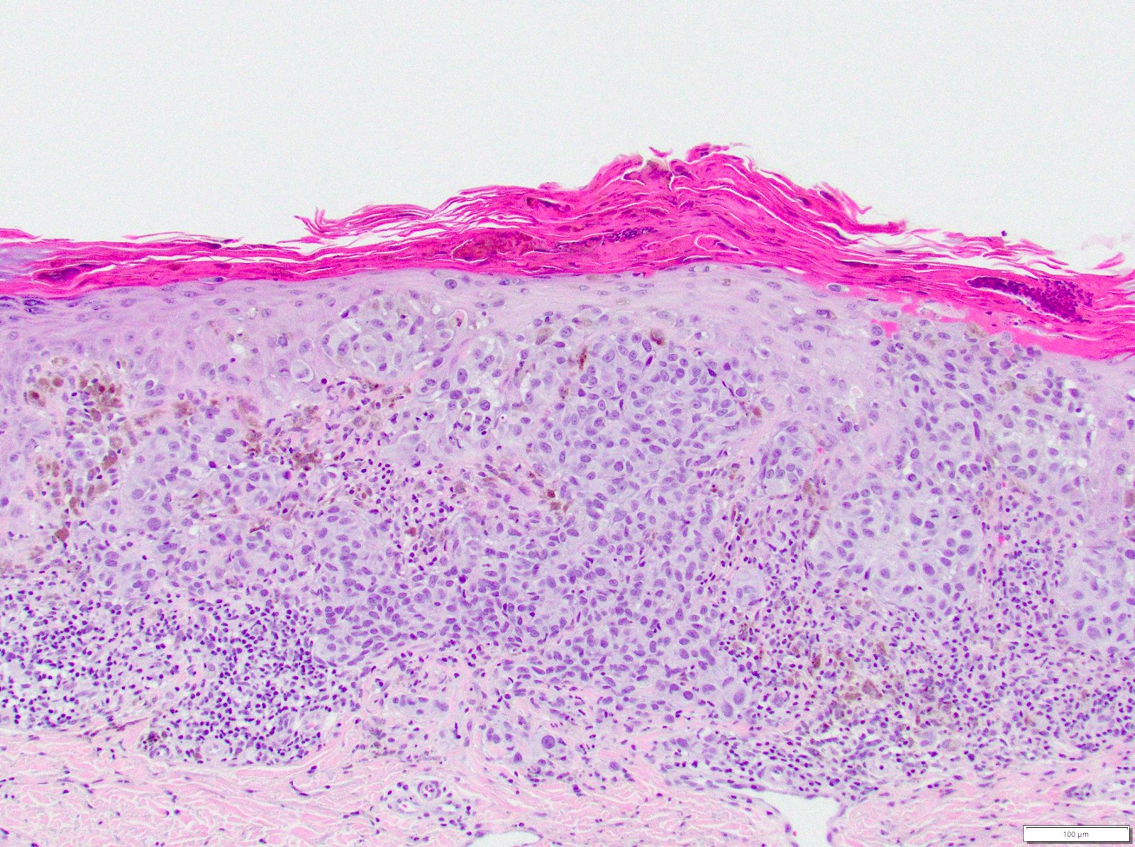
Epithelioid melanocytes

Pagetoid melanocytes

Poorly nested melanocytes

Atypical melanocytes

SOX10 immunostain

HMB45 immunostain
Positive stains
- SOX10 (nuclear)
- MART1, also known as MelanA (cytoplasmic)
- PRAME
- HMB45
- S100
- MITF
- Tyrosinase
- BRAF variable
- Reference: Semin Diagn Pathol 2022;39:239
Negative stains
- Loss of melanocytic markers is possible (Dermatopathology (Basel) 2021;8:359)
- Keratin immunostains
- CK, AE1 / AE3, OSCAR, CAM5.2
- Rare aberrant expression in melanoma (Dermatopathology (Basel) 2021;8:359)
- Neuroendocrine markers
- Neurofilament, glial fibrillary acidic protein, synaptophysin, chromogranin, CD56
- Rare aberrant expression in melanoma (Dermatopathology (Basel) 2021;8:359)
- Muscle specific markers
- Macrophage markers
- CD68, CD163
- Rare aberrant expression in melanoma (Dermatopathology (Basel) 2021;8:359)
- Vascular markers
- CD31, CD34
- Rare aberrant expression in melanoma (Dermatopathology (Basel) 2021;8:359)
- Hematopoietic markers
- CD4, CD20
- Rare aberrant expression in melanoma (Dermatopathology (Basel) 2021;8:359)
- Miscellaneous
- FLI1, GATA3, CEA, calretinin, PAX8, PAX2
- Rare aberrant expression in melanoma (Dermatopathology (Basel) 2021;8:359)
Videos
Superficial spreading melanoma
by Dr. Jerad Gardner
Sample pathology report
- Skin, biopsy:
- Invasive malignant melanoma, superficial spreading subtype, extending to a Breslow depth of ## (see comment)
- Comment: The sections reveal a severely atypical compound melanocytic proliferation composed of atypical epithelioid melanocytes. The melanocytes are poorly nested along the dermal - epidermal junction with confluence and pagetoid scatter. The dermal component is composed of similar appearing melanocytes that fail to mature and disperse with descent. Dermal mitoses are noted.
Differential diagnosis
- Lentigo maligna melanoma:
- High cumulative sun damage
- Significant dermal solar elastosis
- Pagetoid scatter is less prominent
- Radial growth phase
- More common on chronically sun exposed sites of elderly, light skinned adults
- High cumulative sun damage
- Acral lentiginous melanoma:
- Acral sites
- Noncumulative sun damage related
- Nevoid melanoma (Ital J Dermatol Venerol 2021;156:300):
- Resembles intradermal nevus, 2 types
- Papillomatous type
- NRAS mutation most common
- Head, neck, limbs
- Puffy shirt appearance: scanning magnification shows dense cellular aggregates surrounded by bent elongated rete ridges lined up side by side
- Maturing nevoid type: limbs, trunk
- Heterogeneous mutations: BRAF, NRAS
- Nodular melanoma:
- Low or high cumulative sun damage
- No radial growth phase
- Cutaneous involvement of metastatic malignant melanoma:
- History of prior locoregional invasive melanoma
- Features that favor metastases (Arch Pathol Lab Med 2020;144:500)
- Tumor size of < 2 mm
- Absence of tumor infiltrating lymphocytes and plasma cells
- Monomorphism
- Involvement of adnexal epithelium
Additional references
Board review style question #1
Superficial spreading melanoma is most commonly associated with which mutation?
- BRAF
- NRAS
- PTEN
- TERT
- TP53
Board review style answer #1
A. BRAF is the most commonly mutated driver oncogene in low cumulative sun damage melanoma. The most common mutation is the p. V600E. Answers B - E are incorrect because NRAS, TERT, PTEN and TP53 are less commonly mutated in superficial spreading melanoma.
Comment Here
Reference: Superficial spreading melanoma (low CSD melanoma)
Comment Here
Reference: Superficial spreading melanoma (low CSD melanoma)
Board review style question #2
From which of the following pathways does superficial spreading melanoma develop?
- Both high and low cumulative sun damage
- High cumulative sun damage
- Low cumulative sun damage
- No association with cumulative sun damage
Board review style answer #2
C. Low cumulative sun damage. Superficial spreading melanoma arises on sun exposed skin sites with little underlying solar elastosis. Answer B is incorrect since high cumulative sun damage is associated with lentigo maligna melanoma. Answer D is incorrect since superficial melanoma is associated with low cumulative sun damage. Answer A is incorrect because nodular melanoma may be associated with both high and low cumulative sun damage.
Comment Here
Reference: Superficial spreading melanoma (low CSD melanoma)
Comment Here
Reference: Superficial spreading melanoma (low CSD melanoma)
Board review style question #3
Which subtype of melanoma is most likely to arise in association with a precursor melanocytic nevus?
- Acral lentiginous
- Lentigo maligna
- Nodular
- Primary dermal
- Superficial spreading
Board review style answer #3
E. Superficial spreading melanoma is the most frequent histologic subtype of melanoma to be found arising in association with a melanocytic nevus. Other predictors of melanoma arising in association with a nevus include younger age and truncal location (Arch Dermatol 2003;139:1620). Answers A - D are incorrect because these other melanoma subtypes are less commonly documented to arise in association with a precursor melanocytic nevus.
Comment Here
Reference: Superficial spreading melanoma (low CSD melanoma)
Comment Here
Reference: Superficial spreading melanoma (low CSD melanoma)
