

Skin nonmelanocytic tumor
Lymphoma and related disorders (see also Lymphoma chapter)
Histiocytoses
Multicentric reticulohistiocytosis
Author: Nat Pernick, M.D.

Last author update: 1 August 2012
Last staff update: 15 July 2021
Copyright: 2001-2024, PathologyOutlines.com, Inc.
PubMed search: multicentric reticulohistiocytosis [title] skin
Table of Contents
Definition / general | Terminology | Epidemiology | Etiology | Clinical features | Case reports | Treatment | Clinical images | Microscopic (histologic) description | Microscopic (histologic) images | Positive stains | Negative stains | Differential diagnosisCite this page: Pernick N. Multicentric reticulohistiocytosis. PathologyOutlines.com website. https://www.pathologyoutlines.com/topic/skintumormultireticulo.html. Accessed April 26th, 2024.
Definition / general
- Rare disorder of women ages 40 - 50, with widespread cutaneous papules and nodules (eMedicine)
- Often associated with a destructive arthritis and internal malignancy
- Tumor cells are histiocytes and multinucleated giant cells containing abundant eosinophilic cytoplasm with a "ground glass" appearance
Terminology
- Formerly called lipoid dermatoarthritis
Epidemiology
- Usually women 40 - 50 years
Etiology
- Histiocytic origin (Am J Surg Pathol 1990;14:687)
Clinical features
- Skin lesions on the hands, especially at the base of the nails
- Lesions may also be on the face, ears, arms, scalp or mucosal surfaces
- "Coral beads" and vermicular erythematous lesions bordering nostrils are pathognomonic (J Eur Acad Dermatol Venereol 2001;15:524)
- Lesions vary from small papules to lesions several centimeters across, and are usually skin colored, yellow or reddish brown
- Recommended to screen patients for malignancy (Rheumatology 2008;47:1102), since accompanied by neoplasm in 28% of cases
- May be a paraneoplastic process (J Am Acad Dermatol 1998;39:864), or association with neoplasm may be due to reporting bias (eMedicine)
- Associated with destructive arthritis
Case reports
- 6 year old girl (J Rheumatol 1998;25:794)
- 33 year old woman with generalized systemic involvement (Clin Exp Dermatol 2004;29:373)
- 34 year old woman with papular lesions on hands and face (Case of the Week #153)
- 61 year old man with associated liver carcinoma (Case Rep Dermatol 2012;4:163)
- Relapse of multicentric reticulohistiocytosis before relapse of associated neoplasm (Med Pediatr Oncol 1985;13:273)
Treatment
- No consistently reliable treatment (Ryumachi 1993;33:68)
- TNF inhibitors (Arch Dermatol 2008;144:1360), aminobisphosphonates (Arthritis Rheum 2003;48:3538) and immunosuppressive drugs (Dermatol Online J 2009;15:2) have been effective only in individual cases
Clinical images
Images hosted on other servers:

Fingers


Hands

Ear

Various images

Destruction of knee articular surface

Erythematous patches on back
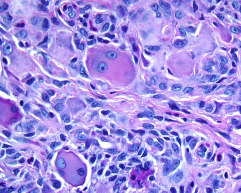
Microscopic (histologic) description
- Prominent oncocytic histiocytes and multinucleated giant cells with eosinophilic, “ground glass” cytoplasm
Microscopic (histologic) images
Case #153
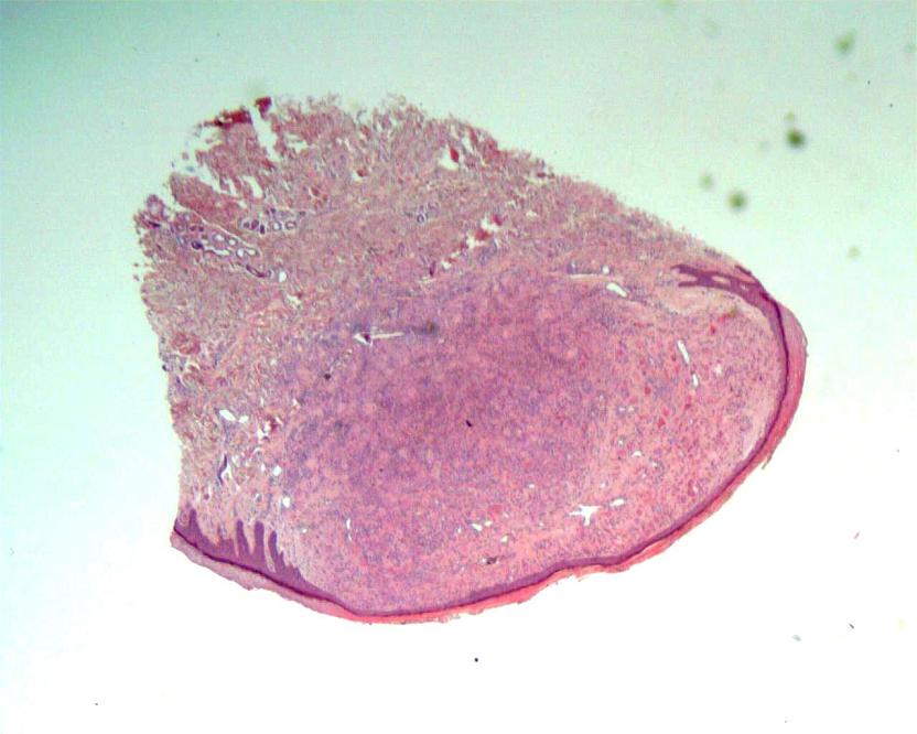
Low power
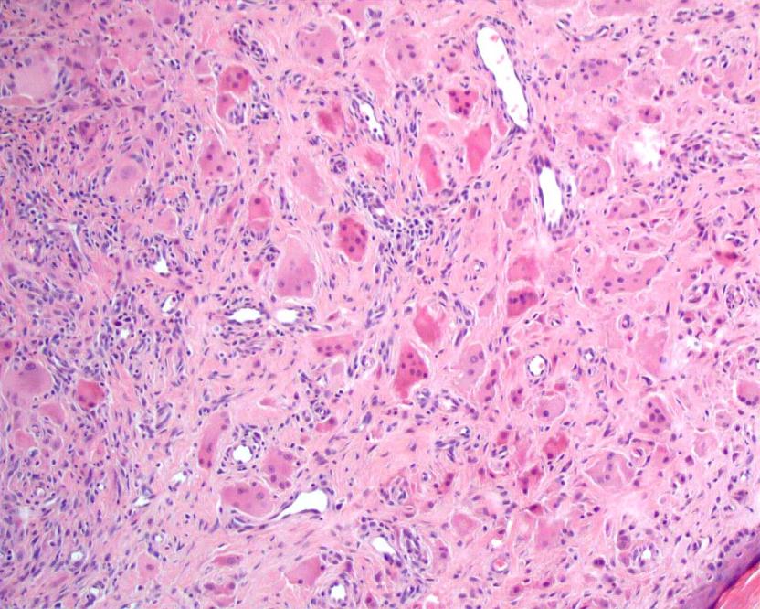
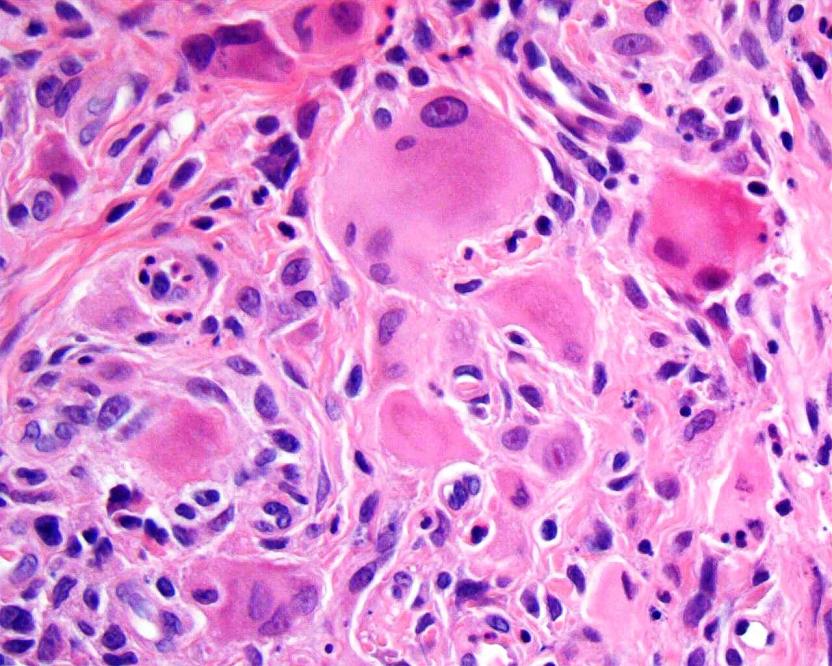
Medium / high power
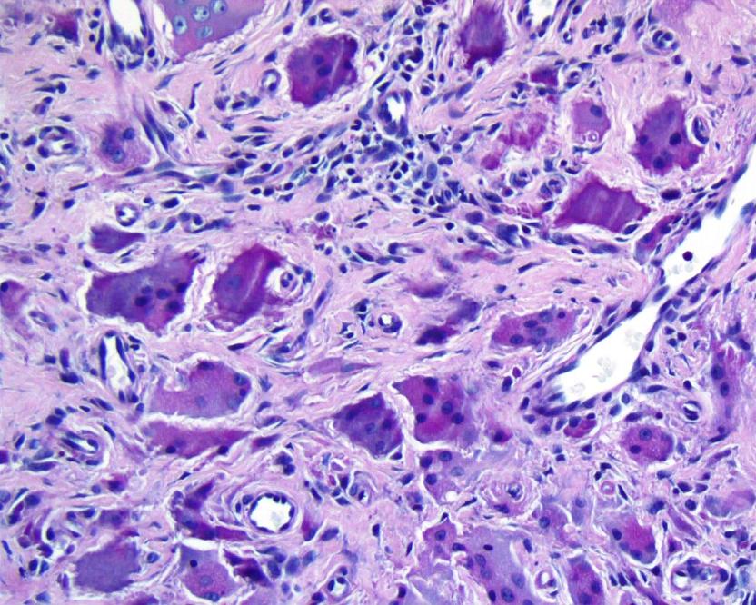
PAS
Images hosted on other servers:

Low power

Medium / high power

H&E, CD68 and vimentin
Negative stains
Differential diagnosis
- Solitary reticulohistiocytoma: younger age, solitary lesions usually not on digits or face (Am J Surg Pathol 2006;30:521)
- Epithelioid fibrous histiocytoma: usually < 1 cm on extremities, usually no giant cells, primarily myofibroblastic, not histiocytic
- Epithelioid sarcoma: deep seated tumor with markedly atypical cells that form granuloma-like clusters with central necrosis; tumor cells are EMA+, keratin+, CD68-
- Granulomatous inflammation: well formed granulomas and prominent lymphocytes, no large epithelioid histiocytes with eosinophilic glassy cytoplasm
- Histiocytic sarcoma: typically forms a large mass of epithelioid histiocytes with significant nuclear atypia and mitotic activity
- Juvenile xanthogranuloma: usually children, has scattered Touton type histiocytic giant cells and numerous eosinophils, but large epithelioid histiocytes are not prominent
- Rosai-Dorfman disease: associated with adenopathy; histiocytes are pleomorphic and S100+
